P- biopsychology
1/149
Earn XP
Description and Tags
Name | Mastery | Learn | Test | Matching | Spaced | Call with Kai | Chat |
|---|
No analytics yet
Send a link to your students to track their progress
150 Terms
phrenology
personality reflected in the lumps and bumps on the skull, reflecting functions of the brain
localisation
specific part of the brain in which a function is carried out
e.g. Broca’s area for speech production
corpus callosum
bundle of fibres that is essentially a communication pathway between two hemispheres
cutting the area can ensure epilepsy is kept to one side of the brain
one side of the brain can also be removed - a hemispherectomy
hemispheric lateralisation
when some physical or psychological functioning is controlled (or dominated) by a particular hemisphere
mostly contralateral, supported by stroke patients
left hemisphere
language processing
Broca’s and Wernicke’s area
areas in the brain (clockwise from front)
frontal
parietal
occipital
temporal
Frontal lobe
motor cortex
Broca’s area
motor cortex
both hemispheres
involved in regulating voluntary movement
sends signals to the muscles
Broca’s area
only left hemisphere
responsible for speech production
Parietal lobe
somatosensory centre
somatosensory centre
both hemispheres
separated from motor by a valley → ‘central sulcus’
adaptable people who read brail have larger area dedicated to their fingers
sensations: touch, pain, temp, pressure
occipital lobe
primary visual cortex
primary visual cortex
receives and process visual information
different regions: colour, shape, depth, movement
contralateral
temporal lobe
Wernicke’s area
primary auditory cortex
Wernicke’s area
only in the left
responsible for speech comprehension
primary auditory cortex
receives and processes auditory information
contralateral
Genie
dichotic listening tasks indicated she was using her right cerebral hemisphere to process language
Lennenburg 1967 suggested critical period for language acquisition (before puberty)
use of right hemisphere may have been direct consequence of not acquiring language in CP
Curtiss 1977 suggested left cerebral hemisphere was no longer available for language acquisition
but he adopted her so he’s biased
Oxana Malaya
raised by dogs, adopted a lot of their qualities
never learned language
claimed critical age was 5
Herasty 1997
females have larger Wernicke’s and Broca’s area than males
gender bias
beta bias → many studies downplay differences between M+F brains
Lebornge (Domanski 201)
suffered from epilepsy, lost ability to speak
Broca conducted post-mortem and found lesion on the left frontal lobe, only visible site of damage
concluded it was area responsible for speech - Broca’s aphasia
preserved and kept in Paris, scanned to confirm area is correctly localised
Wernicke
identified region in left temporal lobe as being responsible for language comprehension - Wernicke’s aphasia
nonsense words as part of speech
brain scans by Peterson 1988, Wernicke’s active in listening, Broca’s when reading
Meyer et al. 2010
when people watch silent films, their primary auditory cortex in both hemispheres will activate if a door is shut with a force - imagining the bang
Case study → GR
developed blindness in RVE, CT scan = haemorrhage
brain damaged in the visual cortex, cortically blind
could not identify letters but expressed awareness, blindsight
Phinneas gage
after incident: hostile, unreliable, rude, swore a lot
Dr Harlow believed that the damaged area housed the planning, reasoning and control of individual
Case study: EB
17yr boy, entire left hemisphere removed at 2.5yrs
surgery to remove huge benign tumour, immediate loss of his language abilities - suggesting that emerging language ability was already left-lateralised
this is the case for 95% of right handers)
intensive rehab, beyond age 5, EB’s language fluency improved, found that right hemisphere compensated
some areas not at expected standard, grammatical, slower at naming objects - loss compensated to a degree
factors: age, trauma, time spent in rehab=less time in school, individual differences
supports functional recovery
Viewing the brain holistically
Karl Lashey 1950 removed areas of the cortex (between 10% and 50% in rat’s brains; the rats were learning to run a maze, he did not find a specific area involved
concluded tat complex tasks are supported by the holistic theory of brain function
Left hemispheric lateralisation
localisation of function, language, is lateralised to the left hemisphere
Right hemispheric lateralisation
dominant in recognising emotions in others (Narumoto et al.)
if photo of face is split so one half is smiling and the other is neutral, left hand side emotion is recognised faster (Heller and Levy)
spatial relations
left hemisphere focuses on details and right processes overall patterns
neurosurgery
can be used in extreme cases of OCD and depression
Dougherty et al. reported on 44 OCD patients who had undergone cingulotomy
involves lesioning of the cingulate gyrus (inability to stop worry-cycles)
post surgical follow up at 32 weeks, 1/3 met criteria for successful response, 14% partial
supports localisation, but not fully as not majority
Brain plasticity
rehabilitation, particularly successful in children
EB case study challenges, but also supports as Hemispheric lateralisation can be compensated for at least to a basic degree
case studies provide evidence to support, not proof
hemispherectomy
half the brain removed, disconnected, or disabled
comisurotomy
corpus callosum severed to reduce symptoms of epilepsy - stops the electrical storm from travelling across
Sperry 1968: AIM
investigate the extent to which the two hemispheres were specialised for certain functions
Sperry 1968: PROCEDURE
compare split brain patients to others w no separation
Visual tasks: words projected into left/right visual field and asked about it
Tactile tasks: hands underneath screen, ‘feel’ only
Sperry 1968: SHOWN IN LVF
would not be able to describe as left visual field goes to right hemisphere which does not have comprehension and language centres
Sperry 1968: SHOWN IN RVF
would be able to describe
Sperry 1968: NORMAL BRAINS
information would be able to travel across corpus callosum to left hemisphere
Sperry 1968: SHOWN IN LVF, FIND OBJ W LEFT HAND
would be able to find as LVF→RH→LeftHand
Sperry 1968: SHOWN IN LVF, FIND OBJ W RIGHT HAND
would not be able to as LVF→ RH does not go to right hand
Sperry 1968: HOLDING OBJ, HIDDEN + TOLD TO FIND
would, but only with same hand, e.g. pen → lefthand → RH → lefthand → pen
Sperry 1968: SHOWN DIF WORDS TO BOTH F, PICK UP W LEFT AND SAY
key → LVF → RH → LeftHand
Ring → RVF → LH → say
Sperry 1968: DRAW PIC SHOWN TO BOTH VF
consistently better when drawn with left hand
right hemisphere was superior at drawing ability
Sperry 1968: CONCLUSIONS
hemispheres process information separately, have different functions, seem to have two separate streams of consciousness with own memories and perceptions
Sperry 1968: EVALUATION
scientific, objective, ethical, opportunity sample, internal validity
11 split brain patients - small sample, all Hx seizures may have a unique effect, control should be epileptic
disconnect greater in some than others, some on drug therapy longer
comparison had no history of drug therapy or epileptic seizures
data artificially produced, in real life corpus callosum can be compensated for
gazzinga 1970
corpus callosum severed shown picture of nude women in LVF, she giggled but couldn’t explain why
Plasticity
ability for the brain to change and adapt it’s structures and processes as a result of experience and new learning
used to limit to infancy and childhood, more research demonstrated the brain continues to create new neural pathways
Machin 2018 found that the fathers planning and problem solving areas became more active
Functional recovery
the way certain abilities of the brain may be moved or redistributed rather than lost following damage/trauma to the brain (RTAs, assaults, falls, strokes → very dependent on level of damage)
much recovery due to anatomical compensation, brought about by intensive rehabilitation, brain learns to compensate for function
plasticity → Maguire et al. 2000
greater volume of grey matter in the posterior hippocampus(spatial) in London black cab drivers than matched control group due to 3yr intensive training
plasticity → Draganski et al. 2006
medical students 3 months before and after final exams, learning induced changes in posterior hippocampus and parietal cortex
Plasticity → Kuhn 2014
supermario at least 30min/day over 2 months, compared to control and found significant differences in the grey matter, particularly in cortex and hippocampus (+spatial navigation, strategy, working memory, motor performance
Functional recovery → Scotty Cramer
professional BMXer, accident in 2016 = paralysis
extremely motivated, regained control within 10 months
ways of functional recovery
axonal sprouting/neural regeneration
growth of new nerve endings will connect with other undamaged nerve cells to form new neural pathways
can be used to strengthen existing connections or repair damaged parts TNS by repairing damaged neural pathways
Reformation of blood vessels, nourishing brain and strengthening connections
Recruitment of homologous areas on the opposite side of the brain to perform specific tasks, after period of time, may shift back
e.g. damage to Broca’s area on left side due to a stroke, right side equivalent carries out function
Evaluation of plasticity and functional recovery - age
decreases with age as greater propensity for reorganisation in childhood as constantly adapting
Bezzola et al. 2012, 40hr gold training = reduced motor cortex activity using fMRI in novice golfers 40-60
Evaluation of plasticity and functional recovery - practical application
neurorehabilitation grown; following trauma, spontaneous recovery tends to slow after number of weeks, so nr may be required to maintain improvements (fixes itself to an EXTENT)
Evaluation of plasticity and functional recovery - cognitive reserve
educational attainment influences how well brain functionality adapts after injury (socially sensitive)
Schneider et al. 2014, time in education=cognitive reserve, increases chance of disability free recovery, 40% achieved DFR >16yrs, 10% <12yrs
Evaluation of plasticity and functional recovery - chronic traumatic encephalopathy
from multiple traumas: professional sports, domestic violence, extreme sport, armed forces
develops slowly with: STM loss, cognitive decline, depression symptoms, poor impulse control
no treatment (aside from for individual symptoms)
diagnosed at post-mortem
methods of studying the brain
post-mortems
fMRI
EEGs
ERPs
Port mortem
analysis of a person’s brain following their death
in research, likely individuals who have a rare disorder and experience unusual deficits in behaviour or mental processes
often compared to neurotypical brains to establish likely cause
post mortem - strengths
informs theories of cognitive functioning
establishes causal relations between impaired brain and patient behaviour
play an essential role in developing understanding of conditions such as schizophrenia
more detailed examination of anatomical and neurochemical aspects
post mortems - limitations
single case studies
damage not usually one area or caused by other factors
modularity theories (localisation of function is a simplistic view of the brain)
have to treat the brain - process takes 2 to 3 working days (treated with formalin)
more invasive than scan
Einstein brain donation, findings unreliable as the(240) brain pieces have been handled a lot, may have effected brain
differences identified by comparing to much younger brain
correlation versus causation retrospective so unknown if differences contributed to intelligence or if intellectual activity caused differences
fMRI
measures energy released by haemoglobin (Hb-protein content of blood that binds with O2)
increase in activity = increase in oxygen = increased blood flow
gives dynamic 3D image of brain, can map involvement of areas (implications for localisation of function)
high spatial resolution: 1-2mm
poor temporal resolution: 5 seconds
fMRI - strengths
non-invasive
no radiation(physical health not at risk) unlike PT
objective and reliable measure of psychological processes
not possible for verbal reports
e.g. lie detection
EEG
skull cap with electrodes placed on scalp of individual to measure electrical activity within the brain
scan recording represents brainwave patterns
low spatial resolution
extremely high temporal - single ms or less
EEG - strengths
diagnosis of conditions(epilepsy) and understanding of ultradian rhythms
much cheaper by does require level if expertise to interpret
measure of brain functioning in real time, active brain, accurately measures task with brain activity associated
EEG - limitations
generalised nature of information received, not useful for pinpointing exact source of neural activity
cannot distinguish between but adjacent locations
cannot reveal deeper regions(e.g. hypothalamus) without invasive procedure
ERPs - event related potential
use same equipment as EEGs but uses statistical averaging techniques, all extraneous filtered out to leave responses to related stimulus
ERPs - strengths
increases understanding of cognitive processes - attention and perception
a lot more specific than EEGs
tests the reliability of self-report answers, especially when area of research is potentially sensitive and open to social desirability bias
ERPs - limitations
not easy to filter out background noise and extraneous material
can only be interpreted by trained professional (requires intensive and expensive training)
used ERP to record responses to nude pictures of both sexes, men said they were aroused by female and women said neutral but ERP showed higher response to opposite sex in both
biological rhythms
a change in body processes or behaviour in response to cyclic time periods, influenced by internal body clock and external environment (endogenous pacemakers and exogenous zeitgebers)
circannual
year e.g. migrations, hibernation
circalunar
month e.g. menstrual, moon affects marine organism
circatidal
twice a day
e.g.
hormone cycles: cortisol-stress, adrenal cortex with HPA in morning
bivalves: muscles/oysters open and close with the tide
circadian
once a day
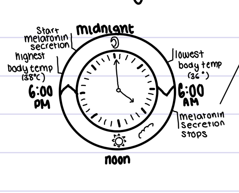
e.g. sleep-wake, core body temperature
why does melatonin secretion stop?
pineal glands respond to light changes, affects serotonin
in winter when there’s less light, can lead to seasonal affective disorder
Sleep-Wake cycle - Siffre 1975
case study, spent 6 months in a cave with no light
given flashlight to navigate
found internal body clock of 25 hours
concluded light source does not impact 24 hour cycle
Sleep-wake cycle - Aschoff and Wever 1976
studied participants living in WWII bunker over a 4-week period, only electrical light, allowed to turn on and off as they wished
eventually free running body clock settled into sleep-wake cycle of 24-25 hours
concluded that we use natural light to entrain, our biological sw-cycle is slightly longer
Sleep-wake cycle individual difference
length of sleep wake cycle can vary between 13-65 hours
Core body temperature
lowest ~04:30 (36 degrees)
highest ~18:00 (38 degrees
Folkard 1977
read children stories at 3pm versus 9am, recall superior for children who read at 3pm
Gupta 1991
improvement on IQ tests at 7pm compared to 2pm and 9am (highest core temperature)
Endogenous pacemakers
internal body clocks that regulate many of our biological rhythms (e.g. hormones such as melatonin, cortisol, metabolic rate, and temperature)
research demonstrates that EPs still function without cues, but may vary as a consequence
Superchiasmatic nucleus (SCN)
most influential EP in the body - bundle of nerve cells in the hypothalamus - found in many mammalian species, regulated by light from environment
Superchiasmatic nucleus - process
retina passes info along the optic nerve to the CNS, the SCN passes info on day length and light to the pineal gland
during night, the pineal gland increases production of melatonin (induces sleep, inhibited during periods of wakefulness)
Superchiasmatic nucleus - research
difficult and unethical in humans, so animal research generalised - controversial
Superchiasmatic nucleus - Decoursey et al. 2000
removed SCN connections in the brain of 30 chipmunks and returned to natural habitat
80 day observation, sleep wake cycle disappeared and by end most were killed by predators
assumed because they were awake and vulnerable
SCN all-important body clock, supported its role in establishing and maintaining the circadian sleep-wake cycle
Superchiasmatic nucleus - Ralph et al. 1990
removed SCN out of genetically abnormal(mutant) hamsters which only had a sleep-wake cycle of 20hours,
transplanted these cells into rats that functioned on the normal 24hour cycle
after transplant, rat’s rhythms shortened to 20hours
suggests SCN is pivotal
ethical issues and extrapolation easier
exogenous zeitgebers
cues from the environment that play an important role in regulating time
act as cues for EPs and help regulate body clock so the individual is synchronised with the environment
e.g. sunlight, noise, seasons, clocks, the moon
exogenous zeitgebers - entrainment
opposite to free running (body does it owns thing) is where EP adjusts in line with environment
e.g. when crossing time zones as EP not synchronised with environment anymore
exogenous zeitgebers - challenge
sun does not set during the summer months in the arctic, still show normal sleep patterns despite prolonged exposure to light
exogenous zeitgebers - support
Visual impairments
Skene and Arendt found that those able to perceive light had normal circadian rhythms, those unable had abnormal rhythms
affect of light on circadian rhythms
Campbell and Murphey
light also detected by skin
introduced light to them during the night at a series of intervals by shining a beam of light on to the back of their knees
their circadian rhythms were disrupted by up to three hours
HEAVILY CRITISISED - disruption of EEG, know they’re being observed, environment was very disruptive
affect of social cues on circadian rhythms
at 6 weeks, circadian rhythms begin
about 16 weeks, most babies are entrained
schedules imposed by parents are likely to be a key influence here, including adult determined meal-times and bedtimes
desynchronisation
practical applications to shift work
Biovin et al. 1996
night workers engaged in shift work experience a period of reduced concentration around 6am
Knutsson 2003
found a relationship between shift work and poor health, shift workers 3x more likely to develop heart disease
ECONOMIC IMPLICATIONS
practical application of research on biological rhythms
use of light therapy as a clinical treatment for depression has now been recognised as an effective and affordable intervention
used alongside drug treatment to max success or as sole treatment
Benedetti et al. 2007 conducted a study examining the effects of light therapy and sleep deprivation on individuals suffering with bipolar
found the use of chronotherapeutics(light therapy) reduced two thirds of the patients’ depression score
Infradian rhythms
cycle LONGER than 24 hours
e.g. the menstrual cycle, governed by the endocrine system, impacted by EZs - light and odours
ultradian rhythms
cycle SHORTER than 24 hours, can happen more than once a day
Affect of light on MC - Reinburg 1967
female ppts spend 3 months in a cave with a small lamp as the only light source
circadian rhythms lengthened to 24.9 hours and her menstrual cycle shortened to 25.7 days
levels of light in the cave could have affected mc
after study, her body took a year to readjust her menstrual cycle back to the original
pheromone
chemical substance produced and released into the environment by an animal, especially a mammal or insect, affecting the behaviour or physiology of others of its species
affect of pheromones on MC - McLintock and Stern 1998
procedure
compounds transferred by the women wiping a pad, which had previously been wiped across the donor’s armpit, above their upper lips
affect of pheromones on MC - McLintock and Stern 1998
at end of cycle
when females received ‘odourless compounds’ from the armpit of women in the latter half of their menstrual cycle
cycle was shortened, presumable by the effects of the other women’s pheromones as they approached the end of their cycle
found 68% of females experienced changes to their cycle, brought them closer to the cycle of their ‘odour donor'
affect of pheromones on MC - McLintock and Stern 1998
at start of cycle
if the compounds were collected from women at the beginning of their cycle, this had the opposite effect, lengthening the cycle of those who had received the compound
affect of pheromones on MC - McLintock and Stern 1998
evaluation
sample mostly women with a history of irregular periods
changes observed were no more likely to appear by chance due to the confounding variables
Trevanthan et al. 1993 - failed to find synchronicity in the menstrual cycle in their female sample