Anatomy and Physiology Lab 1 Practical Review (1)
 Call Kai
 Learn
Learn Practice Test
 Spaced Repetition
 Match
Match Flashcards
 Knowt Play
1/51

There's no tags or description
Looks like no tags are added yet.
Name | Mastery | Learn | Test | Matching | Spaced | Call with Kai | Chat |
|---|
No analytics yet
Send a link to your students to track their progress
52 Terms
Anatomical Position
Body erect (standing upright)
Feet slightly apart
Head and toes forwards
Arms at sides
Palm facing forward
Medial and Lateral
Medial means closer to the middle or center line of the body, while Lateral means farther away from the middle line toward the side
Supine
Lying face up
Prone
Lying face down
Axial
Relating to the head, neck, and trunk
Appendicular
Relating to the limbs and their attachment to the trunk
Anterior
Front
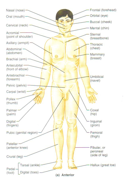
Anterior Body Landmarks
Cephalic, Cranial, Frontal, Ocular/orbital, Nasa, Otic, Buccal, Oral, Mental, Facial, Cervical, Thoracic, Mammary, Abdominal, Umbilical, Axillary, Brachial, Antecubital, Antebrachial , Carpal, Palmar , Manual, Pollex , Femora, Patellar, Crural , Tarsal , Pedal , Hallux , Inguinal , Pubic , Digits
Posterior
Back
Posterior Body Landmarks
Cephalic, Acromial, Olecranal, Dorsal, Lumbar, Gluteal, Popliteal, Sural, Calcaneal, Plantar

Superior
Above/toward head
Inferior
Below/toward feet
Medial
Toward midline
Lateral
Away from midline
Cranial
Toward the head
Caudal
Toward the tail
Dorsal
Backside
Ventral
Belly/front side
Proximal
Nearer trunk
Distal
Farther from trunk
Superficial
At/toward body surface
Deep
Away from body surface
Plane
A three-dimensional axis
Section
A slice parallel to a plane used to visualize internal organization
Sagittal Plane
Oriented parallel to the long axis
Seperates right and left portions
Midsagittal section
Passes through the midline, separating into equal right and left sides
Parasagittal Horizontal
Misses the midline, separating into unequal right and left sides
Transveres (or Horizontal, Cross-sectional) Plane
Separates superior and inferior portions
Dorsal Body Cavity
Canial Cavity: brain
Vertebral (Spinal) Cavity: spinal cord
Ventral Body Cavity
Thoracic Cavity: Contains:
Pericardial cavity: Heart
Pleural cavities: Lungs
Mediastinum
Adominopelvic Cavity:
Abdominal Cavity: stomach, intestine, liver, pancreas, spleen
Pelvic: reproductive organs, bladder, rectum
Other Body Cavities
Oral
Nasal
Orbital
Middle ear
Synovial
The oral, nasal, orbital, and middle ear cavities are located in the head and open to the body exterior
Synovial cavities are found in joints between many bones such as the vertebrae of the spine, and at the knee, shoulder, and hip
Serous Membranes
Parietal serosa: lines body walls
Visceral serosa: covers organs
Peritoneum abdomen: abdomen
Pleura: lungs
Pericardium: heart
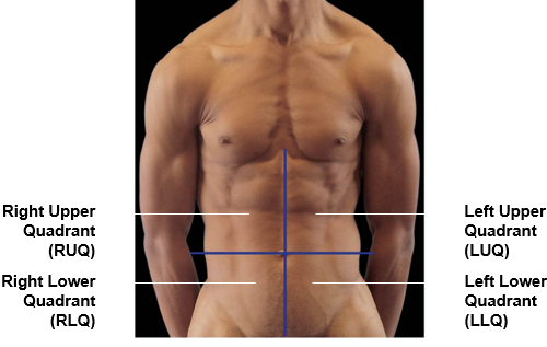
Abdominal Quadrants
RUQ = Right Upper Quadrant
LUQ = Left Upper Quadrant
RLQ = Right Lower Quadrant
LLQ = Left Lower Quadrant
Nine Abdominal Regions
Right hypochondriac region, Right lumbar region, Right inguinal region
Epigastric region, Umbilical cords, Hypogastric (pubic) region
Left hypochondriac region, Left lumbar region, Left inguinal region

Five main classifications of bones (by shape)
Long Bones, Short Bones, Flat Bones, Irregular Bones, Sesamoid Bones

Long Bones
Longer than they are wide; have a thick compact bone exterior.
Examples: Most bones of the appendicular skeleton (limbs), e.g., phalanges, metacarpals, femur, humerus.

Short Bones
Cube-shaped, length and width are roughly equal. Composed of a thick interior of spongy bone covered by a thin layer of compact bone.
Examples: Carpal bones (hand) and Tarsal bones (foot).

Flat Bones
Relatively flat, curved, and thin. Consist of a spongy interior sandwiched by thin layers of compact bone.
Examples: Ribs, sternum.

Irregular Bones
Have no specific or easily classifiable shape or structure.
Examples: Vertebrae

Sesamoid Bones
Small, independent bones that develop within tendons, providing leverage or reducing friction. They are movable.
Example: Patella (kneecap)

Functional Classification of Joints
Synarthorosis, Ampiharthrosis, Diarthrosis
Synarthorosis
immovable joint, no movement in the joint
Example: Skull, Ribs
Ampiharthrosis
Slightly movable joint
Example: Pelvic Bone
Diarthrosis (Synovial)
Freely movable joint
Example: Shoulder, Knee, Elbow
Articular Cartilages
Pad articulating surfaces within articular capsules
Types of Movements
Abduction, Adduction, Circumduction,,
Abduction vs Adduction
Abduction
•Angular motion
•Frontal plane
•Moves away from longitudinal axis
Adduction
•Angular motion
•Frontal plane
•Moves toward longitudinal axis

Circumduction

Types of Movements at Synovial Joints
Pronation, Supination
Pronation and Supination
Pronation
Rotates forearm, radius over ulna
Supination
Forearm in anatomical position

Special Movements
Inversion, Eversion, Dorsiflexion, Plantar flexion
Inversion, Eversion, Dorsiflexion, Plantar flexion
Inversion
•Twists sole of foot medially
Eversion
•Twists sole of foot laterally
Dorsiflexion
•Flexion at ankle (lifting toes)
Plantar flexion
•Extension at ankle (pointing toes)
