Hematology exam 1
 Call Kai
 Learn
Learn Practice Test
 Spaced Repetition
 Match
Match Flashcards
 Knowt Play
1/74

Earn XP
Description and Tags
8/10/26-
Name | Mastery | Learn | Test | Matching | Spaced | Call with Kai | Chat |
|---|
No analytics yet
Send a link to your students to track their progress
75 Terms
components of blood
RBC (O2 transport), WBC (immune system) , plasma (cell transportation), platelets (clotting/hemostasis)
blood collection volume
10%- 20% of total body weight
Where do blood cells originate?”
Liver
microhematocrit tubes
look at cellular vs liquid portions of blood
PCV to determine % RBC
Canine normal PCV
normal is around 45% PCV
Horse and pig normal PCV
42%
Sheep normal PCV
38%
Cat normal PCV
37%
Cattle normal PCV
35%
Goat normal PCV
28%
PCV Icterus
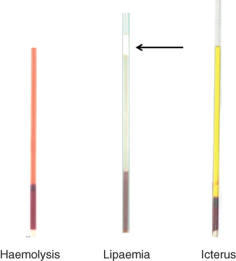
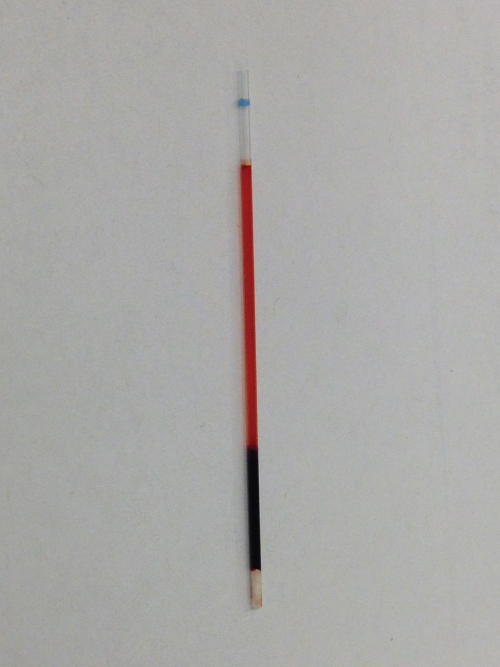
PCV hemolysis
PCV lipemia
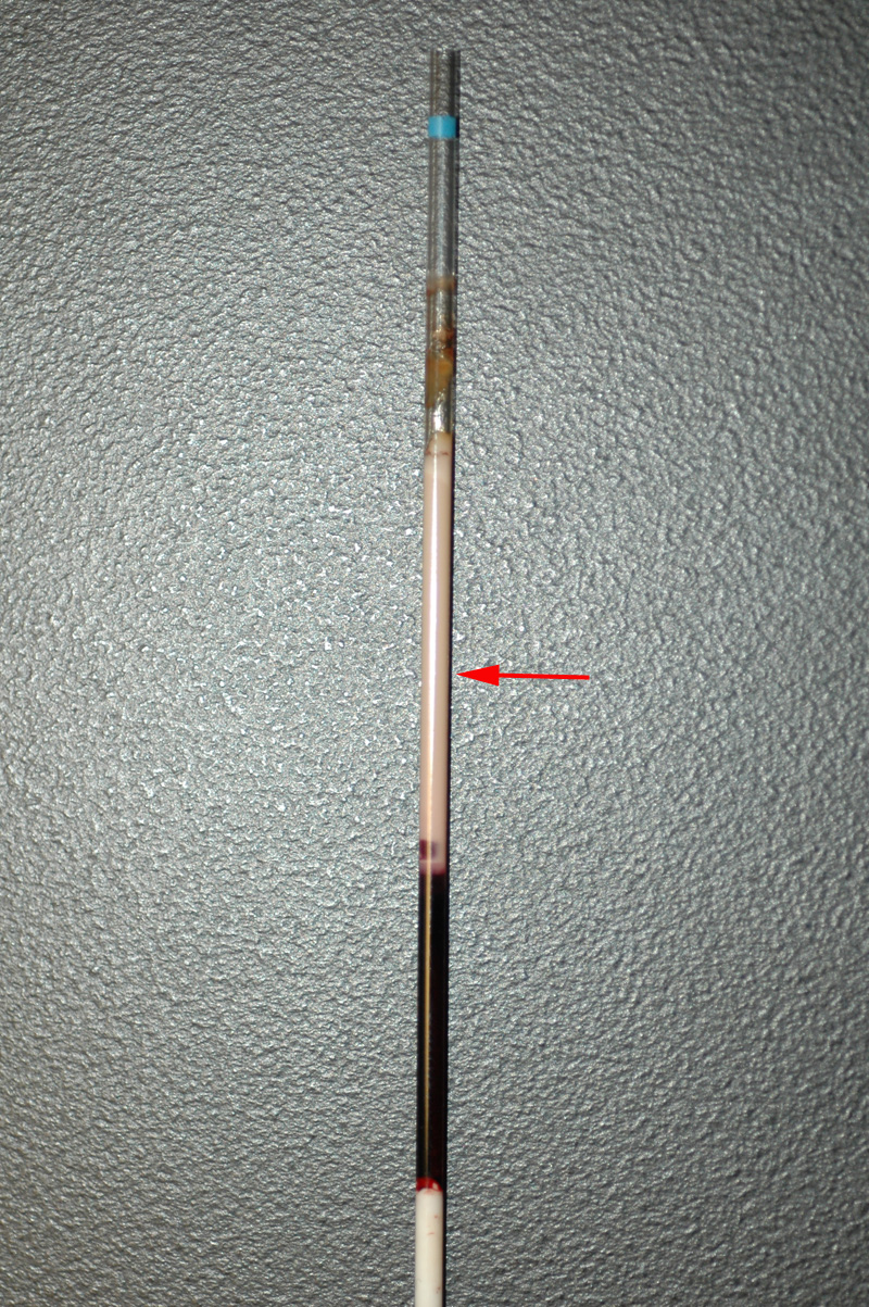
increases in water
decreases protein proportion so more plasmaa
TP
include albumin (majority) and globulins
TP higher in dehydrated animals
chemistry analyzer
measures TP using a chemical reaction
TP - Albumin + Globulins
serum
has no clotting factors
what remains after blood clotted
doesnt contain fibrinogen
Plasma
has clotting factors
liquid portion of unclotted blood
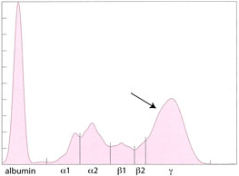
Protein electrophoresis
separates proteins based on charge
Monoclonal gammopathy
cancer
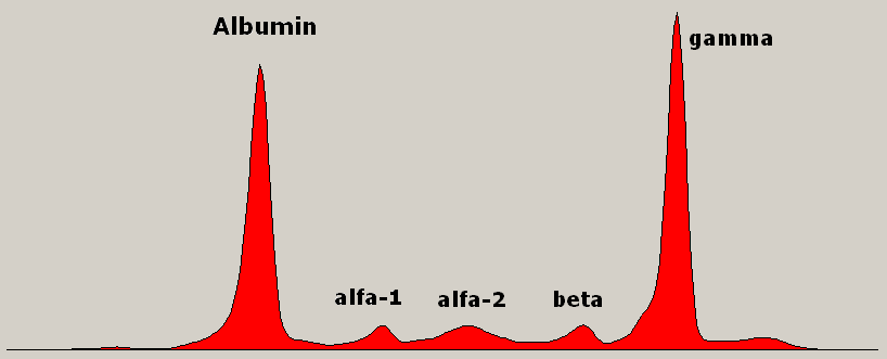
Polyclonal gammopathy
infection
Acute- Phase response
innate immune system
Positive APP
increase inflammation
Fibrinogen (horse and cow)
Serum Amyloid A (horse and cow)
C- Reactive protein (dog and cat)
Negative APP
decrease inflammation
albumin
transferrin
Hematopoiesis
production of RBC, where it occurs depends on animals stage of life-
adult- bone marrow
fetus- liver
embryo- yolk sack
Hematapoietic cells
cells involved in producing the different blood cell lineages
RBC, monocytes, lymphocytes
What can mesenchymal stem cells become
supporting tissues (bone, cartilage, fat)
What can hematopoietic precursors become
blood cells
(all types)
autocrine cell signaling
cell signals to itself
Paracrine cell signaling
cell to cell signaling e
endocrine cell signaling
cell signaling to other cell across blood stream
Growth factor for erythroid cells
EPO (erythroprotein) produced in the kidneys
stimulates RBC production
increased concentrations = increased RBC
G-CSF
granulocyte colony stimulating factor (neutrophil production)
GM-CSF
granulocyte-macrophage colony stimulating hormone
monocytes (macrophage/dendritic cell production)
lymphocytes mature in the
B- bone marrow
T- Thymus
interleukin needed for eosinophil maturiation
IL-5
growth factor for megakaryocytes and platelets
Thrombopoietin (TPO)
stimulates the development of megakaryocytes increasing platelet production
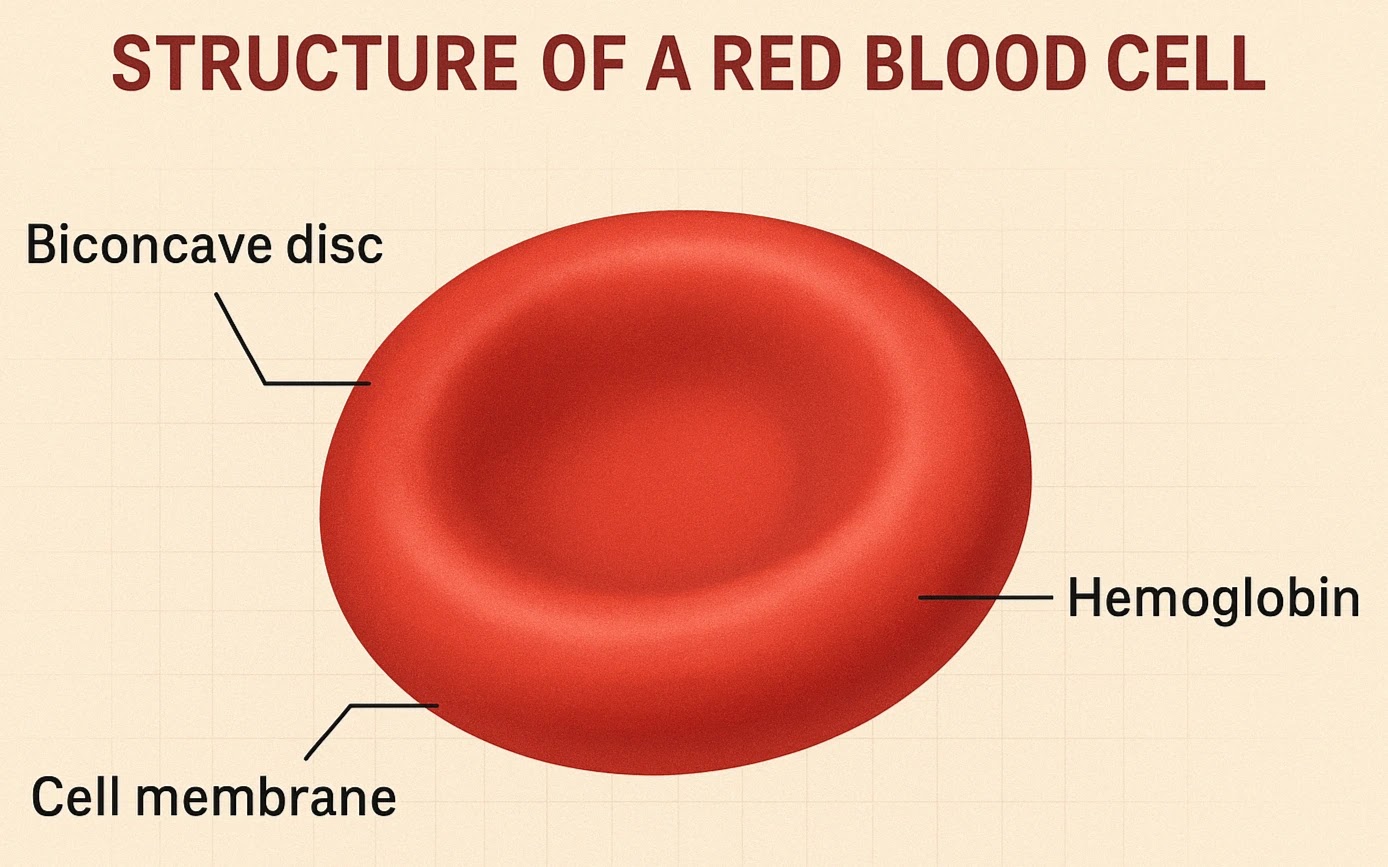
main components of erythrocytes structure and function
lipid bilayer/membrane- outer boundary of RBC, helps with shape
skeletal membrane- form inner lattice to maintain shape
Hemoglobin- carries oxygen, transports CO2
Enzymatic pathways- provide energy and protect from oxidative damage
most animals normal RBC shape
discocytes (biconcave disc)
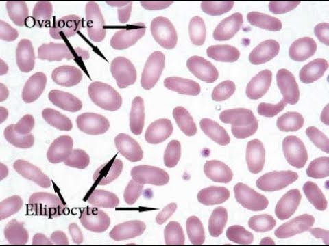
camelids RBC shape
ovalocytes
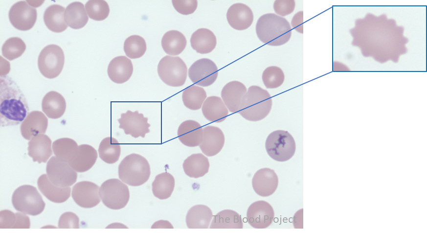
Pig RBC shape
echinocytes
goat RBC shape
poikilocyte
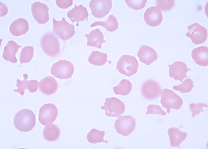
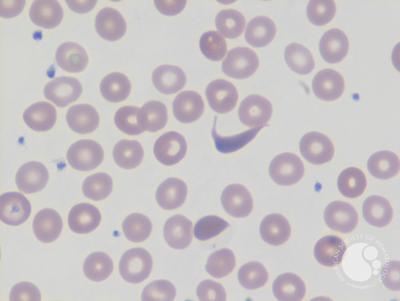
Deer RBC shape
Drepanocyte
birds/fish/reptiles RBC shape
nucleateed oval erythrocytes
erythrocyte antigen for horse
A and Q factors
erythrocyte antigen for dogs
DEA-1, DEA-4, Dal
erythrocyte anitgen for cats
AB group, Mik group
type B cats are PICKY about blood transfusion
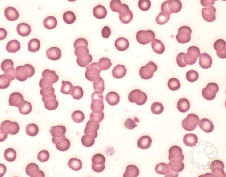
Rouleax association
when RBC stack like coins, normal in cats and horses
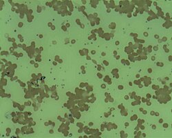
Agglutination association
when RBC clump like grapes bc antigens are sticking, dangerous
Polychromatiaphils
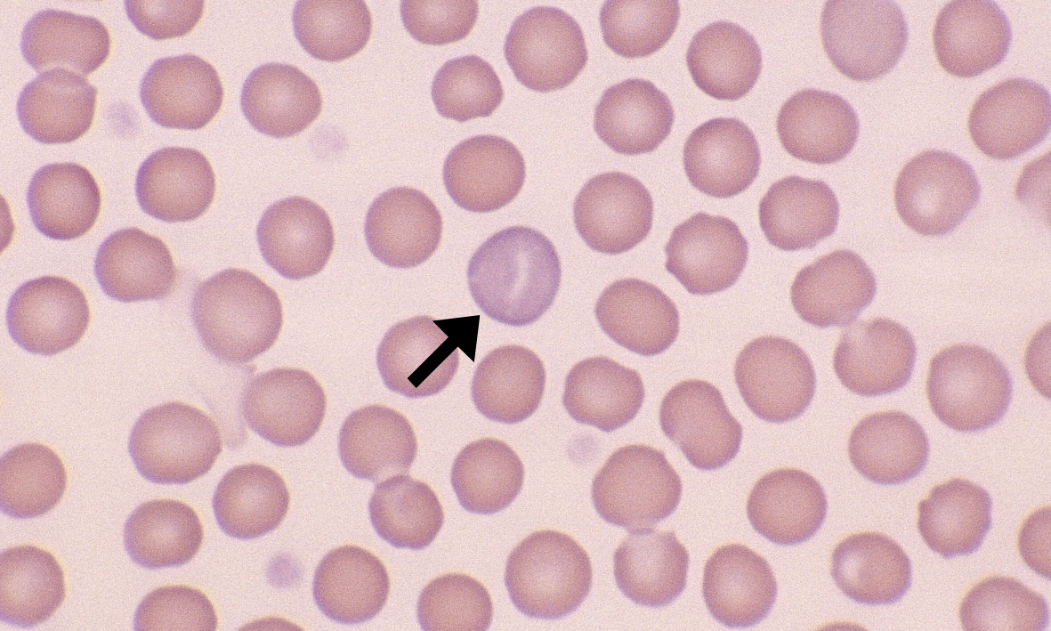
immature RBC that are purple/blue on stain due to increased RNA in structure
Reticulocytes
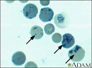
immature RBC that contain ribosomal RNA, have blue dots on stain
Reticulocytosis
an increased number of immature RBC (reticulocytes) in blood- response to anemia trying to regenerate RBC count
caused by Hemorrhage, hemolysis
hormone responsible to RBC production
EPO- erythropoietin
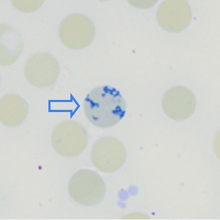
Aggregate reticulocytes
large clumps of RNA which indicates active regeneration
punctuated reticulocytes
have scattered pinpoints of RNA, do not indicate active regeneration (older cells)
What species does not release reticulocytes
horses
lifespan of erythrocytes
2-5 months, die due to oxidative damage, are removed in the spleen, liver and bone marrow
what waste product is formed due to erythrocytes breaking down?
Bilirubin
excreted in bile in the liver
excess bilirubin can cause ictus and hyperbilirubinemia
What factors affect hemoglobin’s oxygen affinity?
pressure (pO2)
higher affinity holds onto oxygen strongly
lower affinity releases oxygen freely
oxygen affinity
how strongly hemoglobin binds to oxygen
Higher affinity
Left shift
less DPG, less T, less CO2, less H+
holds onto O strongly
Lower affinity
Right shift
higher DPG, higher T, higher H+, higher CO2
releases O easily
3 parts of RBC that oxidation can damage
RBC membrane
Heinz bodies
Heme group
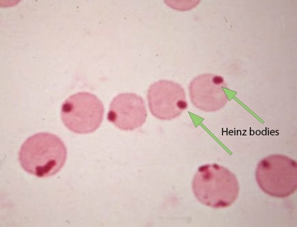
oxidation of heinz body
decreases RBC lifespan and deform cell
oxidation of heme groups
metheme group cannot bind with oxygen which decreases RBC ability to carry O2 turning RBC brown
oxidation of RBC membrane
alters membrane which decreases lifespan
RBC antioxidants
cytochrome, Glutathione, Catalase
RBC metabolism
RBC have no mitochondria so rely on glycolysis for ATP
How does Hepcidin affect iron absorbtion
acts like the stoplight. Will bind to iron which blocks it from exiting transmembrane gates, keeping it in the cell. feedback loop regulation
iron is stored in the form of
ferratin, hemosiderin
How does transferrin affect iron absorbtion
act as ferratin shuttles and moves iron around body
when hepcidin is high
iron is high in the body
what effect does iron deficiency have on erythropoiesis
less iron decreases RBC production, causes smaller RBC, causes pale RBC
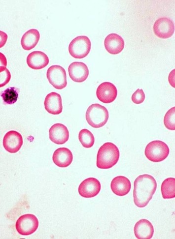
microcytic RBC
smaller than normal
Hypochromic RBC
pale RBC