Physical therapy assistant
 Call Kai
 Learn
Learn Practice Test
 Spaced Repetition
 Match
Match Flashcards
 Knowt Play
1/152

There's no tags or description
Looks like no tags are added yet.
Name | Mastery | Learn | Test | Matching | Spaced | Call with Kai | Chat |
|---|
No analytics yet
Send a link to your students to track their progress
153 Terms
Yergason test
Standing, elbow 90 degree in pronation at the side. Pt must supinate and ex. Rotate. Pain= transverse ligament instability/bicep tendonitis
Speed test
Standing. Arm ext. in and supinate. Pt must resist shoulder flex. Pain=bicepital tendonitis
Neer's impingement test
Siting. PT hand on scapular. Internal rotate arm and bring it into flex. Anterior pain= supraspinatus
Posterior = internal impingement
Supraspinatus test * empty can
Sit/ stand. Pt arm full ext with 90 degree abd. In supination. Have pt pronate arm (like emptying a can). Pain=Tear/impinge supraspinatus tendon or Supra-scapular nerve neuropathy
Clunk test
Pt in supine. PT place fist under scapular. Move arm in full ext. positive test = snapping or clunk soup # glenoid labrum tear
Ant. Apprehension test
Pt in supine, elbow flex and abd. PT does ext. rotation. Pain= ant. Shoulder dislocation
Posterior apprehension test
Pt in supine, elbow flex and abd. PT does horizontal add. Along with medial rotation. Pain= post shoulder dislocation
Adson test
Pt siting arm in ext. PT find radial pulse. Have pt ext. neck and turn head away. Then look back. Positive test= decreased pulse when pt looks back
Tinel sign
Pt in siting. Hand in ext on table. PT tabs along the transverse carpal ligament and distal wrist. Pt numb/ tingling in first 3 1/2 digits. Means positive for carpal tunnel
Pronator teres syndrome test
Pt elbow is flex. PT tries to supinate pt hand. Tell pt don't let me move it (keeping arm probated) while doing that ext. arm putting pronator teres on stretch. Pain in median area of hand= median nerve entrapment not carpal tunnel
Finkelstein test
Pt must put thumb in palm, wrap their 4 fingers around it, put elbow in flex. And wrist in neutral. PT have pt slowly go into ulnar deviation. Pain over abductor polis long us and extensor polis brevity tendon. Positive for deQuervains tenosynovitis
Cozen test "lateral epicondylitis"
Stabilize elbow palmate lateral epicondyle with thumb. Pt make fist pronate and radial deviate and ext wrist. PT gives resistance. Pain at last seal epicondyle = tennis elbow
Medial epicondylitis
Pt stand with elbow flex then supinate forearm ext pt wrist and move elbow into ext. pain along medial epicondyle .golf elbow
Phalen test
Pt does full wrist flex. Hold dorsal side together for 1 minute. Positive test = tingling in thumb index finger middle finger and lateral half ring finger indicate carpal tunnel
Allen test
Open and close hand quick as possible then squeeze hand tightly. Then compress the radial & ulnar artery with thumb. Then ask pt to open hand and release radial. See if blood streams back quick. Repeat with ulnar. Check for vascular compromise
Thomas test
Pt in supine at the edge of table. Have pt ext on leg over the edge of table and hold the opposite leg with both hands with knee in flex. Positive test is extended knee lifting off table = tight hip flexor ( illiopsoas. If pt abd then tight IT band
Ober test
Pt in sideline with knee slightly flex. PT brings leg into abd and ext while stablizing the hip. Lower leg back to table. If leg stays in the air = positive for tight tensor fascia latte/ IT band
Ely test
Pt in prone. Stabilize hip. Flex knee max. If pt hip flex then positive tightness of rectus femoris
Patrick faber
Pt supine. Place affected hip into foot just above knee allowing hip to rotate. Apply gentle pressure. Positive test pain laterally internal or SI
Piriformis test
Pt in supine. Pt hip is passively moved into 90 degree of flex and knee over towards the opposite shoulder. Pt will complain of pain
Craig test
Pt in prone with knee flex. Palpate greater trochanter. Int/ external rotate the hip. This checks for retro (a lot external rotation) and anteversion (a lot internal rotation)
Lachman test
Pt in supine bring test leg into 30 degree knee flex. Fixate femur then slight ext rotate of tibia. Then translate tibia anterior. Soft end feel = ACL rupture
Anterior draw test
Pt in supine. Pt affect leg in knee flex 90 and hip 45. Sit on pt leg. Feel for joint line on knee then move tibia anteriorly explosivly. Test positive if tibia move more 6 mm or soft end feel
Pivot test
Pt in supine leg ext. slight internal rotation of tibia. Move leg from ext to flex. If tibia reduce or jump back or a click sound= positive acl tear
Posterior sag test
Pt in supine hips flex 45 knee flex 90 test
Mcmurray test
Pt in supine hip flex 90 with knee flex then medial rotate/ lateral the tibia. Moving leg from ext to flex. Repeat. If click lock or pain = meniscus damage (lateral tibia rot= testing the medial meniscus vice versa)
Glenohumeral capsular pattern limitations
Greater limitation of external Rotation then abd then internal rotation
Humeroulnar & humeroradial limits
Loss of flex greater than ext
Forearm pattern limits
Equally restricted in supination & pronation
Wrist pattern limits
Fexion = extension
Hip pattern limit
Flexion & internal rotation greater than loss of extension greater than flexion
Tibiofemoral knee
Flexion grossly limited; slight limitation of extension
Talocrural ankle
Hydrocolloid
Absorb exudate partial (epidermis & some dermis) and full thickness (when you see fat)
Hydrogel
Superficial & partial little drainage
Foam
Partial & full (level of exudate vary)
Transparent film
Superficial & partial little drainage
Alginate
Seaweed dressing partial to full. Use with excessive absorption
Superficial wound
only epidermis
minor wounds that affect the outermost layers of skin and include minor cuts and scrapes
heal very well, minimal blood loss,less infection
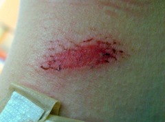
Partial wound
Involve epidermis and may involve dermis. Shallow moist wound bed. No tissue necrosis. No slough
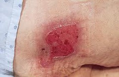
Full thickness wound
Damage goes below epidermis and dermis into subcutaneous tissue. May see tendon muscle and bone
Colles fracture
Fracture at the distal end of the radius that displaces the smaller fragment posteriorly
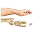
Smith fracture
fracture of distal radius with anterior (palmar) displacement
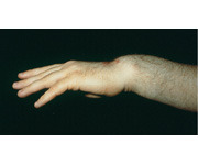
Scaphoid fracture
Most common fracture occurs when person fall onto outstretched UE
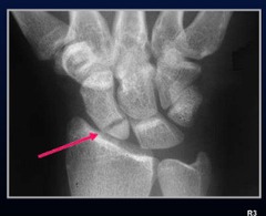
Dupuytrens contracture
Shortening of atrophy of palmar aponeurosis, contracture of 4th and possibly 5th digit. Affect MCP & PIP
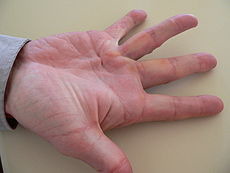
Boutonnière deformity
Observer deformity is ext. of MCP ext. of the DIP along with flex. Of PIP. Common with rheumatoid arthritis with degeneration of central extensor tendon.
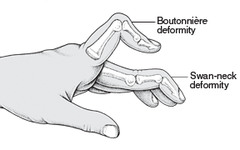
Swan neck deformity
Result from contracture of intrinsic muscles with dorsal subluxation of lateral extensor tendon. Observe deformity is flex of MCP & DIP along with ext. of PIP. Occurs with rheumatoid arthritis following degeneration of lateral extensor tendon
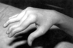
Ape hand deformity
Observed as thenar muscle wasting with first digit moving dorsal until its in line with second digit. Result from median nerve dysfunction
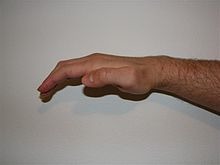
Mallet finger
Injury to distal extensor tendon causing DIP flexion contracture.
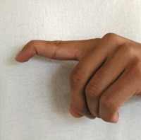
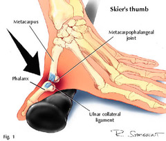
Gamekeeper thumb
Sprain/ rupture of ulnar collateral ligament of MCP joint of first digit. Result in medial instability of thumb. Usually occurs with skiers . Immobilized for 6 wks
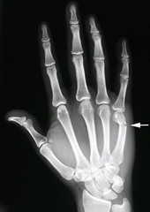
Boxer fracture
Fracture of neck of fifth metacarpal
Trigger finger
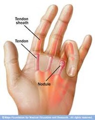
common in DM, stenosing tenosynovitis, nodule at MCP of ring or index finger
Avascular necrosis (AVN) of hip
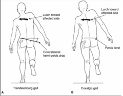
Impaired blood supply to femoral head. Decrease in flex, IR & ABD. Pain in groin/thigh. Coxalgic gait avoid WB on affect side and lateral lurch to opposite side
Coxalgic gait
trunk lurch toward affected side (shortens MA of hip abductors so they have to work less, producing less torque on hip), also called compensated Trendelenburg.
Legg-Calve Perthe (osteochondrosis)
(LCPD) is a childhood hip disorder initiated by a disruption of blood flow to the ball of the femur called the femoral head. Due to the lack of blood flow, the bone dies (osteonecrosis or avascular necrosis) and stops growing.
Slipped capital femoral epiphysis
Disorder in which the capital femoral epiphysis slips over the neck of the femur
seen in children during the growth spurt years
Femoral anteversion
Angle btwn the femoral neck and the acetabulum is >15 (greater) degrees, toe in gait
'Pigeon toed'
Femoral retroversion
Angle less than 0 degrees femoral head rotated backwards in relation to femoral condyle
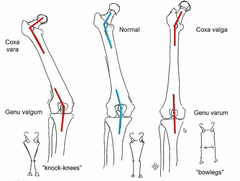
Coxa vara
Angle of femoral neck in relation to shaft is less than 120°. Will give pt genu Valgum
Coxa valgus
Angle between neck & shaft of femur > 135°
Trochanteric bursitis
Inflammation of trochanteric bursa from direct blow, irritation of ITB. Positive for noble test during knee ext & ober test
Piriformis syndrome
Caused by excessive pronation of foot causing femoral to internal rotate. Can lead to compression of sciatic nerve/sacroiliac dysfunction. Pt has restricted internal rotation and pain posterior thigh weak external rotation
Osgood schlatter
disease can cause a painful, bony bump on the shinbone just below the knee. It usually occurs in children and adolescents experiencing growth spurts during puberty.
Q angle
ASIS, midpoint of patella and tibial tuberosity
13 men 18 women
Can result in patella femoral syndrome
Pes anserine bursitis
Knee
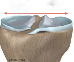
Tibial plateau
Mostly injured combination of valgum & compression force to knee while flex
Femoral condyle
Most injured due to location
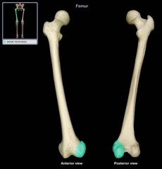
Epiphyseal plate
Injured during WB torsion stress
Fractures at knee joint
Early flexibility to prevent capsular adhesion
Anterior compartment syndrome ACS
Increased compartment pressure resulting in ischemic condition. Symptoms produce by exercise or exertion deep cramping feeling swelling parasthesia severe pain . MEDICAL EMERGENCY STOP TX
Anterior tibial periostitis *shin splits
Muscles included anterior tibialis and extensor hallucis longus. Pain when palpate lateral tibia and anterior compartment. Flexibility ex for anterior compartment and tricep surae
Tricep surae
- Soleus
- 2 head of Gastrocnemius
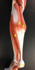
Medial tibial stress syndrome
Overuse of tibial posterior and medial soleus . Excessive pronation. Pain when palpate dorsal posteromedial border of tibia. Extremely tricep surae (flexion)
Lateral ligament sprain
Injury occur when foot is PF and inverted. Ligament usually injured is the anterior talofibular ligament. Use anterior draw test and talar tilt test.
Lateral ligament grade two
Some loss of function with complete tear of anterior talofibular ligament and calcaneofibular ligament
Lateral ligament grade 3
Complete loss of function with complete tear of anterior talofibular ligament and calcaneofibular ligament with partial tear of posterior talofibular ligament
Unimalleolar fracture
Involve medial or lateral malleolus
Bimalleolar fracture
Involve medial and lateral malleolus
Trimalleolar fracture
Involve medial and lateral malleolus and posterior tubercle of distal tibia. Early PROM to prevent capsular adhesion
Tarsal tunnel syndrome
Entrapment do the tibial nerve. Excessive pronation & overuse result in tendinitis of long flexor & posterior tibialis tendon and trauma may cause compromise to tunnel
Tarsal tunnel syndrome
Symptoms include pain numbness paresthesias along the medial ankle to the plantar surface of foot. Use orthoses to maintain neutral alignment of foot
Flexor hallucinating tendonopathy
Identified as tendinitis in acute phase common with ballerina
Pes cavus (hollow foot)
Deformity observe. Is an increase in longitudinal arches, dropping of anterior arch, metatarsal head lower than hind foot plantar flexion and splaying of forefoot and claw toes
Pes planus flat foot
Can be caused by excessive pronation. Deformation is reduce height of medial longitudinal arch. Difficult push o f during gait
Talipes equinovarus club foot
Talipe is an abnormal development of the head& neck of the talus
Club foot
Postural= PF ADD & inverted foot
Talipes = PF at talocrural joint, inversion at subtalar, talocalcaneal, talonavicular, & calcaneocuboid joint. Supination at midtarsal
Equinus
Contracture of gastro and/or soleus. Limited DF. Foot is plantar flex. Pt walks on toes
Hallux valgus
Can be caused by excessive pronation. Deformities = medial deviation of head of first metatarsal from midline of bone. Distal phalanx move laterally.
Metatarsalgia
Tight tricep surae group or Achilles, collapse of transverse arch, short first ray, pronation of forefoot. Structural changes in transverse arch possibly leading vascular and neural compromise in tissue of forefoot. Pt usually complain first and second metatarsal heal
Metatarsus adductus
Cause by muscle imbalance or neuromuscular disease
Charcot-Marie tooth disease
Peroneal muscular atrophy that affects motor& sensory nerve. IntiL affect us le in leg and foot but progress to muscle in hand & forearm
Plantar fasciitis
Irritation of plantar fascia from excessive pronation. Tight tricep surae rigid cavus foot. Limit ROM of MTP AND talocrural joint.work on flex of tricep surae and strength of invertor
Rear foot varus
Inversion of calcaneus when subtalor joint is in neutral.
Rearfoot valgus
Result from genu valgum. Eversion of calcaneus. Owing increases mobility of hind foot
Forefoot varus
Inversion of the forefoot caused by abnormal development of head and neck of talus
Forefoot valgus
Eversion of forefoot
Spondylolysis
Stress fracture to the pars interarticularis
fracture of transverse process of vertebrae "Scotty dog sign"
Spondylolisthesis
forward sliding of lumbar vertebra over vertebra below it
Spondylolisthesis & spondylolysis
Ex focus on dynamic stabilization of trunk with emphasis on abdominal & trunk extension working from a flex position into a neutral. Avoid extension side bending or rotation. Reeducation on posture. Use Boston brace or TLSO
Spinal stenosis
a condition, mostly in adults 50 and older, in which your spinal canal starts to narrow. ... The open spaces between the vertebrae may start to get smaller. The tightness can pinch the spinal cord or the nerves around it, causing pain, tingling, or numbness in your legs, arms, or torso. Pain decrease in spinal flexion and increase in extension. Pain increase with walking and relieved with prolonged rest. Avoid lateral bending and rotation ex. Focus on truck and pelvis dynamic stability