Week 2 - Synaptic Transmission
 Call Kai
 Learn
Learn Practice Test
 Spaced Repetition
 Match
Match Flashcards
 Knowt Play
1/60

Earn XP
Description and Tags
Synaptic Transmission, Autonomic Nervous System, and Cranial Nerves
Name | Mastery | Learn | Test | Matching | Spaced | Call with Kai | Chat |
|---|
No analytics yet
Send a link to your students to track their progress
61 Terms

Locate the different parts on the neurons
Dendrites
Cell body
Axon Hillock
Axon
Axon terminal
How are neurons classified?
By the number of processes that originate from the cell body
Functional classification
By the type of neurotransmitter used
Classification of neurons based on the number of processes from the soma
Unipolar
Pseudounipolar
Bipolar
Multipolar

Unipolar Neuron
Not in humans
Invertebrates

Pseudounipolar Neuron
One projection that splits into 2 axons, no true dendrites
Ex: Somatosensory neurons (dorsal root ganglion)
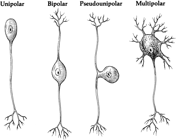
Bipolar Neuron
1 axon, 1 dendritic root
Ex: eye

Multipolar
1 axon, many dendrites
Functional Classification of Neurons
Afferent
Efferent
Interneuronal
Afferent
Convey information into the CNS (usually sensory)
Efferent
Transmit information from CNS to peripheral structures (muscles and glands) (usually motor)
Interneuronal
Send information between neurons
Classificiation: Neurotransmitter used
ACh
Dopamine
Serotonin
GABA
Glutamate
Communication in the Nervous System
Neurons generate electrical signals
Local Potentials
Action Potentials
Local Potentials
Transmit information over short distances
Graded potentials
Action Potentials
Transmit information along an axon by repeated generation of a signal
All or none
Neuroelectrophysiology
Inside (intracellular) and outside (extracellular) the neuron there are charged ions (i.e., Na+, K+, Cl-)
These ions move back and forth between the inside and outside of the neuron
Membrane Potential
The difference in electrical charge (voltage) between the interior and the exterior of the neuron
Resting Membrane Potential
The membrane potential when not transmitting information
There is an unequal distribution of ionic charge across the membrane
Extracellular Charge & Ions
Higher concentration of Sodium (Na+) and chloride (Cl-) than intracellular fluid (positively charged
Intracellular Charge & Ions
Higher concentration of potassium (K+) and anions (A-) (negatively charged)
Where is the electrical signal generated?
In the axon hillock
They travel through the length of the axon to the axon terminal, from where they are transmitted to the next neuron
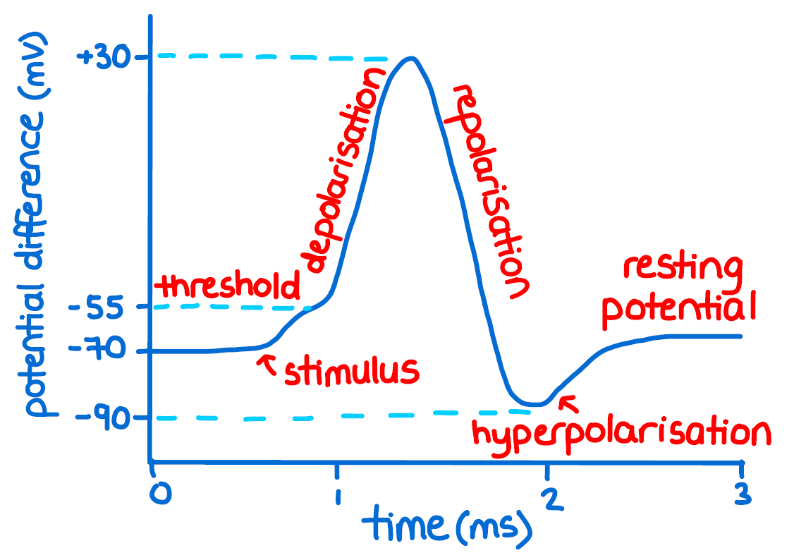
What are the stages of an Action Potential?
Resting membrane potential
Threshold
Depolarization
Repolarization
Hyperpolarization
Returning to resting membrane potential
At ________ _________ ___________ the membrane channels are closed and at -70mV
Resting Membrane Potential
Threshold & Depolarization
Following a stimulus, ions move and cause change in membrane potential
If threshold is reached (~55mV), many voltage-gated Na+ channels open and there is an influx of Na+ into the cell
Neurons become more positively charged inside than the outside
Peaks at about +35mV, Na+ channels close
Repolarization
Na+ channels are closed
K+ channels open, K+ exits the cell
Causes a reversal of the membrane potential
Hyperpolarization
Continued efflux of K+ causes the cell membrane to become even more negative than at rest
At this time, it’s more difficult to initiate another AP (it’s in refractory)
Absolute refractory
Membrane unresponsive to stimuli
Relative refractory
Stimulus must be stronger than normal to elicit an AP
What prevents backwards propagation of an action potential?
Refractory Period
Returning to Resting Membrane Potential
Membrane channels are all closed
Na+K+ pump: actively moves Na+ out of the neuron and K+ back into the neuron
3 Na+ out for 2 K+ into the cell per cycle
Leak channels
Propagation of the Action Potential
Once an AP has been generated, it spreads passively along the axon
Spread of AP along axon is dependent on:
Passive properties of an axon
Diameter
Myelin
Active opening of ion channels (saltatory conduction)
How would a larger diameter of an axon affect the action potential traveling down it?
The bigger the axon the faster
The smaller the slower
How does myelin affect the action potential traveling down it?
Myelinated = faster (saltatory conduction)
Unmyelinated = slower
Nodes of Ranvier
Small unmyelined patches located on myelinated axons
New AP is generated at these nodes
Makes the AP faster
Saltatory conduction: AP appears to “jump” from node to node
Saltatory conduction
AP appears to “jump” from node to node
Synapse
A point where interchange of information occurs between two neurons
Synaptic Terminal
Contains vesicles, which contain neurotransmitters
Neurotransmitters
Enable communication between neurons
Present in the presynaptic terminal
Released into the synaptic cleft
Opens channels to allow ions in or out, possibly resulting in an action potential in the postsynaptic neuron
Must be removed from the synaptic cleft
What are the ways in which a neurotransmitter can be removed from the synaptic cleft?
Reuptake
Absorption by glial cells
Enzymatic breakdown at cleft
Neuromuscular Junction
Where motor neurons synapse with muscle fibers
Voluntary & involuntary muscle movement
Fast-acting obligatory response
Main neurotransmitter: Acetylcholine (Ach)
Etiology
Cause/origin of a disease or abnormal condition
Pathology
What is actually happening in the body
Prognosis
The prospect of recovery from disease
Multiple Sclerosis
Etiology
Autoimmune
Pathology: Demyelination of central nervous system
Causes scar tissue “sclerosis”
Signs and Symptoms
Variable depending on location of demyelination in the body
Sensory: Impaired/atypical sensation
Motor:Weakness
Cranial Nerves: Impaired vision
Cognition: Infrequent or mild (because most of cognition is not myelinated)

Multiple Sclerosis: Graphs
Characterized by periods of relapse and remission
The different types of multiple sclerosis include: (see graph)
Multiple Sclerosis: Prognosis & Treatment
Prognosis: Variable
Currently no cure
OT
Maintain & improve function where possible
Avoid high temperature & excessive exertion
Adaptations for ADLs
Energy conservation & stress reduction techniques
Time management
Home modification
Lifestyle redesign
Guillain Barre Syndrome
Etiology
Autoimmune
Pathology
Demyelination of PNS
Signs/Symptoms
Motor: Weakness, paresis/paralysis
Sensor: atypical sensations, pain
Cranial Nerves: Motor cranial nerves most affected
Guillain Barre Syndrome: Prognosis & Treament
Prognosis
Progressively gets worse for 2-3 weeks then gradual improvement
-Medical intervention
-PT
-OT
Strengthening and regaining functional mobility
Resuming or adapting ADLs
Energy concervation techniques
Myasthenia Gravis
Etiology
Autoimmune (antibodies attack & destroy Ach receptors)
Signs & Symptoms
Motor: Weakness increases with muscle use
Sensory: not impacted
Cranial Nerves: not impacted by skeletal muscles innervated by cranial nerves show fluctuating weakness
Prognosis
With medical treatment >90% survival rate
Anatomical Divisions of the Nervous System
Central Nervous System (CNS)
Brain
Spinal Cord
Peripheral Nervous System (PNS)
Cranial Nerves
Spinal Nerves
Peripheral Nervous System (PNS)
Somatic Nervous System
Spinal Nerves (31 pairs)
Cranial Nerves (12 pairs)
Autonomic Nervous System
Sympathetic Nervous System
Parasympathetic Nervous System
Sympathetic Nervous System
Fight or flight
Stress
Activated during exercise, excitement, emergencies
Parasympathetic Nervous System
Rest and Digest
Peace
Energy conservation & storage
What are the sympathetic and parasympathetic systems trying to maintain in the body?
Homeostasis (balance)
Dynamic balance between autonomic branches
Autonomic Dysreflexia
Spinal cord injury above T6
Sympathetic neurons below te level of lesion are activated due to some noxious stimuli
With lesions above T6, the spinal cord is prevented from receiving signals from the brain that inhibit sympathetic activity
Excessive sympathetic response
The sudden spike in blood pressure can be life threatening
Cranial Nerves
Within the peripheral nervous system, in the somatic nervous system
Innervate face and neck
Can include sensory, motor, or both
Lesion of the Oculomotor Nerve (CN 3)
Diplopia: Double vision
Ptosis: Drooping of the eyelid
Trigeminal Neuralgia (Tic Douloureux)
Pathology
Compression of the trigeminal nerve (CN 5)
Symptoms
Sensory
No loss, but severe sharp stabbing pain
Pain triggered by stimuli which are usually non-noxious (eating, talking, touch, etc)
Begin and end abruptly
Motor: Intact
Prognosis
Variable
Bell’s Palsy
Etiology
Viral infection or immune disorder
Pathology
Lesion to the facial nerve
Symptoms
Unimpaired
Motor: Unilateral paresis/paralysis of the muscles supplied by the facial nerve
Autonomic: Salivation and production of tears affected (severe cases)
Prognosis
In the majority of people, the muscles recover within 2 months depending on the severity of damage
Dysphagia
Difficulty swallowing
Can cause aspiration (food going down the wrong tube)
Continued aspiration can lead to pneumonia
CN
Trigeminal (V)
Facial (VII)
Glossopharyngeal (IX)
Vagus (X)
Hypoglossal (XII)
Dysarthia
Speech disorder resulting in poor articulation (slurred speech)
No difficulty in understanding spoken language or ability to read or write
CN
Trigeminal (V)
Facial (VII)
Glossopharyngeal (IX)
Vagus (X)
Hypoglossal (XII)
Accessory (XI)