biological molecules
 Call Kai
 Learn
Learn Practice Test
 Spaced Repetition
 Match
Match Flashcards
 Knowt Play
1/94

There's no tags or description
Looks like no tags are added yet.
Name | Mastery | Learn | Test | Matching | Spaced | Call with Kai | Chat |
|---|
No analytics yet
Send a link to your students to track their progress
95 Terms
monomers
individual small units that make up a polymer
polymers
large molecules of regular repeating units
all carbohydrates contain
C H O
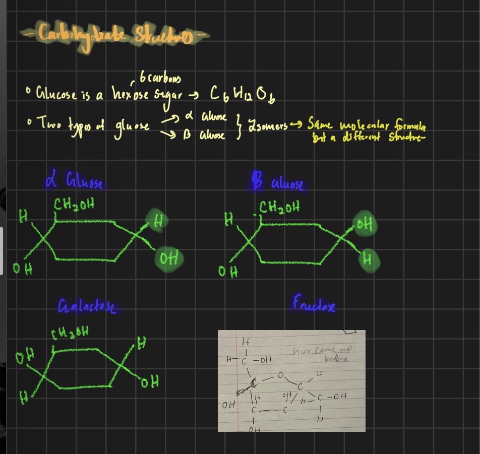
Examples of monosaccharides
alpha glucose
beta glucose
galatacose
fructose
examples of disaccharides
alpha glucose + alpha glucose - maltose
Alpha glucose + galatose - lactose [milk]
Alpha glucose + fructose - sucrose {only non reducing sugar}
Polysaccharides
Starch {excess glucose stored as starch in plants} [ lots of alpha glucose joined togther]
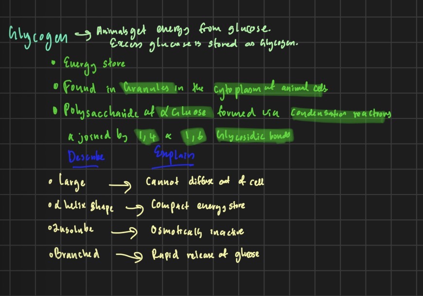
Glycogen {excess glucose in animlas} [ lots of alpha glucose joined togther]
Cellulose {for cell wall strengthening} [lots of beta glucose]
Alpha glucose and beta glucose molecule drawing
What is the test for reducing sugars
Benedicts test
How to do benedicts test for a reducing sugar
Add benedicts
Heat it
Blue to red color chnage
Whats the test for non reducing sugars
If benedicts test is negative we do this - solution stays blue
Heat it with HCL to hydrolyse the disaccharide [ sucrose broken into alpha glucose and fructose]
Neutralise it with sodium hydroxide NaOH - base
Add benedicts again
Blue to red
What is the color spectrum for benedicts test
Blue-green-yellow-orange-red
Properties of starch - polyschhardie made of lots of alpha glucose
Large - cnnot diffuse out of cell
Insoluble - osmotically inactive
Alpha helix shape- compact enegry store
Branched - rapid release of glucose
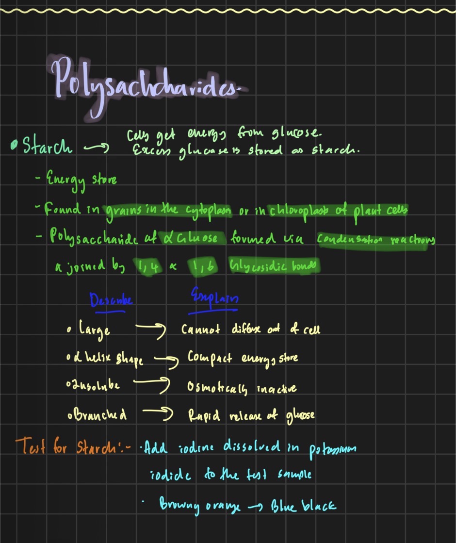
Test for starch
Add iodine dissolved in potassium iodide to the test sample
Browny orange- blue black
Glycogen properties
Large- cannot diffuse ou of cell
Insolube- osmotically inactive
Alpha helix shape- compact negery store
Branched- rapid release of glucose
Properties of cellulose
Forms a straight chain molecule
Forms glycosidic b0nds
Monomers are flipped
Made from beta glucose- allows striaght unbranched chains to join
parallel chains form weak hydrogen bonds[ crosslinks]
cellulose forms microfibrils - adds strength
![<ol><li><p>Forms a straight chain molecule </p></li><li><p>Forms glycosidic b0nds </p></li><li><p>Monomers are flipped </p></li><li><p>Made from beta glucose- allows striaght unbranched chains to join </p></li><li><p>parallel chains form weak hydrogen bonds[ crosslinks] </p></li><li><p>cellulose forms microfibrils - adds strength </p></li></ol><p></p>](https://assets.knowt.com/user-attachments/b6e966ca-5002-422f-850f-5f49776e48fe.jpg)
What is a condensation reaction
A reaction that joins two molecules together with the formation of a chemical bond and involves the elimination of a molecule of water.
What is hydrolysis?
reaction that breaks a chemical bond between two molecules and involves the use of a water molecule.
whats the bond for a carbohydrate
glycosidic bond
Why is cellulose suitable for plant cell walls?
Hydrogen bonds between cellulose chains form strong microfibrils, giving the cell wall high tensile strength.
Why is glycogen suitable for energy storage in animals?
Highly branched structure provides many ends from which glucose can be rapidly released by hydrolysis.
are lipids polymers
lipids are NOT polymers
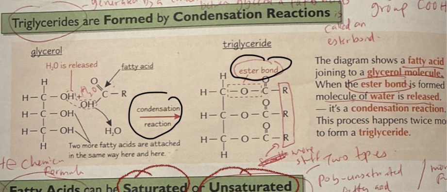
structure of a triglyceride
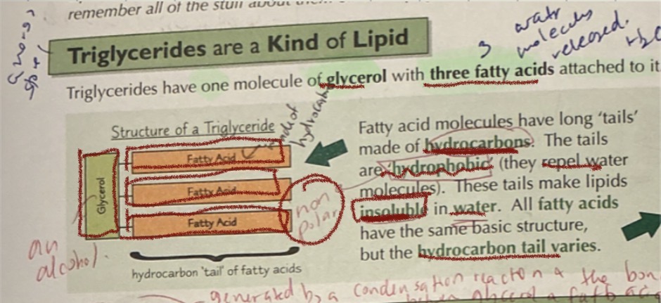
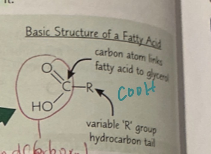
basic strcture of a fatty acid
COOH
WHAT ARE LIPIDS USED FOR
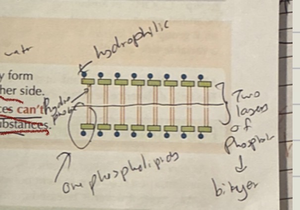
cell membrne - phospholipid bilayer
enegey store
waterproofing - waxy cuticle of leaves is insolublein water
used in insulation- thermal + electrical insulation
protection - covers the heart and kidneys
what are rhe two types of lipids
phospholipids an trigycerides
diagram of triglyceride formation
saturated fatty acids
no double bonds
striaght hydrocarbon chain
Why do phospholipids form bilayers?
Hydrophilic heads interact with water while hydrophobic tails avoid water, causing phospholipids to arrange themselves into a bilayer.
proteins
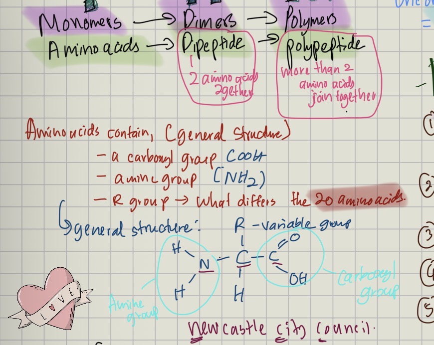
made from long chains of amino acids as monomers
amino acid contain,
a carboxyl group COOH
AMINE GROUP
R GROUP newcastle city council
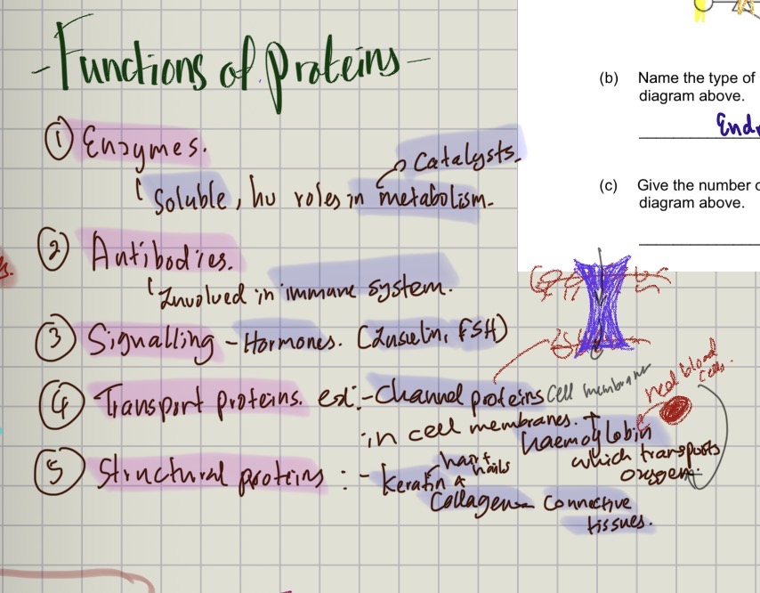
functions of proteins
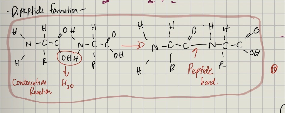
dipeptide formation
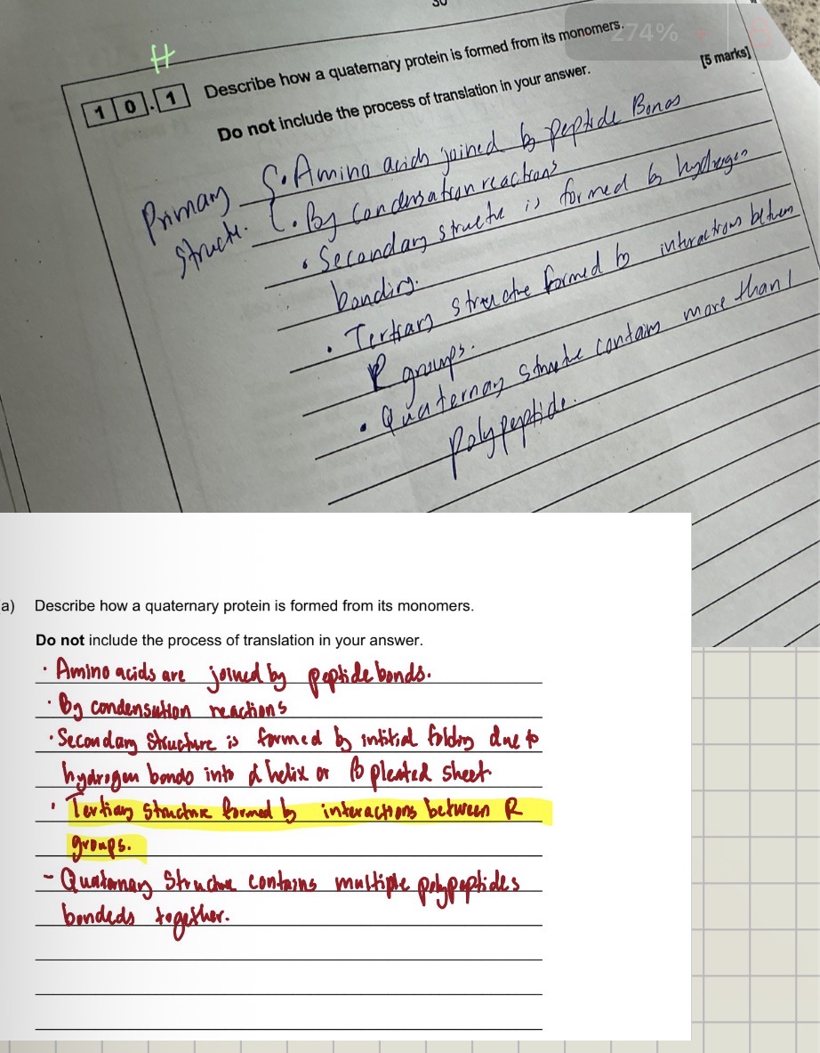
describe how a quaternary protein is formed from its monomers
primary structure
sequence of animo acids
joined by peptide bonds
the sequence s determined by genes
secondary structure of a protein
initial folding due to hydrogen bonding
either coil into an alpha helix or beta pleated sheet
tertiary struture of a protein
3D shape
held togther by hydrogen, ionic, disulfide bonds [strongest bonds]
formed by interactions between R GROUPS
quaternary stricture of a protein
multiple polypeptides folded togther
test for proteins
add biuret
blue- lilac purple
What is a peptide bond?
A bond formed between two amino acids in a condensation reaction.
Dipeptide
Two amino acids joined by a peptide bond.
Polypeptide
Many amino acids joined by peptide bonds.
VERY IMPORTANT
Primary structure determines the final three-dimensional structure of a protein, which determines its function.
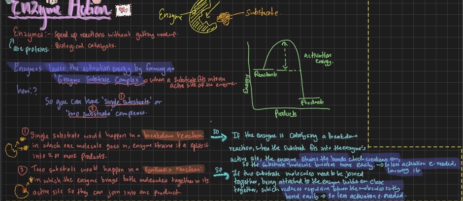
Definition of enzyme
A biological catalyst that lowers the activation energy of a reaction.
Activation energy
The minimum energy required for a reaction to occur.
breakdown and synthesis reaction
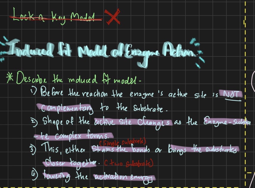
induced fit model
before the reaction the enezymes active site is NOT complemtry to the substarte
shaope of the active site chnages as te enzyme substrate complex forms
this either strains the bonds or brings the substrates closer
lowerinf the acivtation emnergy
factors affcetin enzuymes
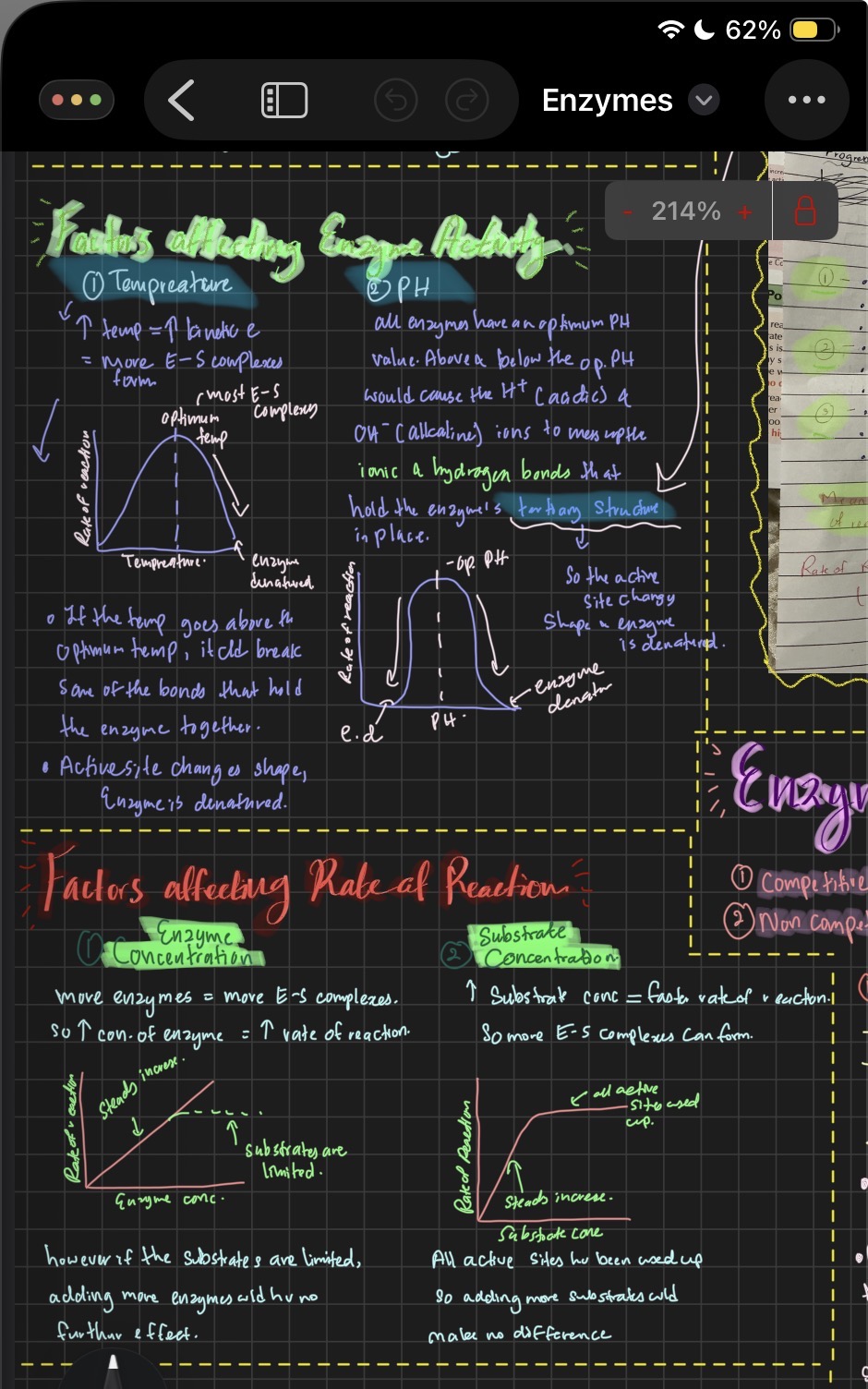
what is the active site of an enzymes shape determined by
the tertiary structure
what can alter the tertiary structure of an enzyme
PH and Tempreature changs
causes the shape of the active site to chnage an the substrate woukld no longer fit in
what is the primary structure of a protein determined by
genes
if theres a mutation in genes the primary structure chnangs so does the tertiary strcure which determines the shape of he active site, leaving the enzyme substrte complex unable to form
what are factors that affcet enzyme activity
tempreture and PH - higher than optimum level ould. cuase the enzyme to denature
what are factors that affect rate of reaction of enzyme subtrate complexes
substrate concentration and enzyme concentration
what are th two types of enzyme inhibitors
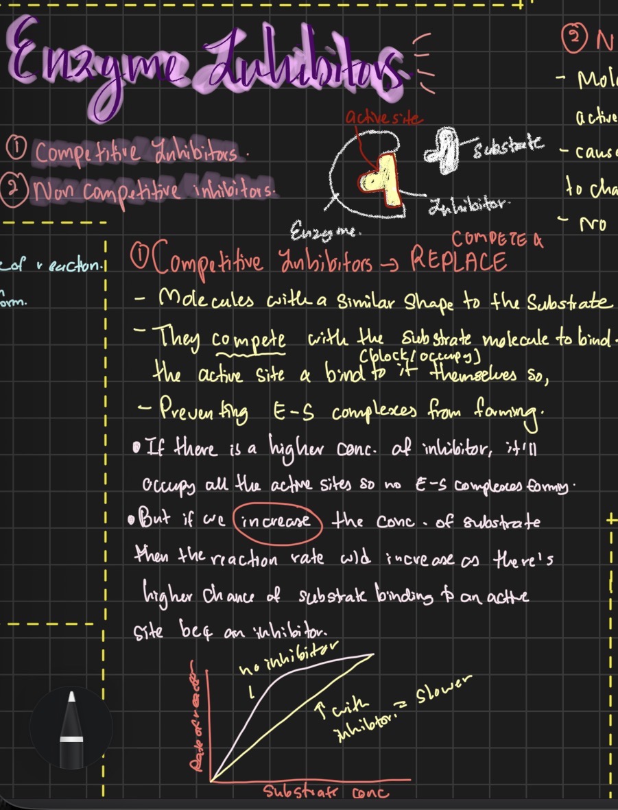
competitive inhibitor
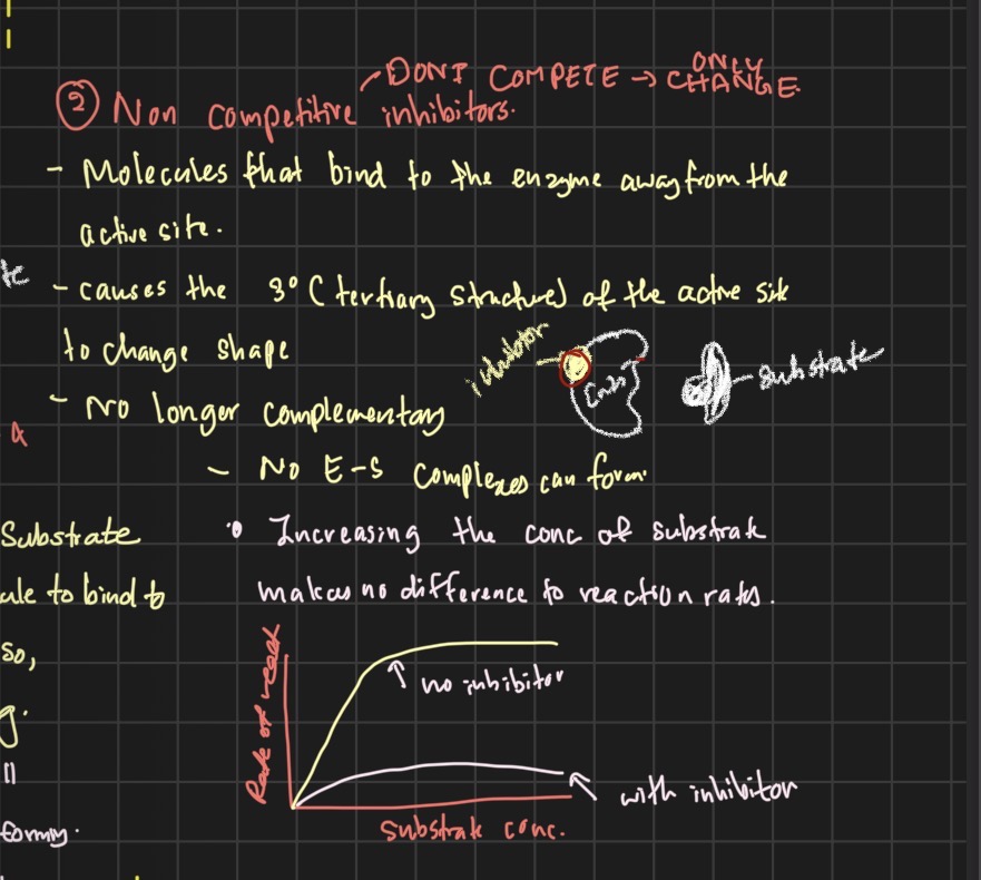
non competetive inhibitor
what does competetive inhibitors do and hw to prevent it
they are molecules with a similar shape to the substartes so they compete with the substrate to bibd to the active site, reducing the amount of e-z complxes able o be fomed
if we increase the substrate concentration theres a higher chnace itll bind to the actiive site faster than the inhibitors
What does non compettive inhibitors do and how to prevent it
Binds to the enzyme away from the active site and chnages the tertiary structure of the active site so the substrate wont fit to the active site anymore, reducing he amount of e-z complexes formed.
Factors affecting enzyme activity
Temperature
Increasing temperature:
→ more kinetic energy
→ more successful collisions
→ rate increases
Above optimum:
→ bonds holding tertiary structure are disrupted
→ active site changes shape
→ substrate no longer binds effectively
→ enzyme denatures
→ rate decreases rapidly.
Important:
Don’t say “enzyme dies.”
Say “enzyme becomes denatured.”
pH
Changing pH changes the concentration of H⁺ ions.
This can disrupt:
ionic bonds
hydrogen bonds
→ changes tertiary structure
→ changes active-site shape
→ reduces enzyme-substrate complex formation.
Substrate concentration
Initially:
→ increasing substrate concentration increases rate.
Eventually:
→ all/most active sites occupied
→ enzyme concentration becomes limiting
→ rate reaches a maximum.
Enzyme concentration
If substrate is in excess:
Increasing enzyme concentration → more active sites → increased rate.
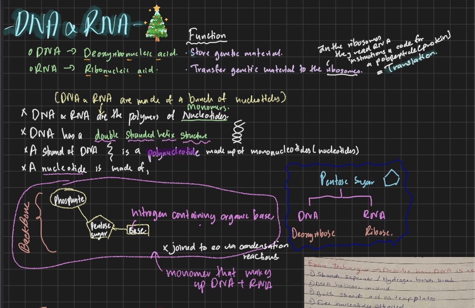
DNA
Deoxyribose nucleic acid
RNA
Ribonucleic acid
Function of dna and rna
Dna- store genetic materil
Rna- transfer genetic material to the ribosomes[in the ribosomes they need genetic material to code for a polypeptide [protein]
What are polymers of dna and rna
Nucleotides
Dna has a
Double stranded helix structure
A strand of dna is a
Polynuclotide made up of mononucleotides
A nucleotide is made of
Pentose sugar- ribose or deoxyribose sugar
Phosphate group
Base- nitrogenous organic base
the bases of nucleotides could be,
Adenine- - thymine they are COMPLEMENTARY BASE PAIRING
cytosine - - - guanine
HYDROGEN BONDS BEETWEN EM
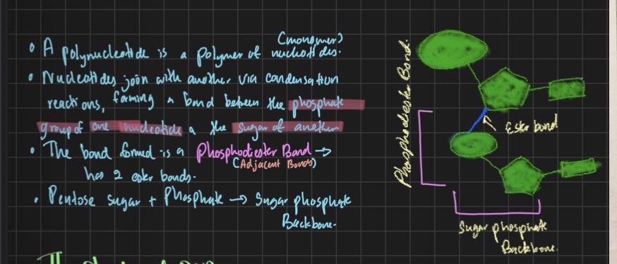
How os a phosphodiester bond formed
Structure of dna
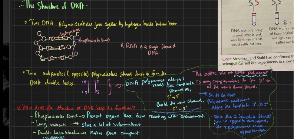
How does the structure of dna help ts function
Phosphodiester bond- prevnets organiic base from reacting with the environment
Large molecule- can store a lot of information
Double helix shape- makes dna compact
Weak hydrpgen bonds - can eaisly be broken down for dna replicaction;+ when a lot of hydrogen bonds are present its very strong so it provides stability
Double stranded- allows replication from template strands
Base sequence- allows info to be stored
Base stackng- makes dna stable
DNA REPLICATION - SEMI CONSERVATIVE REPLICATION
Dna helicase
Causes the strands to sepreate / breaking the hydrogen bonds
Both strands act as template strands
Free floating nucleotides are attached
Hydrogen bonds reform
Complementary base pairing occurs -AT and CG
Dna polymerase joins nucleotides via phosphodiester bonds
Dna has been replicated
Whats the name of dna replicaction
Semi conservative replicaction
What does phosphodiester and hydrofen bonds dop
Hydrogen bonds - joines nitrogous organic bases togther
Phosphodiester bonds - joins nucleotides togther to form a polynuclotide
What does dna helicase and polymerase do
Dna helicase - breakes hydrogen bonds
Dna polymerase- joins nucleotides via phosphodiester bonds
Who did the experiment on semi conservative replication
Melson and stahl
Differene bwteen dna nd rna
DNA | RNA |
Deoxyribose | Ribose |
Thymine | Uracil |
Double helix | Usually single polynucleotide chain |
Long | Relatively short |
Atp
Adenosine triphosphate - 3 phosphates
What is atp
Immediate sruce of energy in a cell
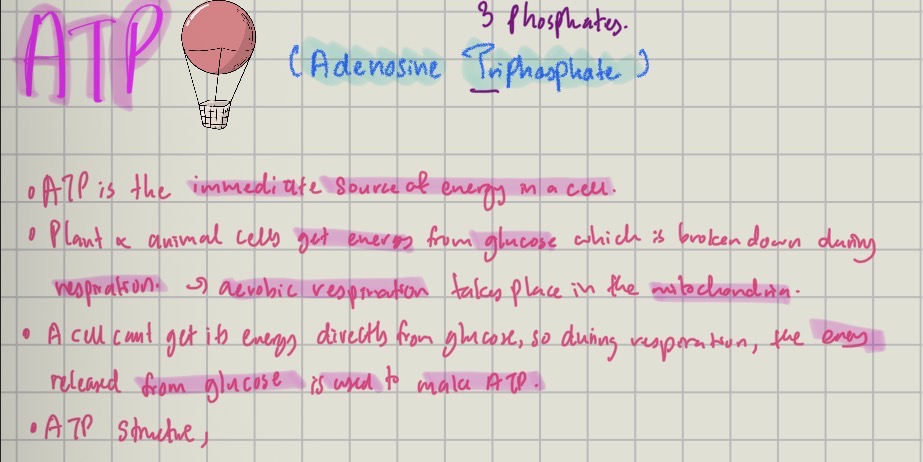
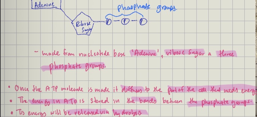
Whats the structure of atp
Adenine base, ribose sugar, 3 phosphte groups
Uses of ATP
Active transport- ions uptake at roots/ glucose absorption
Muscle contraction- movemrnt
Protein synthesis- for growth and repair
Phosphorylation- making molecules more reactive by lowering their activation eergy
In rna the base thymine is swapped with
uracil
unsaturated fatty acids
at least one double bond betwen carbon atoms
caues the chain to kinkk
phospholipids
found on cell surafce membrasnes
phosphate group replaces a fatty acid
phosphate group is hydrophillic [attrats water] fatty acid tails are hydrophobic
![<ol><li><p>found on cell surafce membrasnes </p></li><li><p>phosphate group replaces a fatty acid </p></li><li><p>phosphate group is hydrophillic [attrats water] fatty acid tails are hydrophobic </p></li></ol><p></p>](https://assets.knowt.com/user-attachments/b7c1b664-f6a3-4d6e-a219-639303390324.jpg)
phospholipid syracture
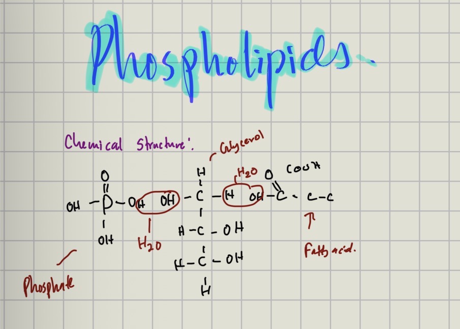
describe phospholipids
hydrophillic head and hydrophobic tail- allows he formation of phsopholipid bilayer
glycolipids can form - allows cell recognition
triglycerides structure
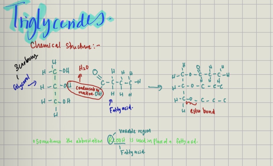
describe an explain tiglycerides
insoluble- osmotically inactive
high ratio of H;O atoms- release water when needed
high ratio of C;H - excellent storage of enegry
kow mass; enegry ratio- dont need to carry a large mass
test for lipids - emulsition
1.grind the sample
add ethanol
shake
add distilled water
white emulsion formed
phospholipids
What is a triglyceride?
A lipid formed by condensation of one glycerol molecule with three fatty acid molecules.
Three ester bonds are formed.
What is an ester bond?
The bond formed between glycerol and a fatty acid during a condensation reaction.