Abdominal Regions & Anterolateral Abdominal Wall (Sloan)
1/72
There's no tags or description
Looks like no tags are added yet.
Name | Mastery | Learn | Test | Matching | Spaced |
|---|
No study sessions yet.
73 Terms
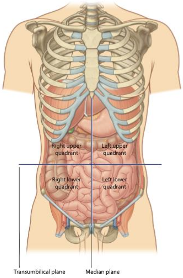
The abdominal cavity is divided using a vertical median plane and a horizontal transumbilical plane.
These planes intersect at the umbilicus.


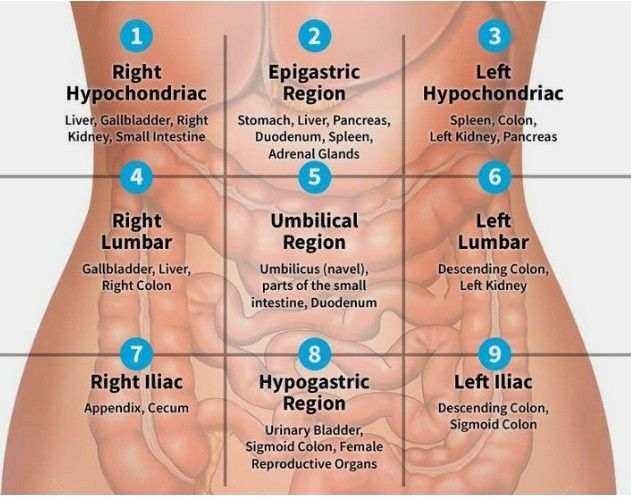
The nine abdominal regions are: Right Hypochondriac, Epigastric, Left Hypochondriac, Right Lumbar, Umbilical, Left Lumbar, Right Iliac, Hypogastric, and Left Iliac.
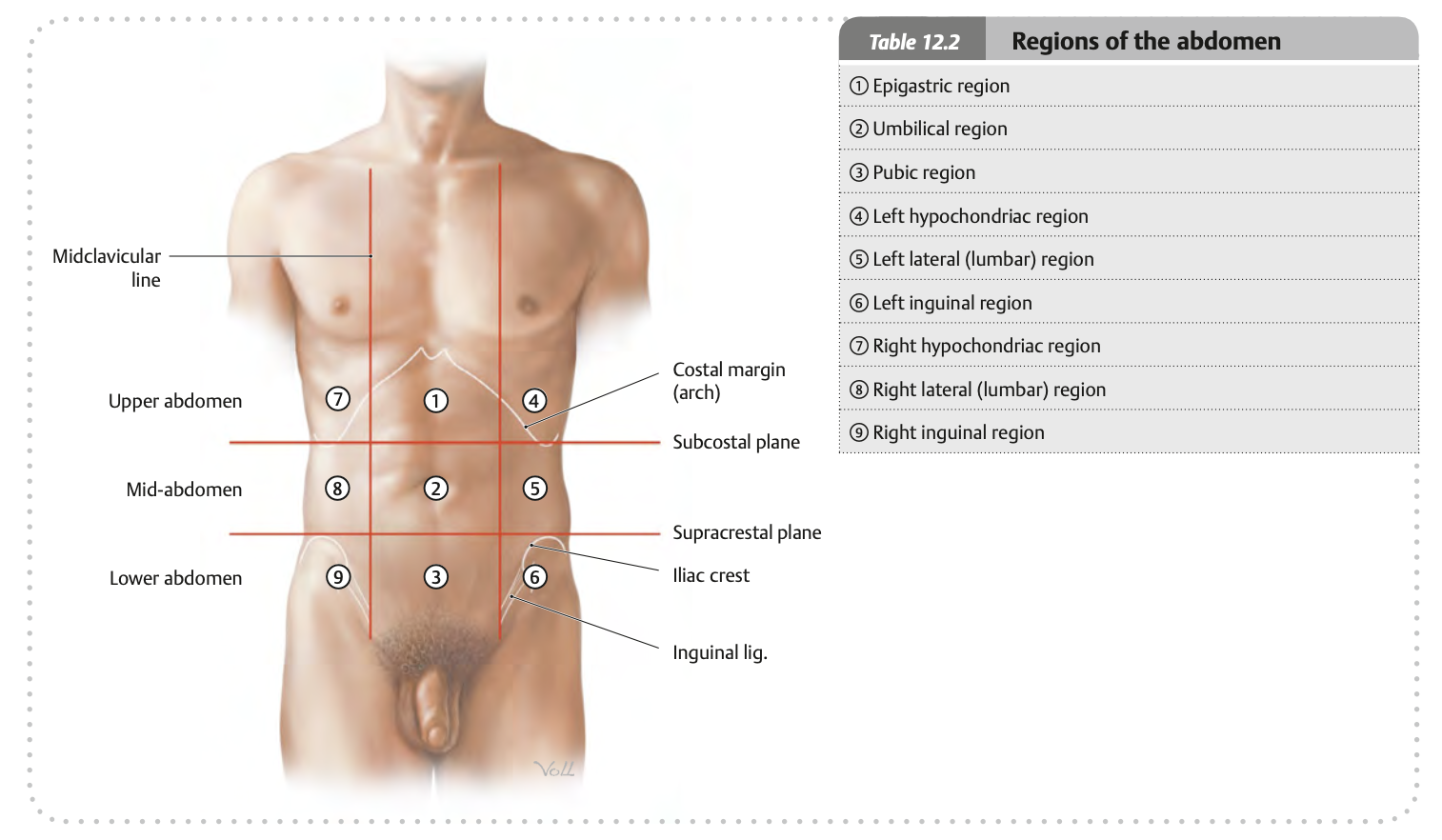


The superior boundary of the abdominal wall is the costal

margin.
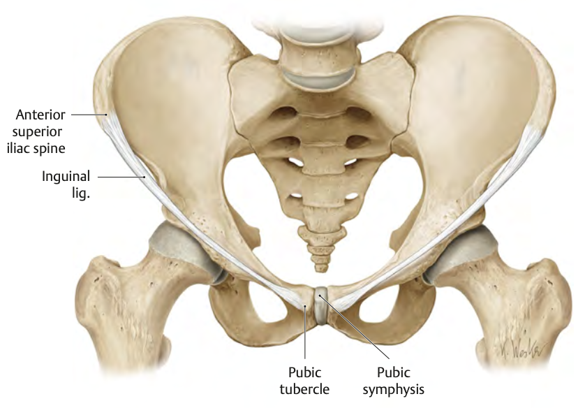
The inferior boundary of the abdominal wall is the anterior superior iliac spine (ASIS), inguinal ligament, and pubic symphysis.
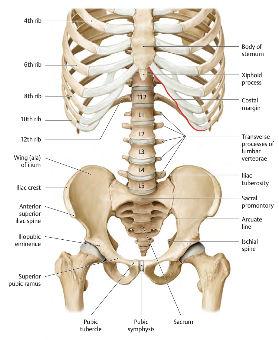
The xiphoid process is located at the inferior end of the sternum, around the T9 vertebra.

The iliac crest is the superior border of the iliac bone and is palpable on the hips.


The functions of the abdominal wall muscles include supporting abdominal viscera, assisting in breathing, protecting internal organs, and aiding in posture and movement.
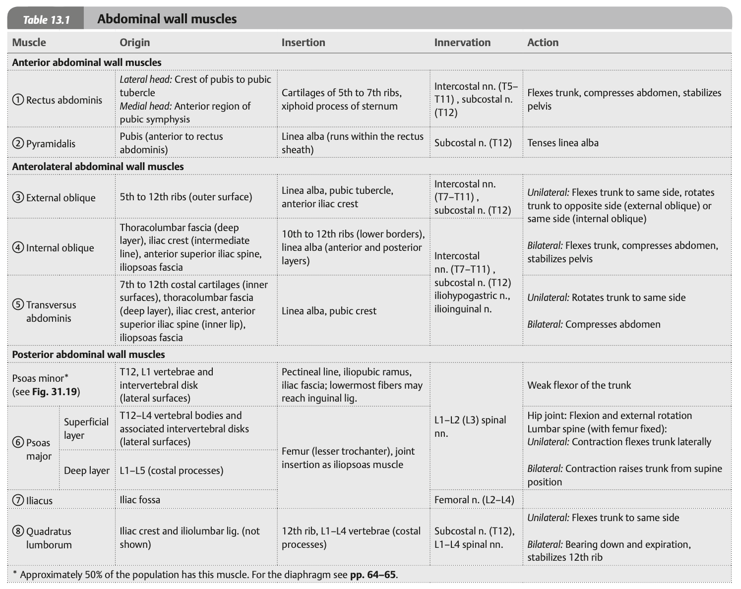
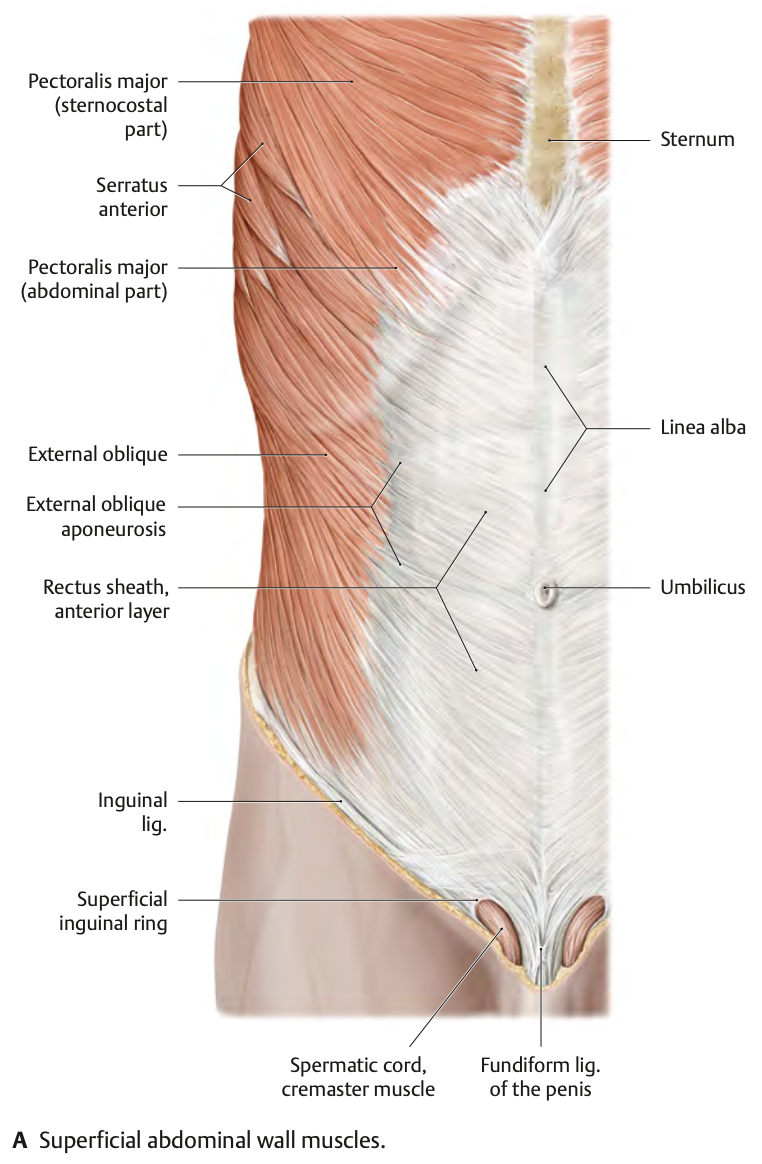
The external oblique muscle is responsible for trunk rotation and compressing the abdomen.

The internal oblique muscle lies underneath the external oblique and assists in trunk rotation and abdominal compression.

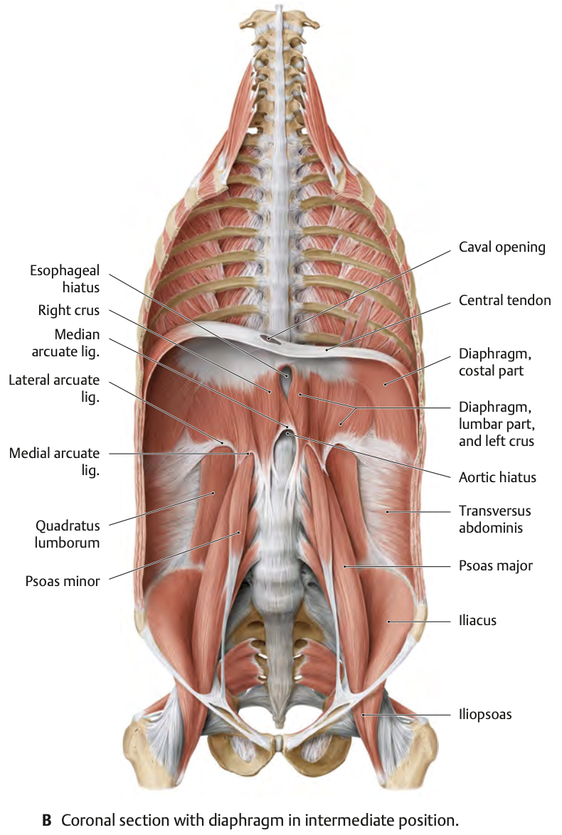
The transversus abdominis is the deepest abdominal muscle and functions to compress the abdominal contents.

The rectus abdominis is located along the midline and functions to flex the vertebral column and compress the abdominal cavity.

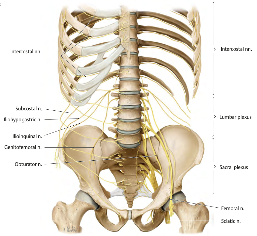
The muscles are innervated by the lower six thoracoabdominal nerves (T7-T11), the subcostal nerve (T12), and the iliohypogastric and ilioinguinal nerves (L1).
The arcuate line is located approximately halfway between the umbilicus and the pubic symphysis. Below this line, the rectus sheath is incomplete, leaving the posterior surface of the rectus abdominis muscle in direct contact with the transversalis fascia.

The arcuate line marks the point of transition where the aponeuroses of the abdominal muscles change, affecting the rectus sheath.
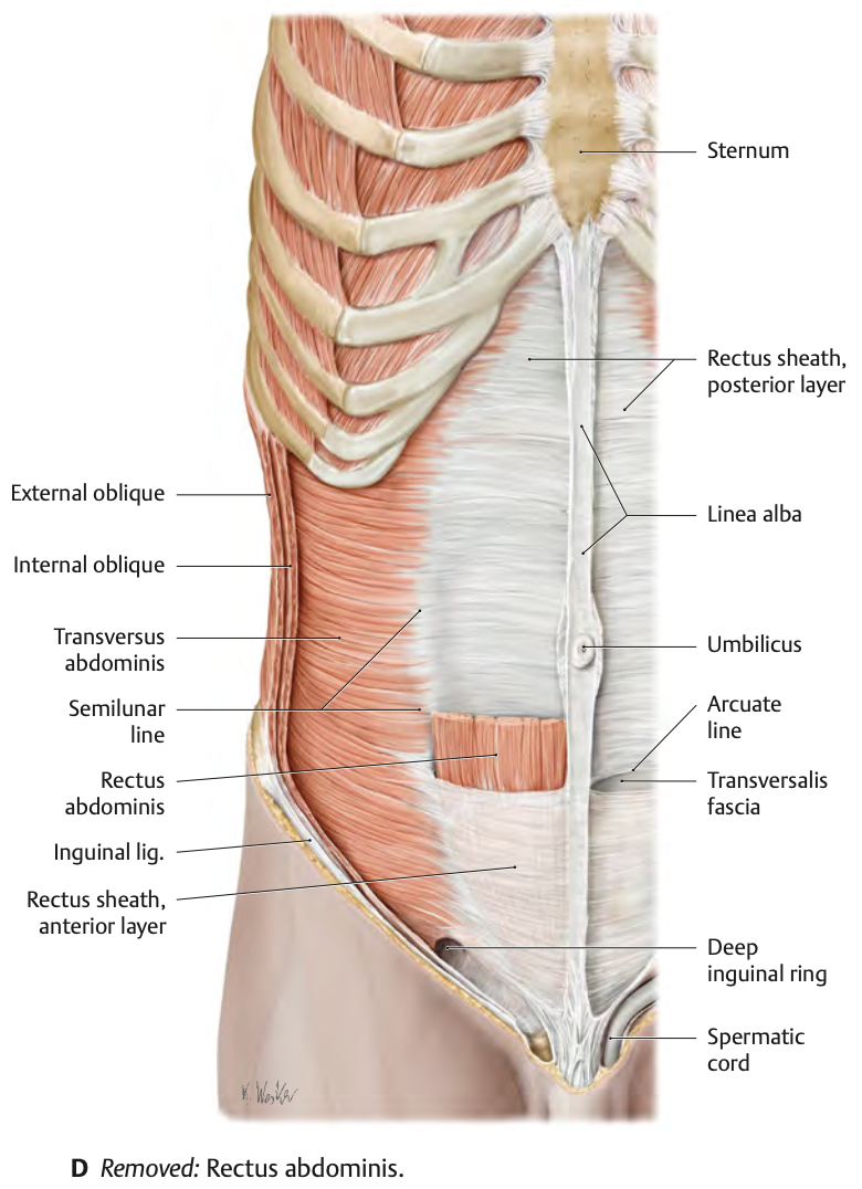
The linea alba is a dense band at the midline of the abdomen, acting as the attachment site for muscles.


The inguinal ligament is a folded border of the external oblique muscle aponeurosis, extending between the ASIS and the pubic tubercle.
The aponeuroses that make up the rectus sheath are bilaminar, meaning each has both a posterior and anterior lamina. These aponeuroses enclose the rectus abdominis muscle.
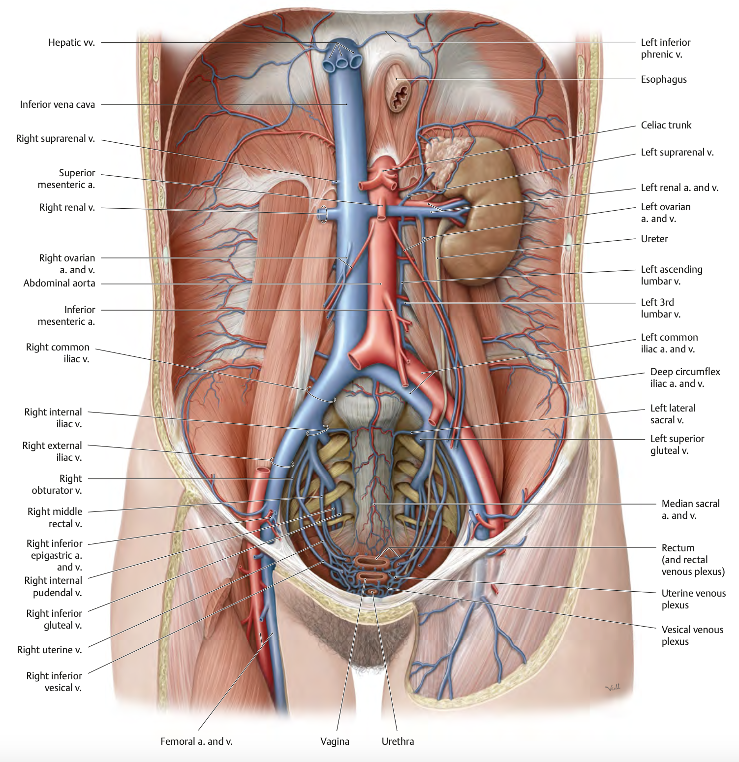
Structures passing through the diaphragmatic apertures include the inferior vena cava (T8), esophagus (T10), aorta (T12), and various nerves. Compression of these structures can lead to problems with blood flow, breathing, and circulation.
The diaphragm is supplied by the superior epigastric artery, superior phrenic arteries, musculophrenic and pericardiophrenic arteries, inferior epigastric artery, and inferior phrenic arteries.
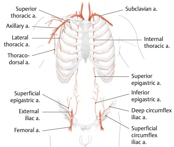
The diaphragm is drained by the phrenic veins, which empty into the inferior vena cava and internal thoracic vein.