6.7 - Response to infection
 Call Kai
 Learn
Learn Practice Test
 Spaced Repetition
 Match
Match Flashcards
 Knowt Play
1/31

There's no tags or description
Looks like no tags are added yet.
Name | Mastery | Learn | Test | Matching | Spaced | Call with Kai | Chat |
|---|
No analytics yet
Send a link to your students to track their progress
32 Terms
What is the immune system?
The body’s defence against disease:
made up of leukocytes (white blood cells) → found in the blood, lymph, tissue fluid & body cavities (e.g. alveoli)
What components make up the blood?
Plasma (aqueous solution): transports nutrients, waste, hormones, proteins & heat
Erythrocytes (red blood cells): transport oxygen & carbon dioxide
Leukocytes (white blood cells): responsible for immune response
Thrombocytes (platelets): responsible for blood clotting
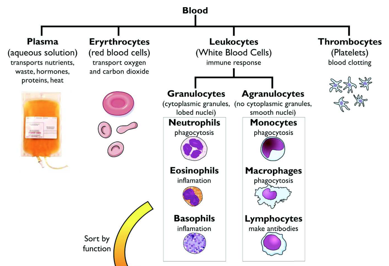
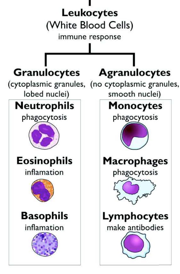
What are the two types of leukocytes (white blood cells) ?
Granulocytes (cytoplasmic granules, lobed nuclei):
neutrophils → phagocytosis
eosinophils & basophils → inflammation
Agranulocytes (no cytoplasmic granules, smooth nuclei):
monocytes & macrophages → phagocytosis
lymphocytes → make antibodies
What are the two types of immune system?
Non-specific immune system & specific immune system
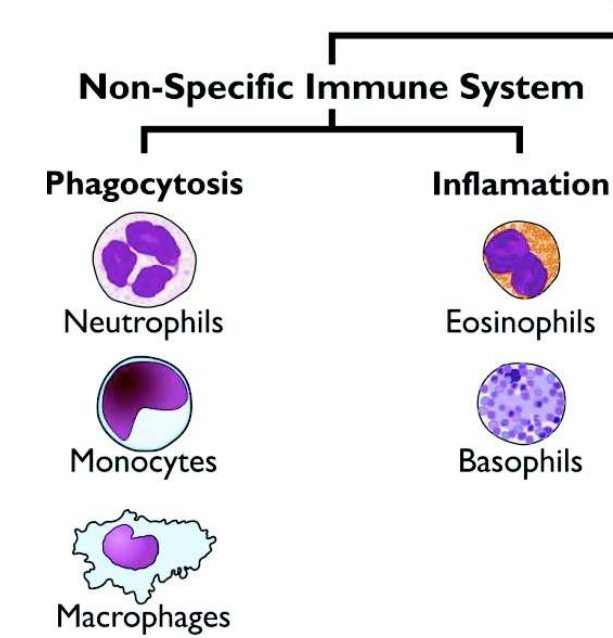
What components make up the non-specific immune system?
Phagocytosis:
neutrophils
monocytes
macrophages
Inflammation:
eosinophils
basophils
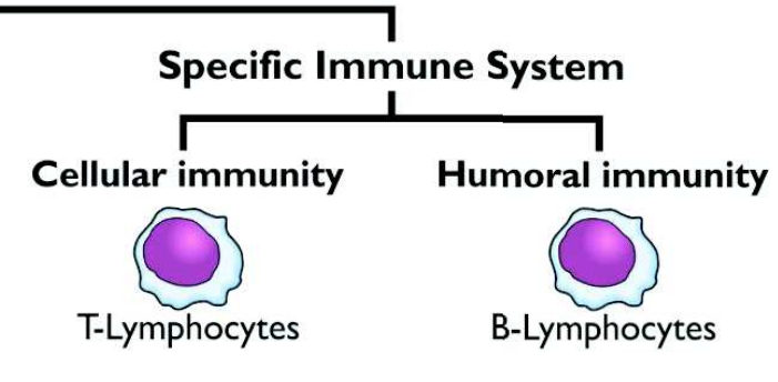
What components make up the specific immune response?
Cellular immunity: T-lymphocytes
Humoral immunity: B-lymphocytes
What is the non-specific immune system?
A collection of general methods of destroying foreign bodies that have entered the body:
main methods → phagocytosis & inflammation
What is phagocytosis?
Digestion of pathogens by phagocytes (neutrophils, monocytes & macrophages):
phagocytes are large, irregularly-shaped white blood cells with a complex cytoskeleton, allowing them to move & change shape
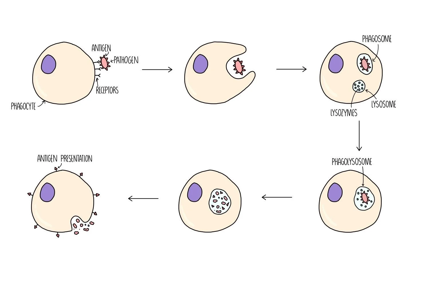
Outline the process of phagocytosis
Chemicals from pathogens attract phagocytes to the infection site
Phagocytes detect non-self antigens on pathogens using receptor proteins on their membrane → receptors bind to antigens on the pathogen’s surface
Phagocyte’s membrane extends around pathogen, forming a phagocytic vacuole (phagosome)
Phagosome fuses with a lysosome, forming a phagolysosome
Lysozymes are released to hydrolyse pathogen (e.g. break down bacterial cell wall)
In macrophages, digested pathogen fragments may be displayed on cell surface to activate specific immune response
Where do the different phagocyte cells work?
Neutrophils: circulate in the blood
Macrophages & monocytes: found in lymph, tissue fluid & lungs where they kill pathogens before they enter the blood
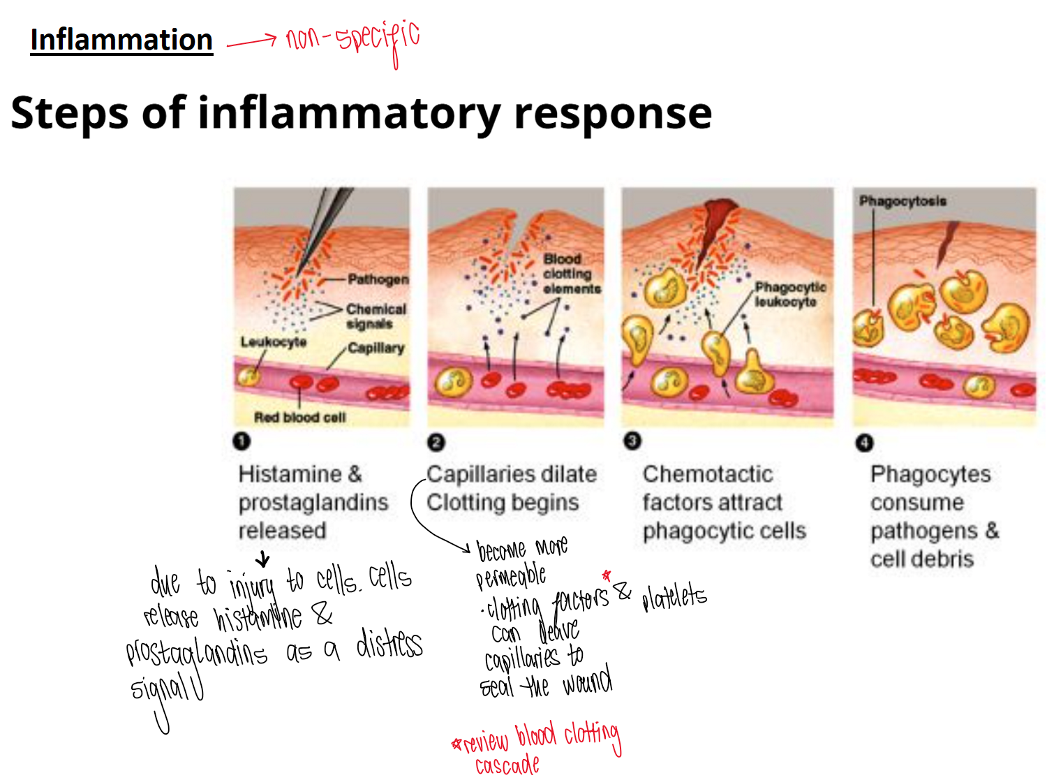
What is inflammation?
Localised response to an injury or infection, driven by granulocyte cells (eosinophils & basophils)
What do granulocytes release during inflammation?
Chemicals (e.g. histamines) which stimulate:
vasodilation → increase blood flow to the area, turning area red
capillary leakage → phagocytes & granulocytes can enter local tissue fluid
sensory neurone impulses → area is tender/painful
blood clotting → seals wound, forming scab
fever → raise body temperature, which is more harmful to pathogens than humans
What is the specific immune system?
Reactions that not only kill invading pathogens, but also leaves a ‘memory’ of the pathogen, so that it can be killed quickly on subsequent infections
What does the specific immune system involve?
Lymphocyte cells (made in bone marrow):
B-lymphocytes
T-lymphocytes
What is the key feature of the specific immune system?
Capable of recognising foreign cells as distinct from its own cells (i.e. self/non-self recognition) → does this by making use of antigens
What are antigens?
Large molecules that bind to lymphocyte cells & trigger a specific immune response
What are antibodies?
A protein molecule that can bind specifically to an antigen (made by B-lymphocytes)
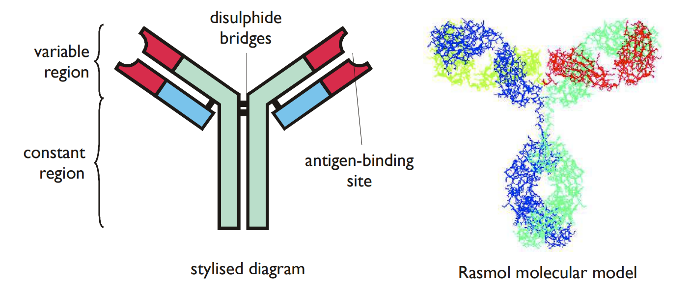
What is the structure of an antibody?
Composed of 4 polypeptide chains (2 heavy & 2 light chains) joined by strong disulphide bonds to form a Y-shaped structure
Stem of the Y is called constant region → all antibodies have same amino acid sequence, so same structure
Ends of the arms are called variable regions → different antibodies have different amino acid sequence, so different structures
Variable regions are where the antigens bind to form a highly specific antigen-antibody complex
Describe IgG & IgM antibodies
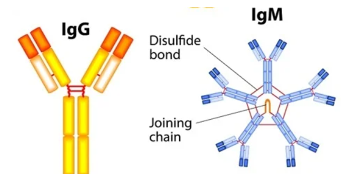
IgG:
most abundant antibody in plasma & mainly involved in secondary (humoral) immune response
the only antibody able to cross the placenta, providing passive immunity to the foetus & new-born baby
plays a crucial role in passive & active immunity, as well as promoting phagocytosis
IgM:
mainly involved in primary immune response & makes up ~5% of serum antibodies
found mainly in the blood & exists as a pentamer → cannot cross placenta
provides rapid initial immune response & plays a role in regulating immune function & maintaining immunological tolerance
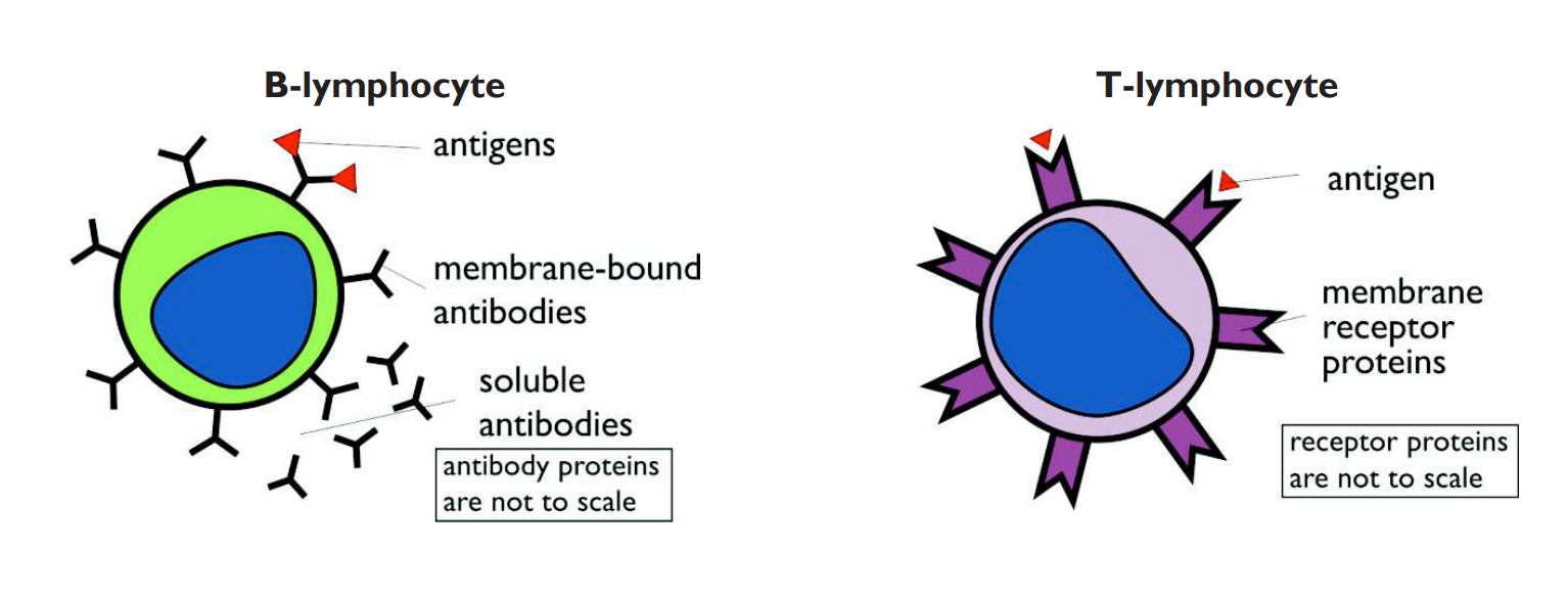
What do T-lymphocytes have on their surfaces?
Receptor proteins:
only have one binding site & are only found on the surface of T-lymphocytes → never free in solution
receptor proteins bind specifically to antigens to form antigen-receptor complexes
Outline the humoral / B cell immune response
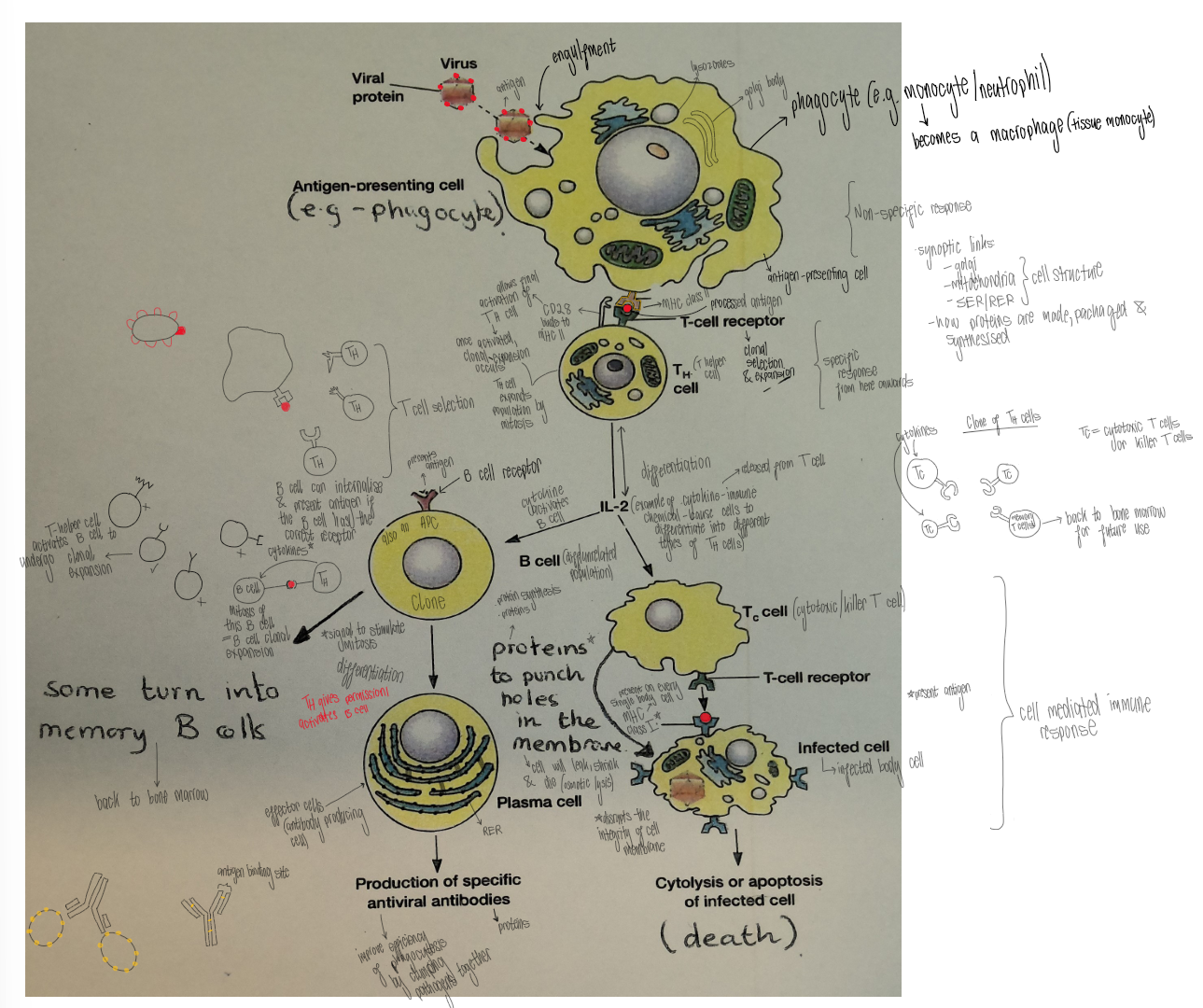
Pathogen is engulfed by macrophage during phagocytosis
Macrophage digests pathogen & processes antigen molecules
Macrophage presents antigen on MHC class II → macrophage acts as antigen-presenting cell
Macrophage encounters T helper cell with complementary T-cell receptor
T-cell receptor binds to antigen-MHC class II complex. CD28 binds to MHC class II, activating T helper cell
Activated T helper cell releases cytokines → causes clonal expansion of T helper cells by mitosis
A B cell with a complementary B cell receptor binds to antigen
B cell engulfs antigen & presents it on its B cell receptor
→ B cell acts as antigen-presenting cellActivated T helper cell uses T cell receptor to bind to antigen on B cell surface → T helper cell releases cytokines, causing B cell to undergo clonal expansion
B cells differentiate into:
plasma cells → produce large quantities of specific antibodies (bind to antigens, enhancing phagocytosis by clumping pathogens together)
memory B cells (remain in the body, ready for second infection with same pathogen → enables faster secondary immune response)
Outline the cell-mediated immune response
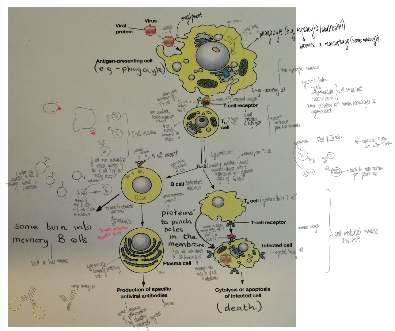
Pathogen is engulfed by macrophage during phagocytosis
Macrophage digests pathogen & processes antigen molecules
Macrophage presents antigen on MHC class II → macrophage acts as antigen-presenting cell
T helper cell with complementary T-cell receptor binds to the antigen-MHC II complex. CD28 binds to MHC class II → activates T helper cell
Activated T helper cell releases cytokines → undergoes clonal expansion by mitosis
Cytokines activate cytotoxic T cells, which identify infected cells
Infected body cell presents antigen on MHC class I (acts as antigen-presenting cell) & cytotoxic T cell binds to the antigen-MHC class I complex by its T-cell receptor
Cytotoxic T cell releases proteins, which punch holes in the cell membrane, causing osmotic lysis & death of infected body cell
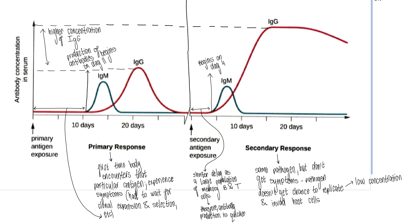
Describe the primary immune response
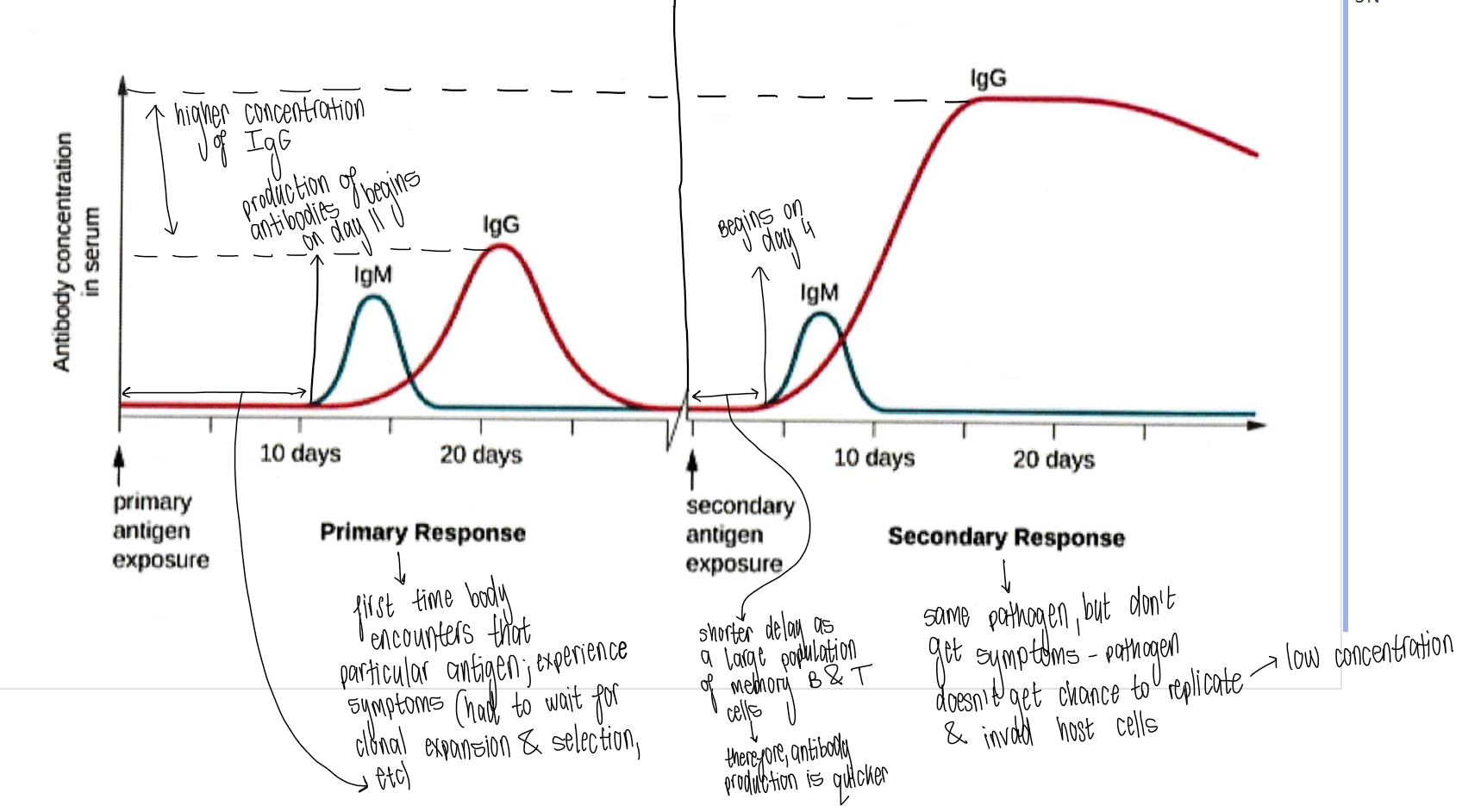
Slow & weak:
when an antigen is initially encountered, there are only a few lymphocyte cells of each kind for the antigen to encounter → can take several days for clonal selection to occur & clone army to be assembled (also tends to be small)
during this period, symptoms of the disease are shown, partly due to toxins & cell death due to the pathogen & partly due to immune response itself (e.g. fever & inflammation)
production of antibodies begins ~day 11
IgM produced first as it can capture 10 antigens simultaneously
Describe the secondary immune response
Faster & greater:
primary response memory cells (T & B lymphocytes) remain in the blood → after a subsequent infection by the same antigen, clonal selection stage can be by-passed (more clone B & T lymphocytes & antibodies are produced)
pathogen is destroyed before it reproduces enough to cause disease (individual is immune to that disease & no symptoms are shown)
production of antibodies begins ~day 4
What is active immunity?
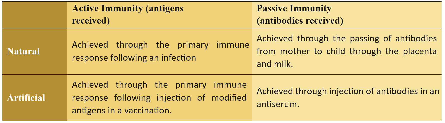
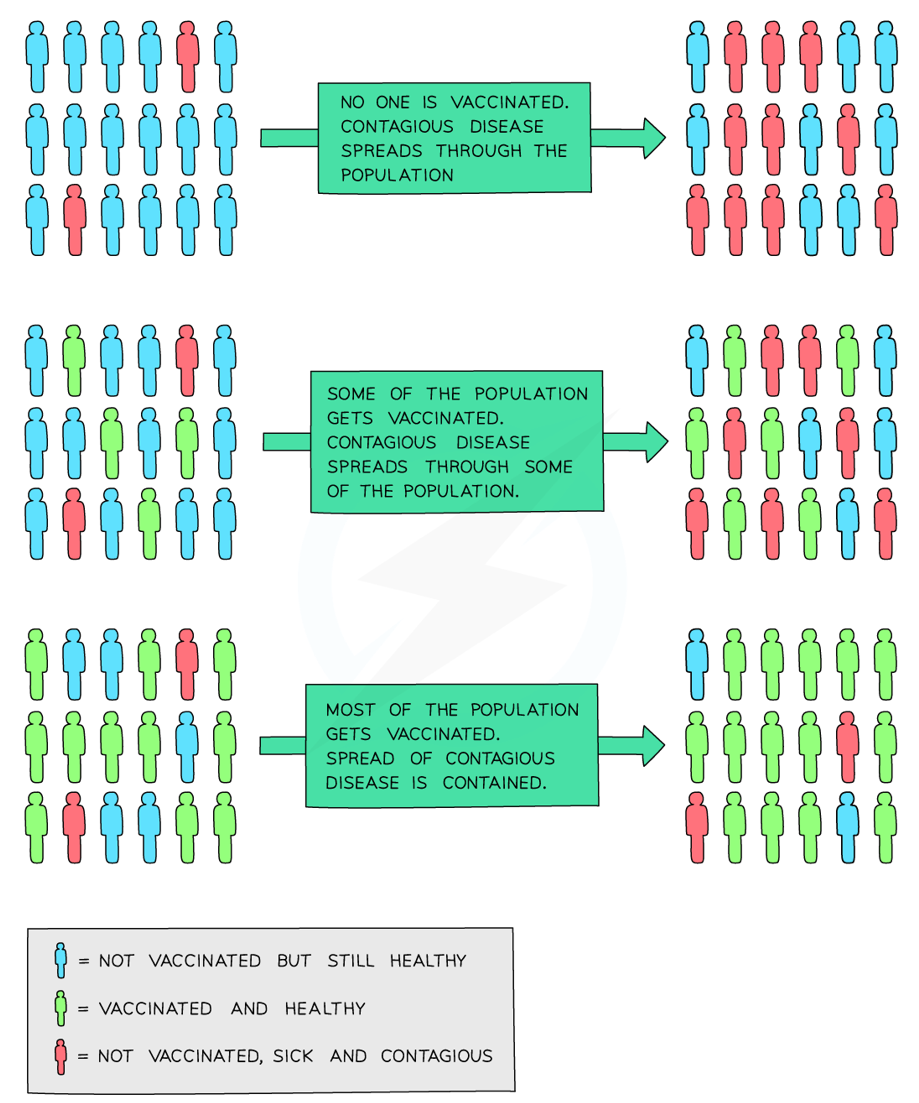
Your own immune system is activated
You make your own antibodies & memory cells
Protection is usually long-lasting
What is the difference between active artificial & active natural immunity?
Active artificial immunity:
gained by vaccination
harmless form of antigen (dead/weakened) is introduced → immune system responds, resulting in production of antibodies & memory cells
results in long-term immunity without getting the disease
Active natural immunity:
gained by natural exposure to a pathogen (e.g. catching chickenpox/infectious disease)
pathogen enters → immune system responds, resulting in production of antibodies & memory cells
results in long-term immunity (often for life)
What is passive immunity?
Antibodies are received, but not made by the body
No memory cells are formed
Protection is immediate but short-lived
The immune response is not stimulated
What is the difference between passive artificial & passive natural immunity?
Passive artificial immunity:
gained by injection of pre-formed antibodies (e.g. antivenom/tetanus antibodies)
antibodies act immediately against the antigen
doesn’t stimulate immune response, but simply supports immune response by binding to antigens
results in immediate, short-term protection (antibodies break down after ~1 month)
Passive natural immunity:
gained by antibodies passing naturally from mother to baby across the placenta (IgG) or through breast milk (IgA)
baby receives ready-made antibodies
results in short-term protection in early life
What is herd immunity?
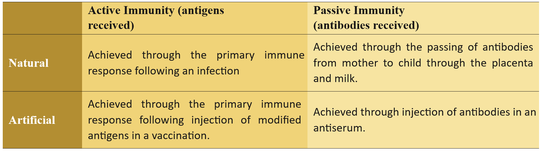
A type of disease immunity that occurs when a large proportion of a population are vaccinated against a disease which prevents the spread of disease to unvaccinated individuals
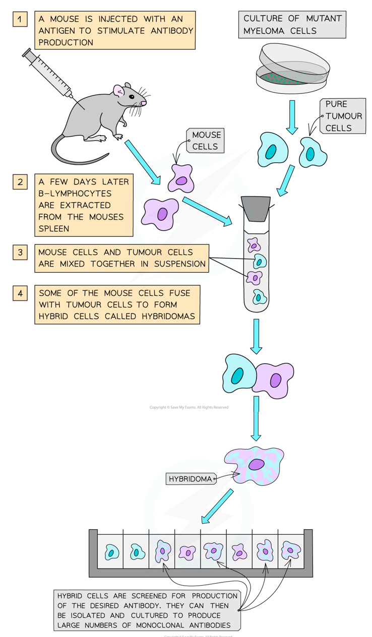
What are monoclonal antibodies & how are they produced?
Identical antibodies produced from a single clone of hybridoma cells, specific to one antigen (produced by the hybridoma method):
A mouse is injected with an antigen to stimulate antibody production
A few days later, B-lymphocytes are extracted from the mouse’s spleen
Mouse cells & tumour cells are mixed together in suspension, where some of the mouse cells fuse with tumour cells to form hybridomas (hybrid cells)
Hybridomas are screened for production of desired antibody, which can then be isolated, cultured & cloned to mass-produce monoclonal antibodies
What are the uses of monoclonal antibodies?
Medical diagnosis: pregnancy tests & pathogen detection (e.g. cancer/HIV)
Treatment: cancer therapy, autoimmune disease, viral infections
Research: protein detection & tagging
What are the limitations/risks of monoclonal antibodies?
Immune response triggered → mouse antibodies seen as foreign & so, rejected
Side effects (e.g. inflammation)
Expensive to produce
Ethical issues (animal use)