Exam 2 NEU 534
 Call Kai
 Learn
Learn Practice Test
 Spaced Repetition
 Match
Match Flashcards
 Knowt Play
1/131

There's no tags or description
Looks like no tags are added yet.
Name | Mastery | Learn | Test | Matching | Spaced | Call with Kai | Chat |
|---|
No analytics yet
Send a link to your students to track their progress
132 Terms
Direct Pathway
“go pathway”
decreases basal ganglia inhibition of thalamus
allows movement
Indirect Pathway
“no go pathway”
increases basal ganglia inhibition of thalamus
blocks movement
D1 receptors
excitatory dopamine recepto
couples with Gs protein to activate adenylate cyclase, increasing cAMP
D2 receptors
inhibitory dopamine receptor
couples with Gi protein to inhibit adenylate cyclase, reducing cAMP
Tyrosine hydroxylase (TH)
converts tyrosine into levodopa (L-DOPA)
L-amino acid decarboxylase (LAADC)
Converts levodopa (L-DOPA) into dopamine
Levodopa (L-DOPA)
Precursor to dopamine
F-DOPA
when used in PET scans of Parkinson’s patients, less uptake = damage to dopaminergic neurons
Primary motor symptoms of parkinson’s
tremor at rest, bradykinesia, rigidity
Bradykinesia
slowness of movement
Inputs to basal ganglia are x, outputs from BG are y
excitatory/glutamatergic, inhibitory/GABAergic
Lewey bodies
found in Parkinson’s and other diseases, contain alpha-synuclein
present in both sporadic and familial Parkinson’s
may play a cytoprotective role
Motor symptoms in Parkinson’s appear after what?
80-90% of substantia nigra par compacta neurons are lost
Prodromal symptom of Parkinson’s
constipation
not diagnostic, often appears in patient history years before diagnosis
SNCA
Genetic risk factor for Parkinson’s
Autosomal dominant
Encodes alpha-synuclein
Aggresomes
segregate and facilitate degradation of damaged/mutated/misfolded proteins
SNc dopamine neurons are particularly vulnerable due to
metabolic stress and oxidative stress
Causes of metabolic stress in SNc
energetically expensive dendritic arbors and long, unmyelinated axons
autonomic pacemaker activity
Causes of oxidative stress in SNc
High levels of dopamine and its metabolites, which produce ROS
Increased oxidative stress combined with mitochondrial dysfunction → impairment of lysosomal autophagy system
Lysosomal autophagy system (LAS)
Involved in removal of alpha-synuclein
MPTP
contaminant of MPPP (synthetic opioid)
causes symptoms of parkinson’s
selective inhibitor of ETC complex 1
Rotenone
Herbicide
Linked to parkinson’s
ETC complex 1 inhibitor
Oxidative stress
Production of reactive oxygen species (ROS) outweighs removal by antioxidant enzymes
Superoxide dismutase
an antioxidant enzyme
can turn superoxide radical into hydrogen peroxide
Most common oxygen free radicals
hydroxyl radical and superoxide radical
hydrogen peroxide
not a free radical, but reactions can lead to production of free radicals
Catalase
Converts hydrogen peroxide into water and oxygen
Dopamine-o-quinone
Oxidized from dopamine
Reactive intermediate
Can be turned into 6-hydroxydopamine
6-hydroxydopamine
Damaging compound that is derived from dopamine-o-quinone, a reactive intermediate of dopamine
Generates significant superoxide radical by inhibiting ETC complex 1
Neuromelanin
gives SNc its pigmentation
sequesters toxic byproducts of dopamine metabolism
alterations contribute to processes that lead to PD
ETC and ROS
inevitable electron leakage during oxidative phosphorylation
ETC complexes 1 & 3 → diffusion into mitochondrial matrix and intermembrane space
generate superoxide radical
Factors that can contribute to electron leakage
Advancing age, environmental toxins (MPTP, rotenone), or misfolded proteins (alpha-synuclein)
cytochrome c
pro-apoptotic factor
mice injected with alpha-synuclein had less what?
tyrosine hydroxylase positive neurons in the substantia nigra
N-terminal region of alpha-synuclein
Several familial Parkinson’s mutations (A53T, E46K, and A30P) found in this location
Central region of alpha-synuclein
High tendencies to produce beta-pleated sheets that are prone to aggregation
C-terminal region of alpha-synuclein
rich in proline and negatively charged residues (common characteristic of intrinsically disordered proteins)
Alpha-synuclein represents what % of the total protein population?
1
Under physiological conditions, alpha synuclein exists in what state?
Soluble random coil state
What prevents misfolding of alpha-synuclein?
chaperones, proteasomes, phagosome/lysosome systems
What aspect of the aging brain contributes to the accumulation of misfolded alpha synuclein?
A progressive decline in proteolytic defense mechanisms
Amyloid fibrils
Neuronal death in parkinson’s disease
excess oxidative stress
misfolding of alpha-synuclein
misfolded alpha-synuclein is degraded by proteosomes
misfolded alpha-synuclein is sequestered into Lewey bodies
misfolded alpha-synuclein increases ROS production in mitochondria
mitochondria become dysfunctional, cant produce ATP
impaired ATP and increase in ROS leads to toxicity and death
Other contributing factors include dopamine oxidation
What happens to normal fetal cells transplanted into the SN of Parkinson’s patients?
they develop Lewey bodies
Misfolded alpha-synuclein can do what to normal alpha-synuclein?
seed
Cell-to-cell spread
misfolded alpha-synuclein can spread between neurons
Alpha-synuclein oligomers can cross what?
the blood-brain barrier
Braak hypothesis
possible mechanism for spread of misfolded alpha-synuclein from gut to brain
Carbidopa
Prevents levodopa from being converted into dopamine in the PNS
Deep brain stimulation
changes firing rate and pattern of individual neurons in the basal ganglia
promotes release of local neurotransmitters
increases blood flow
stimulates neurogenesis
Potential mechanism of DBS
may reduce hypersynchronized circuits
DBS electrodes record what?
local field potentials
Beta-sheet rich secondary structure
shared by all disease causing proteins
cross beta structure
beta sheets are perpendicular to the long axis of the fiber
amyloidgenesis
Conversion from normal alpha-helical rich conformation to pathological beta-sheet rich
Tau and amyloid beta
implicated in Alzheimer’s
Prion proteins
implicated in CJD, mad cow disease, kuru, chronic wasting disease, scrapie
Prion
proteinaceous infectious particle
proteins that acquire alternative conformations that become self propagating
Protein only hypothesis
prions are misfolded proteins that are infectious in the absence of nucleic acids
PRNP gene
encodes cellular prion protein
mutations implicated in familial Creutzfeldt-Jakob disease
Transmissible Spongiform Encephalopathies
Accumulation of prion protein causes translational repression of global protein synthesis that leads to neuronal death
Mechanism of neuronal death in Transmissible Spongiform Encephalopathies
may involve oxidative stress and excitotoxicity
Sporadic Creutzfeldt-Jakob disease
no mutations in PRNP gene
New cases of sCJD annually
1 million world wide, 350 in the united states
Median survival time after onset of CJD symptoms
4 months
Deletion of PRNP in mice
Leads to immunity to prion disease
Inheritance of fCJD
autosomal dominant
Iatrogenic
disease caused by medical intervention
Cadaveric human growth hormone
used to treat children with short stature from the 50s through 1985
contamination with prions led to CJD in some patients
some patients also developed amyloid-beta deposits
mice expressing human amyloid beta injected with contaminated chGH
developed amyloid beta plaques
vCJD
occurs when prions from cows with bovine spongiform encephalopathy spreads to human
231 cases reported as of 2018
PrP Sc
disease causing form of prion protein
has different biochemical properties than PrP c - for instance, it is protease resistant
Prion disease species barrier
four residues in protein are implicated - Asn108, Met112, Met129, and Ala13
PrP c
“normal” form of prion protein
glycosylphosphatidylinositol-anchored plasma membrane glycoprotein
may function within lipid rafts to regulate various signal
transduction pathways
Small oligomeric assemblies of PrP Sc
Thought to be primary causative agent of prion disease pathology
protein folding “fast track”
alpha and beta domains fold concurrently and form intermediate conformation
protein folding “slow track”
chain becomes trapped in long-lived intermediate with persistent structure only in the alpha domain
further folding requires transition over high energy barrier or partial unfolding
unfolded state of protein
has high conformational entropy
nucleation is [ ] for most proteins in physiological settings
extremely unlikely
lag phase
prion monomers self-assemble into amyloid fibers after this
it is eliminated by addition of preformed prion oligomer seeds
primordial polypeptide conformation
may be represented by beta-sheet-rich amyloid fibers
self replicating protein conformations in early life forms
amyloid conformations of fibrin-derived peptides
activate tissue type plasminogen activator
Sup35
yeast protein that regulates translation
propogates as a prion
Hsp104
hexameric yeast protein that
has two ATPase domains
Low concentrations of Hsp104
promotes Sup35 fiber assembly in two ways
binds ATP, Hsp104 catalyzes the formation of amyloidogenic Sup35 oligomers, eliminates lag
couples ATP hydrolysis to the severing of a small subset of short Sup35 fibers, which generates extra surfaces for conformational replication, accelerates fiber assembly
High concentrations
couples ATPase activity to the disassembly of amyloidogenic oligomers and fibers of Sup35
might minimize the half-life of amyloidogenic structures
promotes cell survival after exposure to environmental stress
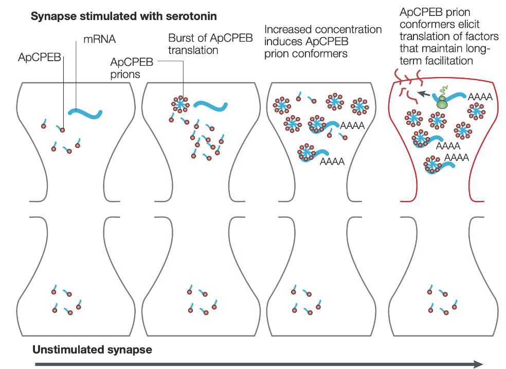
Aplaysia CPEB (ApCPEB)
regulates translation of mRNA
shown to have prion like properties
participate in maintenance of long-term potentiation
ApCPEB and long term potentiation
Sensory transduction
detection of stimulus energy and translation into electrical signal
Amplitude coding
size of electrical response in sensory receptor cell increases with strength of stimulus
receptor potentials (type of graded potential)
Frequency coding
stimulus intensity is coded by frequency of action potentials
Sensory adaptation
response of receptor cell diminishes or disappears in response to sustained stimulus
Sensory receptors respond best to what?
Changes in stimuli
Receptive field
Sub-region of a sensory surface to which an individual sensory receptor cell responses
Four types of somatic sensation
1. Touch – mechanotransduction
2. Proprioception – mechanotransduction
3. Temperature – thermotransduction
4. Nociception – multimodal: mechano-, thermo- & chemotransduction
Receptive fields in hands are?
smaller
Two-point discrimination
the ability to discern two closely spaced stimuli
Lateral inhibition
carried out by lateral inhibitory neurons at the second-order level
allows for two-point discrimination when receptive fields overlap
The difference between the directly stimulated regions on the skin and the immediately adjacent
neighboring areas is larger in the secondary neurons than in the primary sensory neurons themselves
Pacinian corpuscles
large receptive fields
very sensitive to vibration
respond best to stimulus onset and offset
Meissner’s corpuscles
small receptive fields
very sensitive to vibration
respond best to stimulus onset and offset