Pathophys Exam 1
 Call Kai
 Learn
Learn Practice Test
 Spaced Repetition
 Match
Match Flashcards
 Knowt Play
1/190

There's no tags or description
Looks like no tags are added yet.
Name | Mastery | Learn | Test | Matching | Spaced | Call with Kai | Chat |
|---|
No analytics yet
Send a link to your students to track their progress
191 Terms
L1
pathology
the causes of disease and the changes in cells, tissues, and organs that are associated with development of disease; same pathology does not mean same etiology/pathogenesis
etiology
the origin of a disease, including the underlying causes and modifying factors; can be multiple
ex: hypoxia, ischemia, toxins, infections, abnormal immune reactions, genetic abnormalities, nutritional imbalances, physical agents
pathogenesis
steps in disease development
ex: biochemical changes, structural changes
homeostasis
steady state of steady internal physical and chemical conditions
adaptation
a new steady state that preserved viability and function
reversible injury
changes in function and morphology that cells can recover from if damaging stimulus is removed
irreversible injury
inability to restore mitochondrial function even after removal of stimulus
altered structure and loss of function of plasma and intracellular membranes
loss of structural integrity of DNA and chromatin
leads to cell death
types of cellular stress
oxidative stress
ER stress and UPR
disruption of calcium homeostasis
oxidative stress
cellular damage induced by the accumulation of reactive oxygen species (ROS; form of free radicals)
unstable and “attack” cellular components
peroxidation of membrane lipids
crosslinking and modification of proteins
DNA damage
types of injury that cause oxidative stress —> by inc rate of production or dec clearance
chemical and radiation injury
hypoxia
cellular aging
tissue injury by inflammatory cells
ischemia-reperfusion injury
**normal cell functions can produce ROS so it needs to be maintained in a balance
cleared enzymatically or by antioxidants → block formation of free radicals or by scavenging
enzymes: glutathione peroxidase, catalase
chaperones
proteins in ER that ensure proper folding of newly synthesized proteins
Unfolded protein response (UPR)
adaptive response
inc chaperone expression → fix misfolded proteins & sense cellular stress
reduces protein synthesis
inc protein degradation
misfolded proteins in ER activate UPR via sensors (ex: IRE1)
ER stress
causes of intracellular accumulation of misfolded proteins
abnormalities that inc production of misfolded proteins
reduced ability to eliminate misfolded proteins
mutations in protein that is misfolding or UPR pathway
aging → dec adaptive capability w age
viral infections
intracellular pH and redox state
hypoxia
ischemia
high levels of misfolded proteins can trigger apoptosis via mitochondrial intrinsic pathway
injury leads to problems w protein folding
misfolded proteins can cause disease by
creating a deficiency of an essential protein - loss of function
inducing apoptosis
gaining a toxic function
proteasome
endopeptidase protein complex; cell’s trash can
Ubiquitin-proteasome system
proteins tagged by ubiquitin ligases
UPS proteolyzes ubiquitinated proteins
disruption of UPS plays role in disease
polyglutamine repeat disease
parkin in Parkinson’s disease
disruption of calcium homeostasis
intracellular Ca2+ → LOW
extracellular Ca2+ → HIGH
having high intracellular Ca2+ disrupts signaling pathways and activates enzymes (proteases, phospholipases) that damage cellular components (plasma membrane, cytoskeleton)
ischemia and certain toxins inc cytosolic Ca2+ due to release from intracellular stores in mitochondria and ER and later from across a damaged plasma membrane
cellular adaptations
hypertrophy
hyperplasia
atrophy
metaplasia
cellular adaptations to stress
reversible changes in the number, size, phenotype, metabolic activity, or functions of cells in response to changes in their environment; physiologic and pathologic
physiologic adaptation
the responses of cells to normal stimulation by hormones or endogenous chemical mediators, or to the demands of mechanical stress
pathologic adaptations
the responses to stress that allow cells to modulate their structure and function and thus escape injury, but at the expense of normal function
hypertrophy
inc cell size and organ size; NO inc in cell number
often in response to inc workload
induced by growth factors produced in response to mechanical stress or other stimuli
occurs in tissues with a limited capacity to divide
can progress to cell injury if the stress is not relieved or if it exceeds the adaptive capacity of the tissue
ex:
physiologic enlargement of uterus during pregnancy → caused by estrogen stimulus (hypertrophy + hyperplasia)
pathologic hypertrophy of the heart in response to inc workload (hypertrophy)
hyperplasia
inc cell number; inc proliferation in response to hormones and other growth factors
occurs in tissues whose cells can divide or contain abundant tissue stem cells
often occurs concurrently w hypertrophy in response to the same stimulus
stops when initial stimulus is removed
ex:
physiologic
hormonal → proliferation of the glandular epithelium of the female breast at puberty and during pregnancy
compensatory → residual tissue grows after removal or loss of part of an organ
pathologic
endometrial hyperplasia → inc uterine epithelial proliferation due to inc estrogenic stimulation
benign prostatic hyperplasia → disruption of androgens and estrogens leads to hyperplasia in the prostate
bone marrow cells
epidermal cells
epithelial cells
atrophy
dec cell and organ size
associated w dec synthesis of cellular building blocks and inc breakdown of cellular organelles
combo of dec protein synthesis + inc protein degradation (UPS)
accompanied by inc autophagy
may undergo apoptosis as atrophy worsens
both pathologic and physiologic causes
dec workload
loss of innervation
diminished blood supply
inadequate nutrition
loss of endocrine stimulation
aging
metaplasia
change in which one adult cell type is replaced by another adult cell type
often due to reprogramming of stem cells in response to chronic irritation
epithelial metaplasia → replacement of normal columnar epithelial cells of trachea w tough, stratified squamous epithelial cells - in chronic smokers
chronic gastric reflux → normal stratified squamous epithelium of the lower esophagus undergo metaplastic transformation to gastric or intestinal-type columnar epithelium
makes cells better able to withstand the stress
may result in reduced functions or inc propensity for malignant transformation
always pathological
high risk of malignant transformation and development of cancer
causes of cell injury
ex: hypoxia, ischemia, toxins, infections, abnormal immune reactions, genetic abnormalities, nutritional imbalances, physical agents
mechanisms of cell injury/death
cell response to injuries depends on type of injury and duration and severity
depends on type of cell and its metabolic state, adaptability, and genetic makeup
survival without oxygen of
skeletal muscle (2-3 hrs)
cardiac muscle (20-30 mins)
neurons (1-2 mins)
genetic diff in cytochrome p450 pathways
injury results from functional and biochemical abnormalities in one or more essential cellular components
morphological changes in reversible cell injury
cellular swelling
inc cell size
swollen organelles
accumulation of degenerated organelles and lipids within injured cells
morphology → pallor (bc of compression of capillaries), turgor, inc organ weight
microscopic changes → hydroponic change, vacuolar degeneration
common during hypoxia
fatty changes
lipid vacuoles in cytoplasm
common in organs inv w lipid metabolism (liver)
mitochondrial dysfunction and damage
due to hypoxia, ischemia, mitochondrial toxicants, radiation, high intracellular calcium
dec atp generation which leads to
reduced activity of plasma membrane atp-dependent sodium pumps → cellular swelling → dilation of ER
compensatory inc in anaerobic glycolysis → inc lactic acid accumulation → dec intracellular pH → dec activity of cellular enzymes
prolonged atp depletion leads to structural disruption of protein synthetic apparatus
detachment of ribosomes from rough ER
dissociation of polysomes
reduction in protein synthesis
formation of mitochondrial permeability transition pore (mPTP)
mitochondrial fragmentation
production of ROS → oxidative stress
prolonged damage leads to apoptosis
membrane damage
inc permeability → necrosis
damage due to inc ROS production, loss of ATP, and inc intracellular calcium
mitochondrial membrane
formation of mitochondrial permeability transition pore (mPTP) → reduction of atp production due to loss of membrane potential and pH changes
plasma membrane
loss of osmotic balance
influx of fluids and ions
loss of cellular contents
lysosomal membranes
leakage of enzymes into cytosol and break down necessary cellular components
these enzymes only function at low pH
DNA damage
exposure of cells to radiation or chemotherapeutic agents, intracellular generation of ROS, and acquisition of mutations may all induce DNA damage which may trigger apoptotic death
basic mechanism of cancer
sentinel proteins detect DNA damage and transmit signals that lead to p53 accumulation
p53 causes cell cycle arrest for DNA to repair itself before replication
if DNA damage is excessive → p53 triggers apoptosis via mitochondrial intrinsic pathway
TP53 (gene for p53) is mutated in more than 50% of tumors
intracellular depositions
abnormal accumulations caused by dec removal and degradation or inc production of endogenous substance or deposition of abnormal exogenous material
fatty change (steatosis)
abnormal accumulation of triglycerides within parenchymel cells; common in liver
cholesterol and cholesteryl esters → occur due to inc intake or dec catabolism of lipids; as seen in artherosclerosis
proteins
caused by inc uptake or synthesis, misfolding, dec degradation
glycogen
abnormalities in metabolism of glucose or glycogen
pigments
colored substances that may be exogenous (carbon) or endogenous (melanin)
extracellular deposits
pathologic calcification
dystrophic calcification
metastatic calcification
pathologic calcification
seen in a wide variety of disease states; result of abnormal deposition of calcium salts (fine white granules or clumps and gritty deposits)
dystrophic calcification
deposition of crystalline calcium phosphate in membrane bound vesicles
found in areas of necrosis
seen in plaques in atherosclerosis and necrotic cells in tuberculosis
can cause dysfunction
often incidental finding of past cell injury
metastatic calcification
associated w hypercalcemia; affects interstitial tissues of vasculature, kidneys, lungs, and gastric mucosa; does not cause clinical dysfunction
inc secretion of parathyroid hormone
destruction of bone
high vit D levels
renal failure
aging
progressive decline of physiologic, cellular, and molecular homeostatic mechanisms after the reproductive years
mechanisms of cellular aging
DNA damage → mutations in mitochondrial and nuclear DNA accumulate w age and cause
telomere dysfunction and cellular senescence (proliferative arrest)
mitochondrial dysfunction
specific signaling pathways → certain environmental stresses, such as calorie restriction, alter signaling pathways that influence aging, IGF-1, and mTOR signaling
persistent inflammation → due to accumulation of damaged cell components
telomeres
short repeated sequences of DNA at ends of chromosomes
ensure complete replication of chromosome ends
protect chromosome ends from fusion and degradation
progressively shortened w each division of somatic cells
when completely eroded → cell cycle arrest
telomerase
maintains telomere length in germ cells and stem cells; but not somatic cells
reactivated in many cancer cells
telomeropathies
inherited deficiencies in telomerase activity include aplastic anemia, pulmonary liver fibrosis, premature graying of hair, and characteristic changes in skin pigmentation and nails
mechanisms of cellular aging
environmental and metabolic insults → (ROS) accumulation of mutations in DNA → (defective DNA repair) dec cell functions, cell loss
telomere shortening → dec cellular respiration → cell loss
abnormal protein homeostasis → dec proteins, damaged proteins → dec cell functions
signaling pathways → (dec TOR, dec IGF signaling) altered transcription → dec cell functions
persistent low-level inflammation → chronic diseases (atherosclerosis, type 2 diabetes)
slowing aging
exercise and physical activity
caloric restriction (30% dec in food intake without malnutrition or deprivation of essential nutrients)
stress can accelerate aging
necrosis characteristics
cell size
enlarged
nucleus
variable fragmentation and condensation
nuclear membrane rupture
plasma membrane
disrupted
cellular contents
enzymatic digestion
may leak out of cell
adjacent inflammation
frequent
physiologic/pathologic role
invariably pathologic
always pathologic
apoptosis characteristics
cell size
reduced
nucleus
chromatin condensation and DNA fragmentation into nucleosome-sized fragments
nuclear fragmentation into membrane-bound apoptotic bodies
plasma membrane
intact
altered structure, especially orientation of lipids
cellular contents
intact
may be released in apoptotic bodies
adjacent inflammation
absent
physiologic/pathologic role
often physiologic
means of eliminating unnecessary cells
may be pathologic
necrosis cellular morphology
cytoplasmic changes
glassy, homogenous appearance
vacuolated cytoplasm
breakdown of plasma and organelle membranes → everything spills out
swelling (dilation) of mitochondria w large amorphous deposits
disruption of lysosomes; enzymes spill out and break things down in cell
intracytoplasmic myelin (shape around them) figure; deposition of phospholipids
nuclear changes
breakdown of DNA and chromatin
pyknosis
karyorrhexis
karyolysis
pyknosis
DNA condensation and nuclear shrinkage
karyorrhexis
fragmentation
karyolysis
digestion of DNA
apoptosis cellular morphology
allows regulation without post-death response
programmed pathway
cells degrade their own DNA and proteins causing cellular and nuclear fragmentation and chromatin condensation
membrane blebs
intact plasma membrane
no inflammatory reaction (bc fragments are cleaned up)
membrane blebs
fragments of apoptotic cells break off and form apoptotic bodies; membrane-bound fragments
apoptosis conditions and mechanisms
physiologic
during normal development of organism
highly proliferative and hormone-responsive tissues undergo cycles
determined by levels of growth factors of survival signals
immune system - apoptosis removes excess leukocytes, B lymphocytes in germinal centers, and lymphocytes that recognize self antigens
pathologic
eliminates cells w certain types of irreparable damage
ex: severe DNA damage after exposure to radiation and cytotoxic drugs
infectious agents, specifically some viruses
mitochondrial (intrinsic) pathway
most physiologic and pathologic situations
permeability is controlled by Bcl-2 proteins (some are pro-apoptotic and some are anti-)
release of pro-apoptotic proteins from mitochondria
cytosolic cytochrome-c leads to activation of caspase cascade
environmental conditions releases BH3-only proteins
causes release of BCL-2 family effectors
BAX/BAK dimerize to form channel
cytochrome c (inside intermembranous space of mitochondria) and other proapoptotic proteins released from channel into cytosol
activate caspase-9
Extrinsic (death receptor) pathway
controlled by death receptors (TNF receptor family and Fas)
when ligand binds, receptors cross-link via death domain and bind adapter proteins
leads to activation of caspase cascade
external signal causes cross-link between Fas and FasL
Fas binds to adaptor protein
activates caspase-8
caspase cascade
procaspase —> caspase (cut up to activate and activates other enzymes)
degradation of cellular proteins and nuclear fragmentation
apoptotic cells recruit phagocytes that clearance of apoptotic bdoies
no inflammatory response
caspase activates enzymes (incl endonuclease)
causes nuclear fragmentation
causes breakdown of proteins and cytoskeelton
causes secretion of soluble factors by apoptotic cells
membrane alteration and formation of “eat me” signals for phagocytes
pyroptosis
mediated by the inflammasome
necroptosis
induced by TNF w necrotic and apoptotic features
ferroptosis
dependent of cellular iron levels
autophagy
self-eating
lysosomal digestion of a cell’s own components
recycling mechanism during nutrient deprivation; allows a starved cell to survive
autophagosome fuses w lysosome where enzymes digest cellular components
can trigger apoptosis if too severe
autophagosome formation
nutrient depletion causes cytoplasmic sensors to initiate creation of Atg proteins
elongates and matures into autophagosome
fuses w enzymes from lysosome and enzymes digest cellular components
hypoxia/ischemia —>
necrosis
radiation, other injurious agents —> [mitochondria] dec E and inc ROS —> dec E-dependent functions and inc damage to lipids, proteins, nucleic acids —> cell injury —> necrosis
ROS —>
necrosis
ROS, other injurious agents → [cell membranes] damage to lysosomal membranes and plasma membranes —> leakage of enzymes and impaired transport functions, leakage of cell contents —> necrosis
DNA damage —>
apoptosis
radiation mutations —> [nucleus] DNA damage —> cell cycle arrest OR activation of BH3-only sensors —> apoptosis
ER stress —>
apoptosis
mutations, cell stress, infections —> [ER] accumulation of misfolded proteins —> unfolded protein response OR —> apoptosis
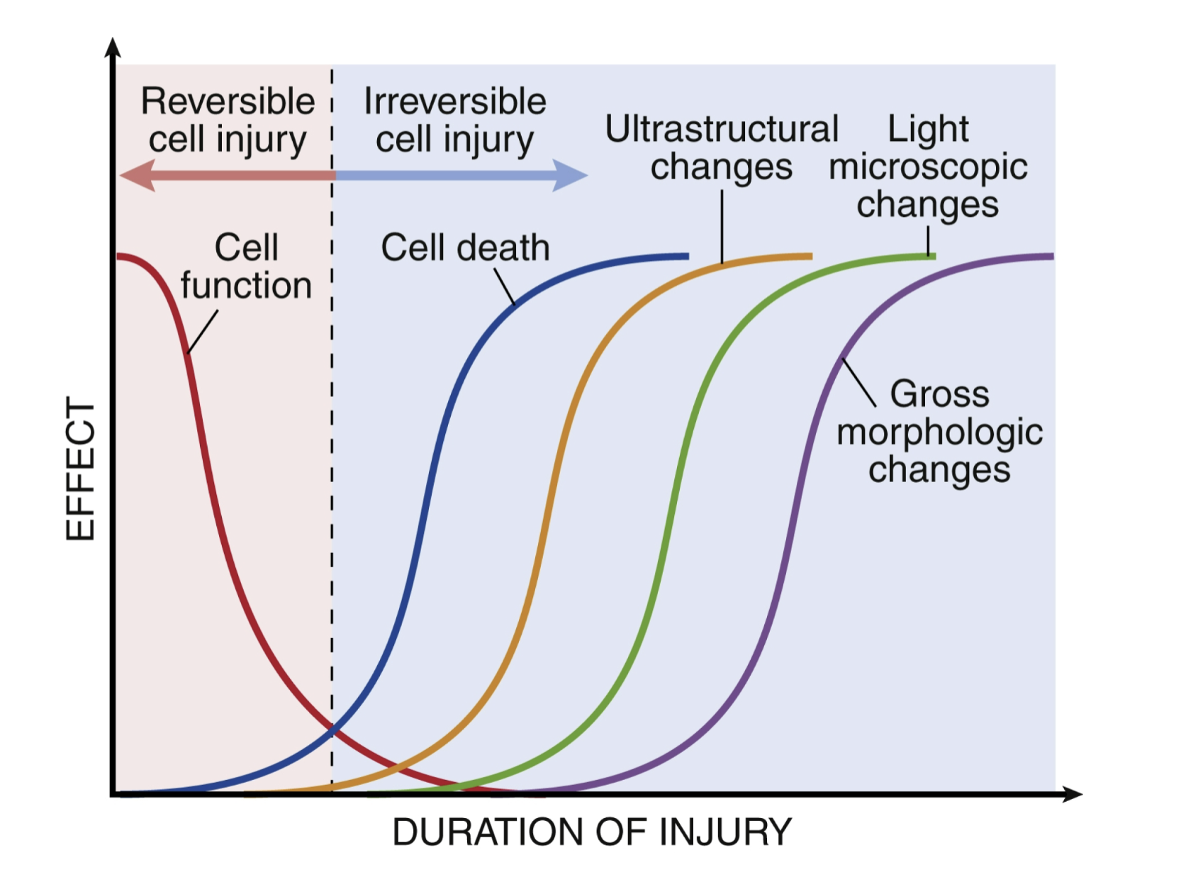
injury and death are a continuum
changes in function are only apparent wayy after cell function starts degrading
need to find markers where dysfunction exists but isn’t fully shown to make it reversible
hypoxia
low oxygen levels
blood flow is maintained
anaerobic metabolism ONLY
ischemia
reduced or blocked blood flow
NO anaerobic metabolism
causes more rapid and severe cell and tissue injury
hypoxia and ischemia
cells activate compensatory mechanisms to help it survive —> reversible adaptations
ex: inc growth factors, stimulate uptake of glucose
persistent conditions lead to depletion of ATP
failure of plasma membrane Na/K pump
dec of intracellular pH → changes activity of enzymes
inc ROS production
reduction in protein synthesis
ER swelling, membrane damage, mitochondrial damage
inflammation is triggered
severe conditions leads to necrosis
ischemia-reperfusion injury
restoration of blood flow to ischemic but viable tissues results in inc cell injury and necrosis
contributes to tissue damage esp after stroke
inc ROS production during reoxygenation → too much too soon as they adapted to low O2 conditions
influx of calcium disrupts calcium homeostasis
inflammation-induced by ischemic injury may inc w reperfusion due to influx and activation of leukocytes
direct-acting cell injury
caused by toxins and toxicants; combine directly to cellular proteins or organelles
ex:
mercury poisoning from seafood
mercury binds to cell membrane proteins, inhibiting ATP-dependent transport and inc membrane permeability
chemotherapy drugs
directly damage DNA to induce cell death
latent cell injury
caused by toxins and toxicants; chemical must be converted to a reactive metabolite that acts on target cells to exert toxic effect, usually by cytochrome p450 in the liver
ex:
acetaminophen poisoning
high doses saturate metabolic pathways leading to the production of a highly hepatotoxic metabolite
L2
human genome
3.3 billion DNA base pairs
22 pairs of autosomes, 1 pair of sex chromosomes
85% of genome is transcribed into RNA (not all goes to protein)
80% of genome is devoted to regulation of gene expression
non-protein coding sequences
promoters and enhancers
non-coding regulatory RNAs
transposons
telomeres and centromeres
promoters and enhancers
bind transcription factors
binding sites for DNA-binding proteins that organize and maintain chromatin structure
non-coding regulatory RNAs
genes that are transcribed but not translated
includes microRNAs, long non-coding RNAs
transposons
mobile genetic elements; “jumping genes”
telomeres and centromeres
structural regions of DNA
epigenetic mechanisms
stably but reversibly control gene expression without changing the sequence; mimic a mutation
covalent modifications of DNA
5-methylcytosine (cancer)
5-hydroxymethylcytosine
post-translational modifications of histones
3D chromatin structure
important in cancer
single nucleotide polymorphisms (SNPs)
variants at single nucleotide positions in coding and non-coding regions
non-coding SNPs → may alter regulation of gene expression
coding SNPs → may change the sequence of a protein
neutral SNPs → have no effect on gene function or phenotype; can be used as biomarkers
wide variation of frequency amongst diff populations
weak disease susceptibility due to individual SNPs
copy number variations (CNVs)
variation in number of large continuous stretches of DNA
abt 50% of CNVs involve coding sequences
ex: duplications, triplications, deletions, inversions
mutations in protein-coding genes
permanent changes in DNA sequence
point mutations
frameshift mutations
trinucleotide repeat mutations
structural variations
point mutations
substitution of a single nucleotide base by a diff base
missense point mutation
changes an amino acid
nonsense point mutation
creates a stop coding
frameshift mutation
insertion or deletion of 1-2 base pairs alters the reading frame of DNA strand
trinucleotide repeat mutation
amplification of repeated sequence of 3 bases
causes lower protein expression
structural variations
genomic rearrangement leads to amplifications, deletions, or translocations of chromosomal segments
single genes to entire chromosomes
intra- or inter-chromosomal
cause CNVs → linked to higher risk of disorder
gene editing (CRISPR)
clustered regularly interspaced short palindromic repeats
form of bacterial immunity
uses artificial guide RNAs that target a DNA sequence of interest to introduce mutations
first CRISPR/Cas9 based therapy approved in 12/2023 for sickle cell disease
epigenetic and genetic mechanisms in cancer
inactivation of tumor suppressor genes (promoter hypermethylation)
activation of oncogenes (promoter demethylation)
genetic disorders
disease-causing mutations
mendelian disorders
cytogenetic diseases
non-mendelian genetic diseases
disease-associated mutations
complex disorders
mendelian disorders
result from mutation in single genes
hereditary
high penetrance
show phenotypic effects
cytogenetic diseases
arise from changes in chromosomal number or structure
ex: CML, down syndrome
non-mendelian genetic diseases
arise from single gene mutations w atypical pattern of expression
ex: trinucleotide repeat, mitochondrial DNA disorder
complex disorders
involve multiple genes as well as environmental influences
affect risk of disease
very common
do not cause disease on their own
ex: hypertension
autosomal dominant
one copy is sufficient to cause disease, disease manifests in heterozygous state
does not skip a generation
affected offspring have affected parents
male and female offspring are equally likely to be affected

autosomal recessive
two copies are needed to cause disease, disease manifests in homozygous state
typically skips a generation
affected offspring may have unaffected parents
male and female offspring are equally likely to be affected

sex-linked
most are X-linked
heterozygous females can transmit only to sons
daughters may be carriers
affected males cannot transmit the disorder to sons

pleiotropy
a single gene mutation has many phenotypic effects
genetic heterogeneity
multiple genes cause the same trait
penetrance
the proportion of individuals w the mutation that exhibit clinical symptoms among all individuals w such mutation
incomplete penetrance → BRCA1 mutations