Citology 1 Cell Structure Flashcards
1/58
Earn XP
Description and Tags
Flashcards about general cell biology and structure
Name | Mastery | Learn | Test | Matching | Spaced | Call with Kai |
|---|
No analytics yet
Send a link to your students to track their progress
59 Terms
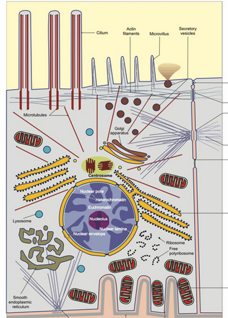
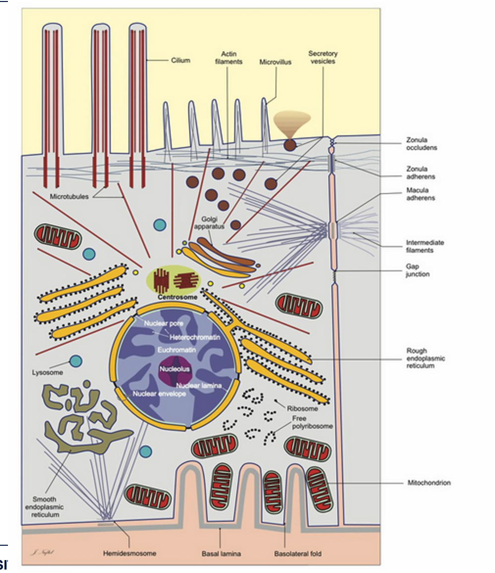
Examples of cell structures include __.
Microtubules, Lysosome, Smooth endoplasmic reticulum, Actin filaments, Microvillus, Golgi apparatus, Centrosome, Nuclear pore, Heterochromatin, Euchromatin, Nucleolus, Nuclear lamina, Nuclear envelope, Secretory vesicles, Ribosome, Free polyribosome, Hemidesmosome, Basal lamina, Basolateral fold, Zonula occludens, Zonula adherens, Macula adherens, Intermediate filaments, Gap junction, Rough endoplasmic reticulum, Mitochondrion
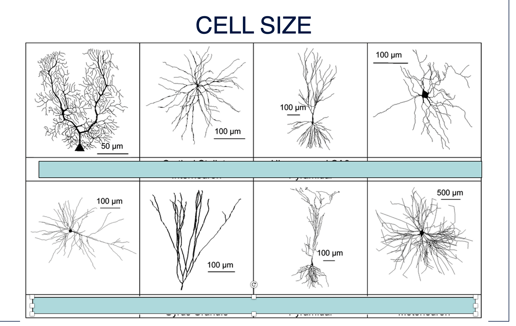
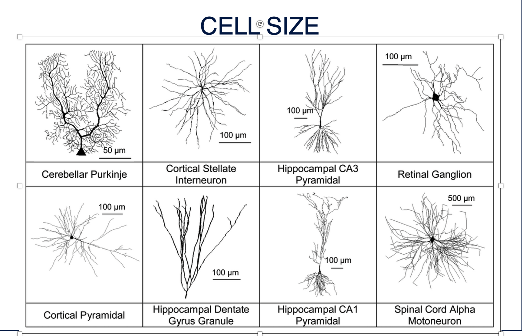
Examples of cells, varying in size, include __.
Cerebellar Purkinje, Cortical Pyramidal, Cortical Stellate Interneuron, Hippocampal CA3 Pyramidal, Retinal Ganglion, Hippocampal Dentate Gyrus Granule, Hippocampal CA1 Pyramidal, Spinal Cord Alpha Motoneuron
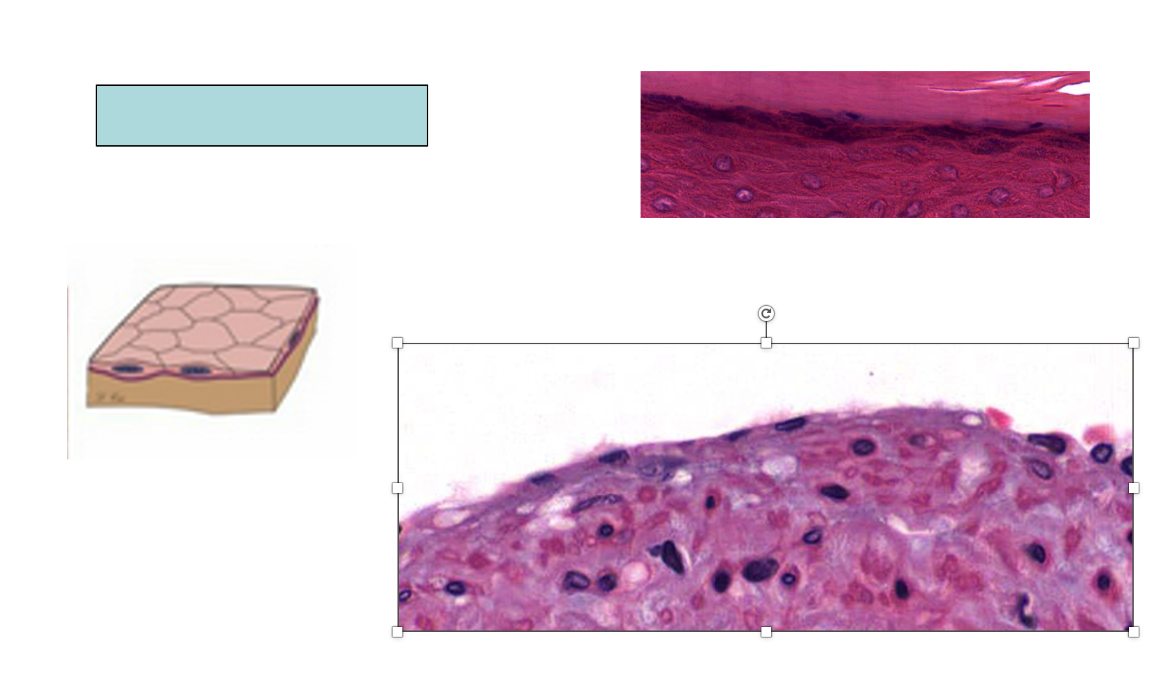
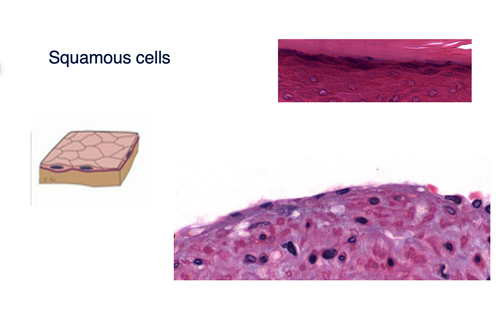
An example of a type of cell is __.
Squamous cells
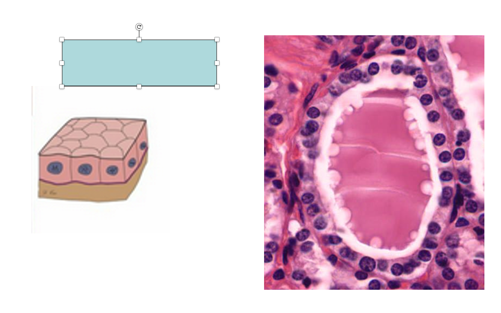
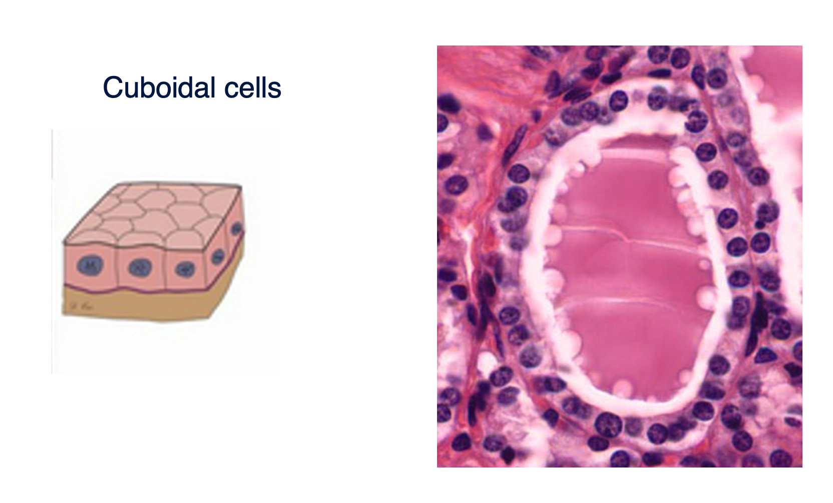
An example of a type of cell is __.
Cuboidal cells
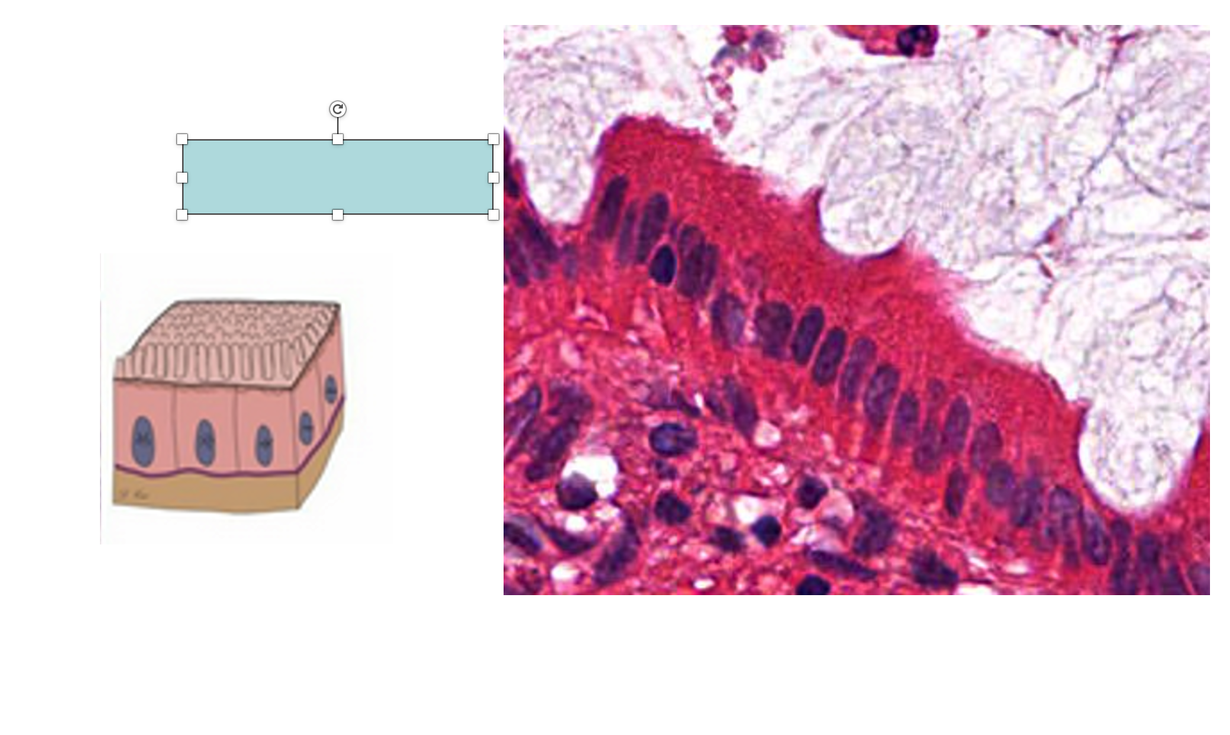
An example of a type of cell is __.
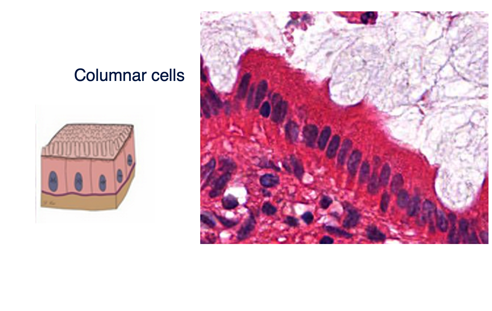
Columnar cells
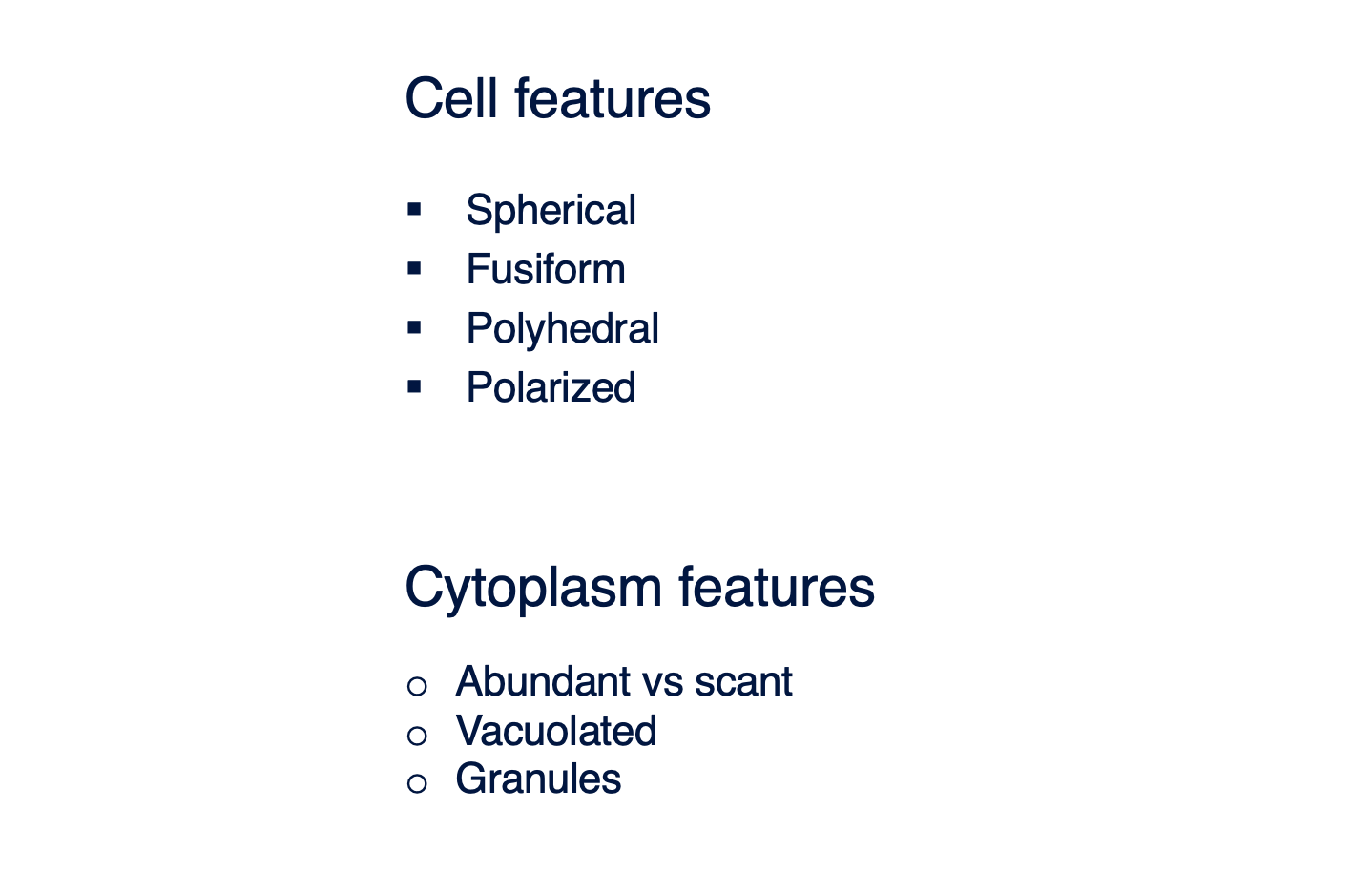
Examples of cell features include __.
Spherical, Fusiform, Polyhedral, Polarized

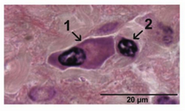
Examples of Cytoplasm features include __.
Abundant vs scant, Vacuolated, Granules
About Stomach Anatomy:
What are the main regions of the stomach shown in the diagram?
Where does the esophagus connect to the stomach?
What part of the stomach connects to the duodenum?
Can you identify the different parts of the pylorus?
About Gastric Gland Structure:
What are the main structural components of a gastric gland?
Where are the gastric pits located in relation to the gastric glands?
What is the significance of the "isthmus (neck)" and "body" regions of the gastric gland?
About Specific Cell Types and Their Secretions/Functions:
What are the different types of cells found in the gastric glands?
Which cell type is responsible for producing mucus on the surface of the stomach?
What do mucous neck cells secrete, and where are they located?
Which cells produce hydrochloric acid (HCl) and intrinsic factor? Why is intrinsic factor important?
What is the primary secretion of chief cells?
What are neuroendocrine cells, and what hormones/substances do they secrete (e.g., G cells, D cells, ECL cells)?
How does the structure of each cell type relate to its function (e.g., abundance of mitochondria in parietal cells)?
Trace the path of food through the stomach, highlighting the relevant anatomical structures.
Comparative/Integrative Questions:
How do the different cell types in the gastric gland work together to contribute to stomach digestion?
Are all these cell types found uniformly throughout the stomach, or do their distributions vary by region?
How do the secretions from these cells affect the pH within the stomach lumen?
About Stomach Anatomy:
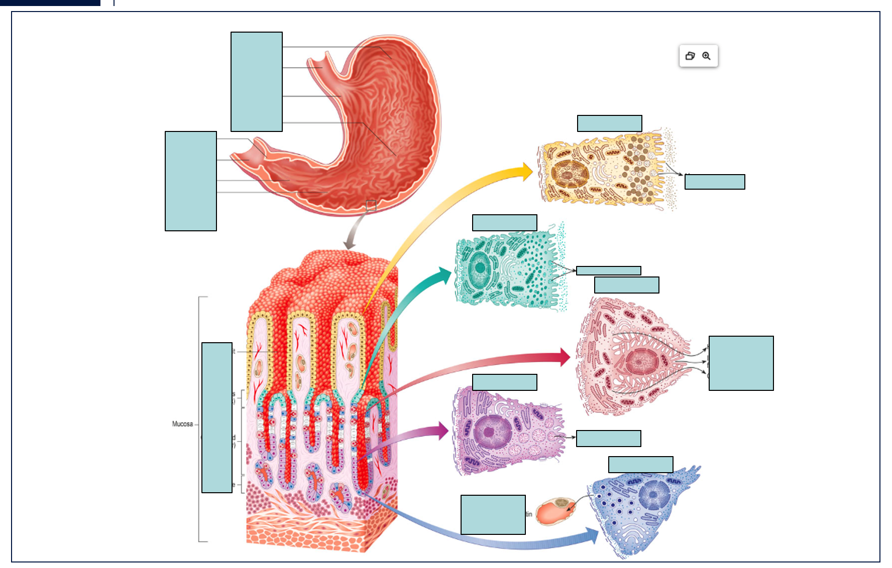
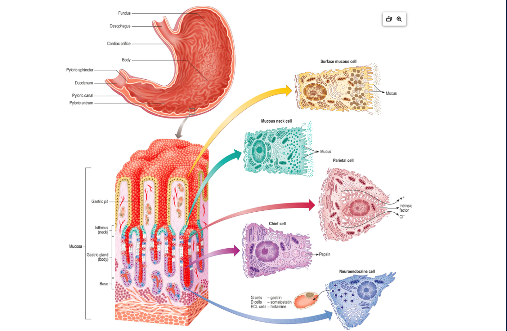
What are the main regions of the stomach shown in the diagram? The main regions of the stomach shown are the Fundus, Esophagus, Cardiac orifice, Body, Pyloric sphincter, Duodenum, Pyloric canal, and Pyloric antrum.
Where does the esophagus connect to the stomach? The esophagus connects to the stomach at the Cardiac orifice.
What part of the stomach connects to the duodenum? The Pyloric sphincter connects the stomach to the Duodenum.
Can you identify the different parts of the pylorus? Yes, the parts of the pylorus identified are the Pyloric antrum, Pyloric canal, and Pyloric sphincter.
About Gastric Gland Structure:
What are the main structural components of a gastric gland? The main structural components of a gastric gland shown are the Gastric pit, Isthmus (neck), Mucosa (which contains the Gastric gland body), and the Base.
Where are the gastric pits located in relation to the gastric glands? Gastric pits are invaginations from the surface of the mucosa, leading down into the gastric glands. They are essentially the openings of the gastric glands onto the stomach lumen.
What is the significance of the "isthmus (neck)" and "body" regions of the gastric gland? The "isthmus (neck)" and "body" regions of the gastric gland represent different zones where specific types of cells are predominantly located, contributing to the specialized secretions of the stomach. For instance, mucous neck cells are found in the neck, while parietal and chief cells are more abundant in the body.
About Specific Cell Types and Their Secretions/Functions:
What are the different types of cells found in the gastric glands? The different types of cells found in the gastric glands, as shown, are Surface mucous cells, Mucous neck cells, Parietal cells, Chief cells, and Neuroendocrine cells.
Which cell type is responsible for producing mucus on the surface of the stomach? The Surface mucous cells are responsible for producing mucus on the surface of the stomach.
What do mucous neck cells secrete, and where are they located? Mucous neck cells secrete mucus. They are located in the "isthmus (neck)" region of the gastric glands.
Which cells produce hydrochloric acid (HCl) and intrinsic factor? Why is intrinsic factor important? Parietal cells produce hydrochloric acid (HCl) and intrinsic factor. Intrinsic factor is crucial for the absorption of vitamin B12 in the small intestine. Without it, vitamin B12 deficiency (and pernicious anemia) can occur.
What is the primary secretion of chief cells? The primary secretion of chief cells is Pepsinogen, the inactive precursor to pepsin.
What are neuroendocrine cells, and what hormones/substances do they secrete (e.g., G cells, D cells, ECL cells)? Neuroendocrine cells are specialized cells that secrete hormones and other substances that regulate digestive functions. As indicated in the diagram, they include:
G cells: Secrete Gastrin.
D cells: Secrete Somatostatin.
ECL cells: Secrete Histamine.
How does the structure of each cell type relate to its function (e.g., abundance of mitochondria in parietal cells)?
Parietal cells: Their large size and prominent intracellular canaliculi with abundant mitochondria reflect the high energy demand for actively pumping H+ ions (for HCl production).
Chief cells: Their basophilic cytoplasm (due to extensive rough endoplasmic reticulum) and apical secretory granules are characteristic of protein-secreting cells (pepsinogen).
Mucous cells (surface and neck): Their cytoplasm is rich in mucin granules, which swell and release mucus upon secretion, providing protection.
Neuroendocrine cells: They contain dense-cored granules characteristic of cells that store and release peptide hormones.
Trace the path of food through the stomach, highlighting the relevant anatomical structures. Food enters the stomach from the Esophagus, passing through the Cardiac orifice. It then enters the main body of the stomach (Fundus and Body). After digestion, the partially digested food (chyme) passes through the Pyloric antrum, Pyloric canal, and finally exits the stomach through the Pyloric sphincter into the Duodenum.
Comparative/Integrative Questions:
How do the different cell types in the gastric gland work together to contribute to stomach digestion? The different cell types work synergistically:
Mucous cells protect the stomach lining from acid.
Parietal cells produce HCl for protein denaturation and activation of pepsinogen, and intrinsic factor for B12 absorption.
Chief cells produce pepsinogen, which is activated by HCl into pepsin for protein digestion.
Neuroendocrine cells release hormones that regulate the activity of other gastric cells (e.g., gastrin stimulates HCl secretion, somatostatin inhibits it, histamine enhances it). This coordinated effort ensures efficient chemical digestion and protection of the stomach wall.
Are all these cell types found uniformly throughout the stomach, or do their distributions vary by region?The distribution of these cell types varies by region. For example:
Surface mucous cells cover the entire luminal surface.
Parietal and Chief cells are most abundant in the glands of the fundus and body of the stomach.
Neuroendocrine cells (like G cells) are more concentrated in the pyloric antrum. Mucous neck cells are found in the neck region of glands throughout.
How do the secretions from these cells affect the pH within the stomach lumen? The primary factor affecting the pH is the large amount of HCl secreted by the Parietal cells, which makes the stomach lumen highly acidic (pH typically 1.5-3.5). The mucus secreted by Surface mucous cells and Mucous neck cells helps to buffer this acidity at the epithelial surface, protecting the stomach lining from self-digestion, but the bulk lumen remains very acidic.
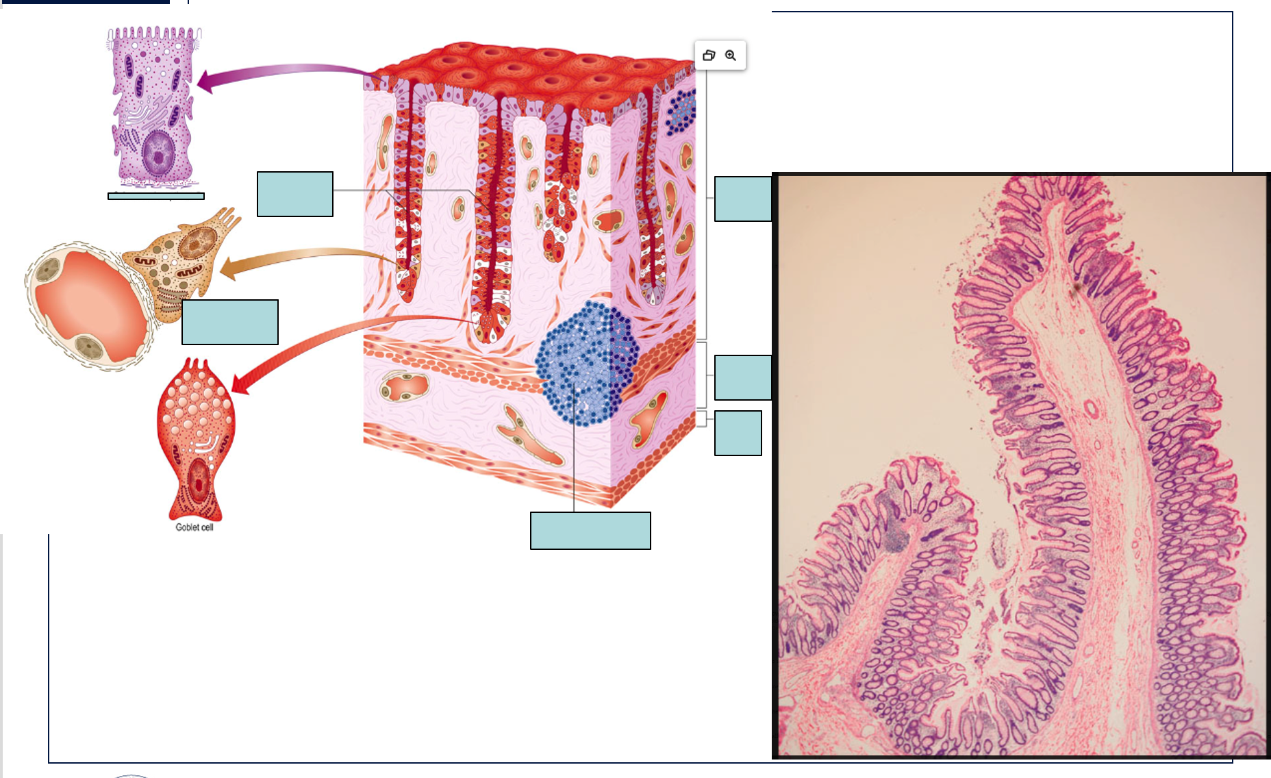
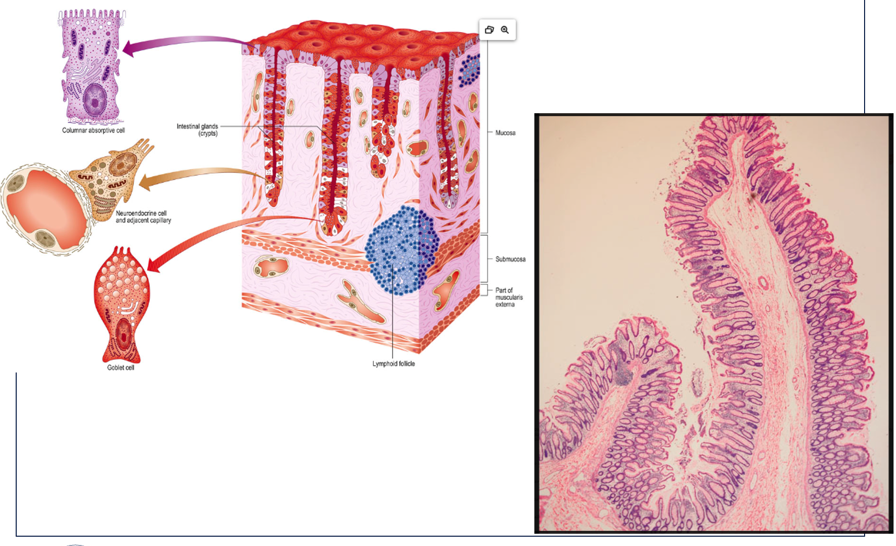
About the Diagram (Left Panel):
What are the main layers of the intestinal wall shown in the diagram?
What specific structures are found within the Mucosa layer?
What are the characteristic invaginations of the intestinal lining called?
Identify the different cell types depicted in the intestinal glands.
What is the primary function of the "Columnar absorptive cell" in this region?
What is the primary function of the "Goblet cell" and what does it secrete?
Where are "Neuroendocrine cells" located and what is their general role?
What is the significance of the "Lymphoid follicle" in the submucosa?
What is the role of the "Part of muscularis externa" shown in the diagram?
How does the arrangement of cells in the intestinal glands differ from the surface epithelium?
About the Microscopic Image (Right Panel):
What anatomical structure is likely represented in the microscopic image? (Hint: based on the features shown).
Can you identify the crypts (intestinal glands) in the microscopic image?
What is the prominent cell type visible lining the crypts and surface, and what features help identify it?
What are the clear, vacuolated cells often seen in abundance in this tissue, and what do they represent?
How does the microscopic image correlate with the structural features shown in the diagram?
Comparative/Integrative Questions:
How does the histology of this region (colon) differ from that of the small intestine (e.g., presence/absence of villi, abundance of goblet cells)?
What are the key adaptations of this intestinal region for its specific digestive/absorptive functions, as suggested by its histology?
How do the various cell types shown contribute to the overall function of the large intestine?
About the Diagram (Left Panel):
What are the main layers of the intestinal wall shown in the diagram? The main layers of the intestinal wall shown are the Mucosa, Submucosa, and a "Part of muscularis externa".
What specific structures are found within the Mucosa layer? Within the Mucosa layer, we can see Intestinal glands (or crypts), and various cell types lining them, including Columnar absorptive cells and Goblet cells. The diagram also indicates Neuroendocrine cells and an adjacent capillary within the gland.
What are the characteristic invaginations of the intestinal lining called? The characteristic invaginations of the intestinal lining are called Intestinal glands, also known as crypts of Lieberkühn.
Identify the different cell types depicted in the intestinal glands. The different cell types depicted are Columnar absorptive cells, Neuroendocrine cells (with an adjacent capillary), and Goblet cells.
What is the primary function of the "Columnar absorptive cell" in this region? The primary function of the Columnar absorptive cell in the large intestine is the absorption of water and electrolytes.
What is the primary function of the "Goblet cell" and what does it secrete? The primary function of the Goblet cell is to produce and secrete mucus. This mucus lubricates the passage of fecal material and protects the intestinal lining.
Where are "Neuroendocrine cells" located and what is their general role? Neuroendocrine cells are located within the intestinal glands. Their general role is to secrete hormones and signaling molecules that regulate various digestive functions, such as gut motility and secretion, in response to luminal contents.
What is the significance of the "Lymphoid follicle" in the submucosa? The Lymphoid follicle in the submucosa is part of the gut-associated lymphoid tissue (GALT). Its significance lies in its role in immune surveillance and defense against pathogens entering the digestive tract.
What is the role of the "Part of muscularis externa" shown in the diagram? The muscularis externa is primarily responsible for the movements of the intestine, including peristalsis (propulsion of contents) and segmentation (mixing of contents).
How does the arrangement of cells in the intestinal glands differ from the surface epithelium? The diagram shows that the surface epithelium is primarily composed of columnar absorptive cells with interspersed goblet cells. The intestinal glands (crypts) are invaginations where goblet cells become more numerous, especially deeper in the crypts, and various specialized cells (like neuroendocrine cells) reside, which are typically less abundant on the surface.
About the Microscopic Image (Right Panel):
What anatomical structure is likely represented in the microscopic image? (Hint: based on the features shown). The microscopic image likely represents the large intestine (colon), characterized by its straight, unbranched intestinal glands (crypts) and the lack of villi.
Can you identify the crypts (intestinal glands) in the microscopic image? Yes, the crypts (intestinal glands) are clearly visible as the numerous, long, test-tube-like invaginations extending downwards from the surface epithelium into the lamina propria.
What is the prominent cell type visible lining the crypts and surface, and what features help identify it? The prominent cell type lining the crypts and surface is the columnar absorptive cell. It is identified by its tall, columnar shape and often a brush border (though not clearly resolved at this magnification) at its apical surface.
What are the clear, vacuolated cells often seen in abundance in this tissue, and what do they represent? The clear, vacuolated cells, which appear as "empty" spaces within the epithelial lining (especially prominent in the crypts), are Goblet cells. Their clear appearance is due to the mucin they contain, which washes out during histological preparation.
How does the microscopic image correlate with the structural features shown in the diagram? The microscopic image strongly correlates with the diagram. The diagram is a simplified, idealized representation, while the micrograph is a real tissue section. Both show the characteristic straight tubular intestinal glands, the columnar epithelial lining, and the presence of goblet cells. The layers of the wall (mucosa, submucosa) are also visible in the micrograph, consistent with the diagram.
Comparative/Integrative Questions:
How does the histology of this region (colon) differ from that of the small intestine (e.g., presence/absence of villi, abundance of goblet cells)?
Villi: The colon (large intestine) lacks villi, which are prominent finger-like projections found in the small intestine to increase surface area for nutrient absorption. The colon has a relatively flat surface with deep crypts.
Goblet cells: Goblet cells are much more abundant in the large intestine compared to the small intestine. This reflects the large intestine's primary role in water absorption and the need for copious mucus to lubricate fecal material.
What are the key adaptations of this intestinal region for its specific digestive/absorptive functions, as suggested by its histology? The key adaptations are:
Abundant Goblet Cells: Provide lubrication for the passage of solidifying fecal material and protection against mechanical abrasion and bacterial action.
Deep Intestinal Glands/Crypts: Provide a large surface area for the specialized cells, particularly those involved in water and electrolyte absorption, and mucus secretion.
Prominent Columnar Absorptive Cells (without villi): Optimized for water and electrolyte absorption, which is the primary absorptive function of the colon, rather than nutrient absorption.
Presence of Lymphoid Follicles: Essential for immune defense against the vast bacterial population in the large intestine.
How do the various cell types shown contribute to the overall function of the large intestine?
Columnar Absorptive Cells: Are responsible for the primary function of the large intestine: absorbing water and electrolytes, leading to the formation of solid feces.
Goblet Cells: Secrete mucus, which lubricates the intestinal contents, protects the epithelium from friction, and aids in bacterial binding and elimination.
Neuroendocrine Cells: Secrete hormones that regulate colonic motility and secretion, ensuring proper transit time and absorption.
The lymphoid follicles provide immune protection against the dense microbial flora of the large intestine.
Cell types found in Intestinal glands include __.
Columnar absorptive cell, Neuroendocrine cell, Goblet cell, Lymphoid follicle
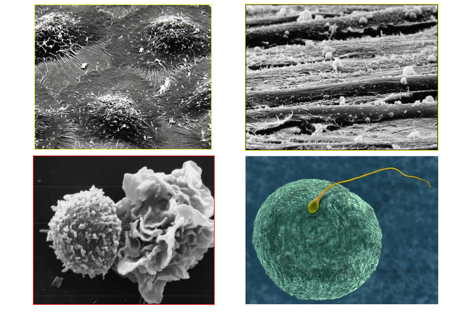
Name
Epithelial cells, muscular, Lymphocytes, human egg and sperm

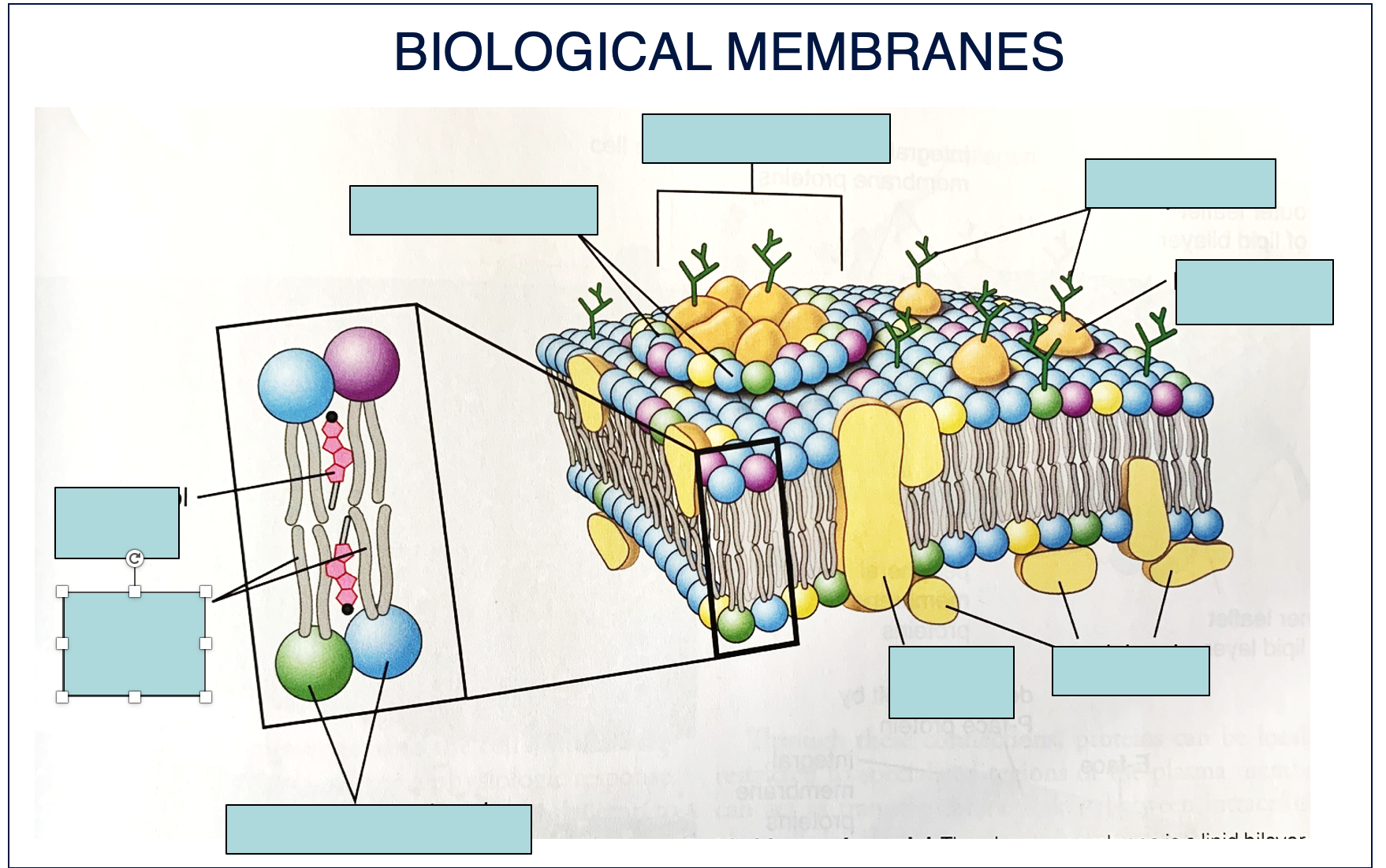
Describe
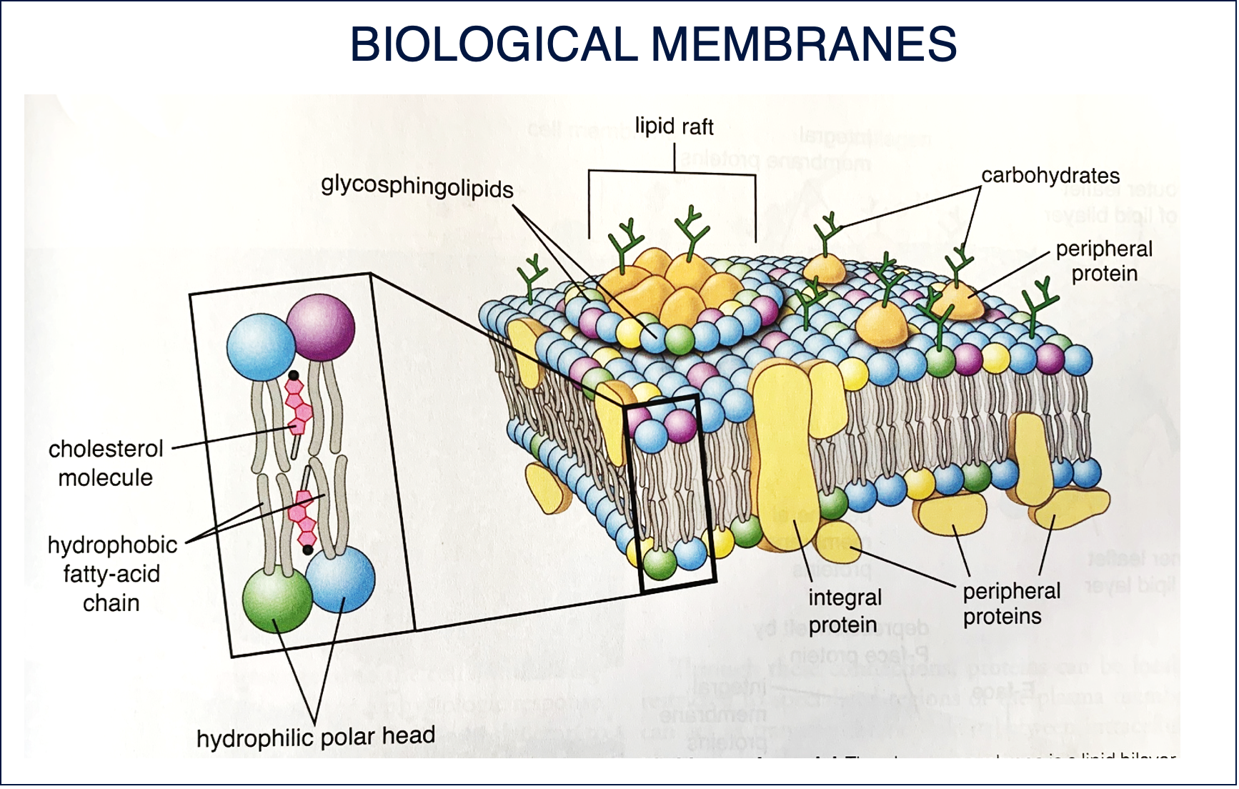
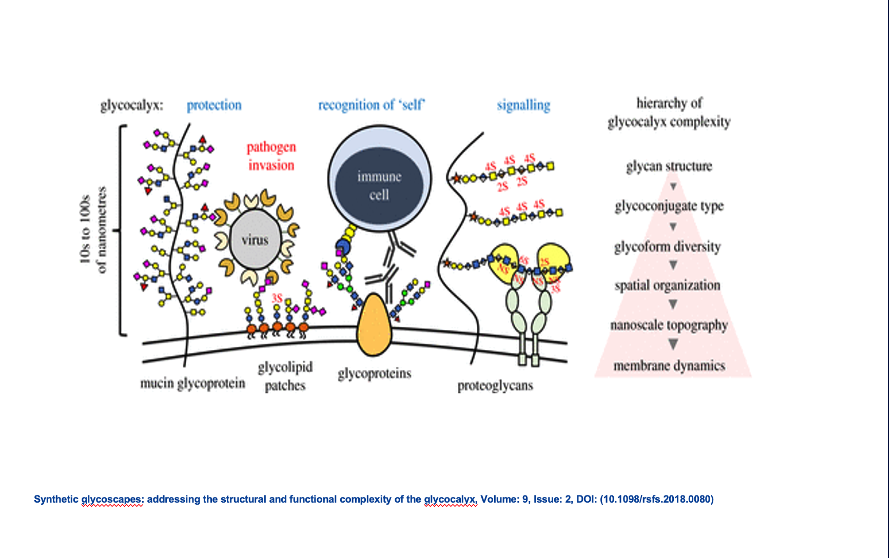
The plasma membrane is a lipid bilayer consisting of phospholipid molecules, cholesterol and proteins; integralmembrane proteins and peripheral membrane proteins; carbohydrates associated to extracellular face (glycoproteins and glycolipids). Cholesterol molecules are incorporated within the gaps between phospholipids equally on both sides of the membrane. Lipid rafts contain high conecentrations of cholesterol and glycosphingolipids. Lipid rafts are signalingplatforms due to the close proximity of interacting proteins. These platforms can be used by bacteria like Shigella or Salmonella which hijack the rafts with their signaling mechanism and use them to support their own entry into the cell.
them to support their own entry into the cell. Many bacteria use the rafts to escape phagocytosis. They form vacuoles made of rafts components, not detectable by phagocitic compartment, so bacteria can be transported across the cell.

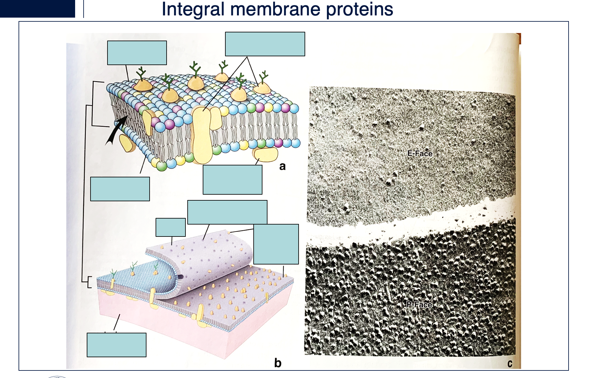
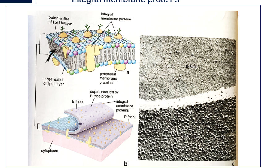
label and and say how integral membrane can be visualized
Integral membrane proteins can be visualized with the Freeze-fracture technique. Membranes typically split or cleavealong the hydrophobic plane to expose two interior faces of the membrane, an E-face and a P-face. The surfaces of the cleaved membrane are coated, forming replicas; the replicas are separated from the tissue and examined by Electron Microscopy (EM). Proteins appear as bumps. Note the paucity of particles in the E-face compared to P-face.

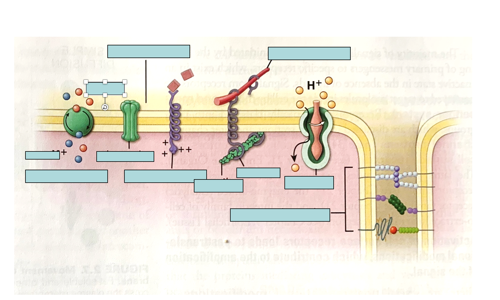
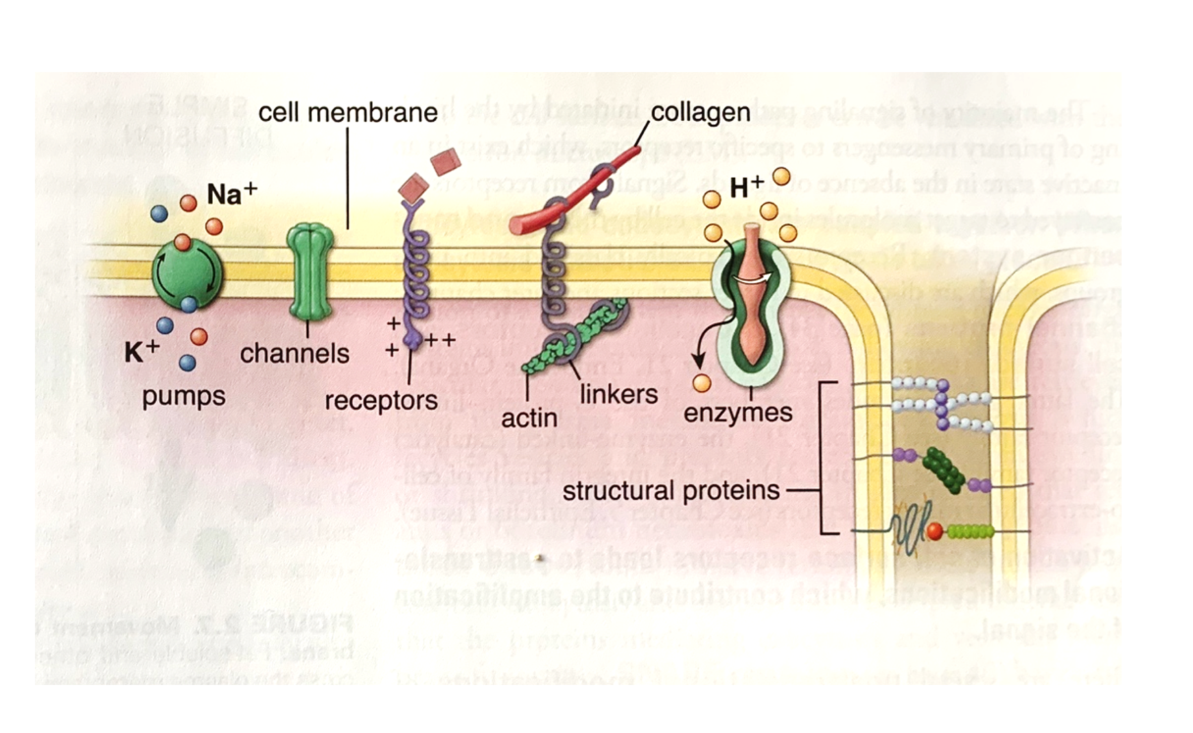
Pumps, channels, receptors, linkers, enzymes, structural proteins
LABEL
Molecular movement across the plasma membrane use__.
LABEL.
The different types of proteins shown are: pumps, channels, receptors, linkers, enzymes, and structural proteins
WHAT ARE THEIR FUNCTIONS?
So we have talk about specialization of different membrane portion, and this specialstic domains are characterized principally by the presence of specific proteins integrated in the bilayer. Six broad categories of membrane proteins have been defined in terms of their function :
Pumps – active transport of ions, also metabolic precursor such as amino acids and sugars. - passage of small ions, molecules, water in either directions (passive); present also in gap junctions where THEY allow the passage of molecules involved in signalling from one cell to adjacent.
Receptors- allow recognition and localized binding of ligands in processes such as hormonal stimulation, antibody reactions,…
Linkers – anchor the intracellular cytoskeleton to the extracellular matrix (i.e. Integrins).
Enzymes – have a variety of roles. ATPase have a specific role in ion pumping; ATP synthase is the major protein of the inner mitochondrial membrane, and digestive enzymes are integral membrane proteins.
Stuctural proteins – Specialized junctional complexes in polarized cells suchas epithelial cells.
Channels are transmembrane proteins that facilitate the passive, selective movement of specific ions or small molecules across the cell membrane, often regulated by various stimuli.
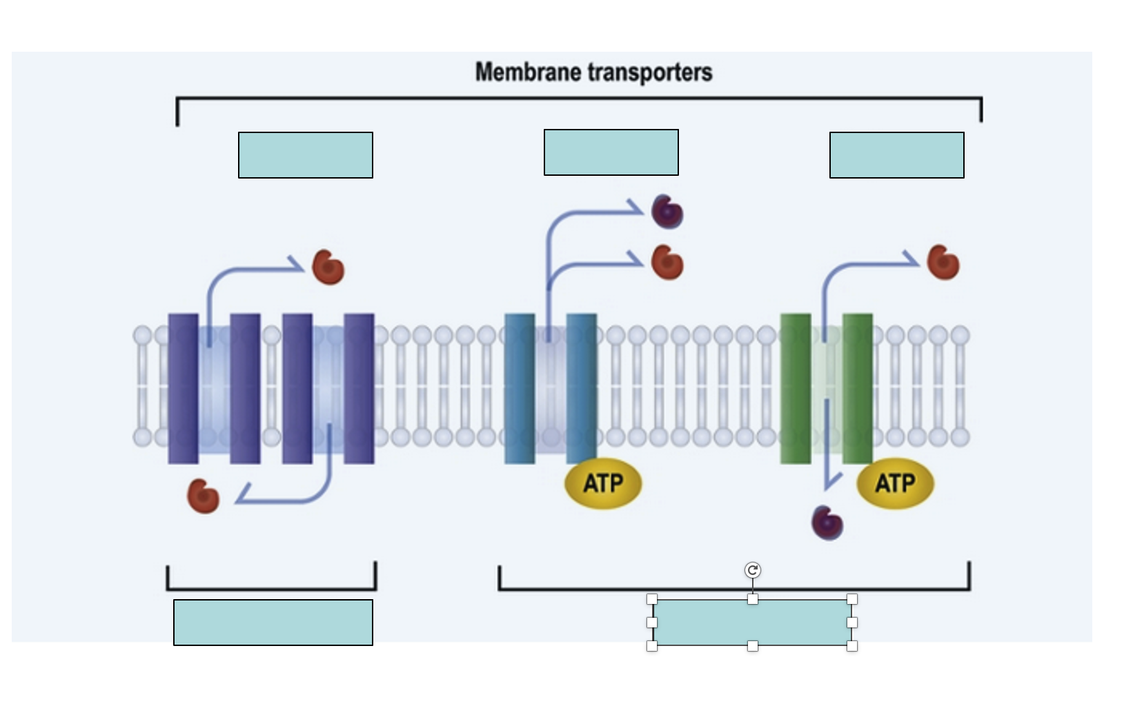
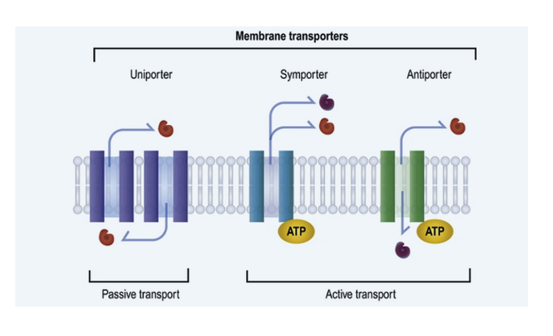
LABEL what are the 3 primary types of membrane transporters
This slide illustrates three primary types of membrane transporters—uniporters, symporters, and antiporters—categorized by their mechanism of transport (passive vs. active) and the directionality and co-transport of molecules across the cell membrane.
ATP (adenosine triphosphate) is the primary energy currency of the cell; in membrane transport, it provides the energy required for active transport mechanisms, enabling the movement of molecules against their concentration gradient.
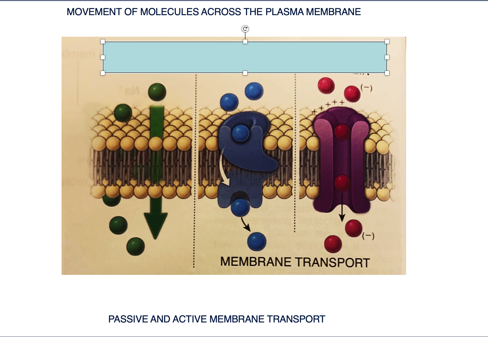
DESCRIBE THE MOVEMENT OF MOLECULES
There are 2 classes of transport proteins describe them
Fat soluble and other small, uncharged molecules cross the plasma membrane by diffusion (passive) down their concentration gradient. All other molecules require membrane transport proteins to provide them with individual passage across the plasma membrane. There are 2 classes of transport proteins:
Carrier proteins - transfer small water soluble molecules. They are highly selective. After binding the protein they undergo two conformational changes and release the molecule on the other side of the membrane. Some such as pump Na/K require energy for active transport, other such glucose carriers participate in passive transport.
Channel proteins - transfer small water soluble molecules, but they have a different structure. Generally channels are made by transmembrane protein with several membrane spanning domains that create hydrophilic channels through the plasma membrane. Channel protein transport can be regulated by membrane potentials (i.e. voltage-gated ion channels in neurons), neurotransmitters (i.e. ligand-gated ion channels such as actylcholine receptors in muscle cells), or mechanicalstress (mechanically gated ion channels in the internal ear.

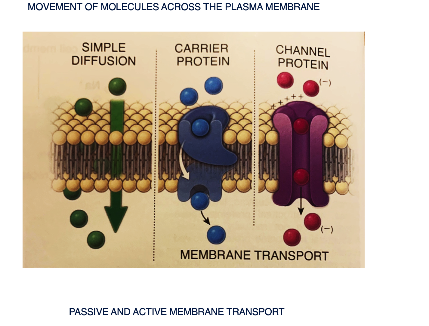
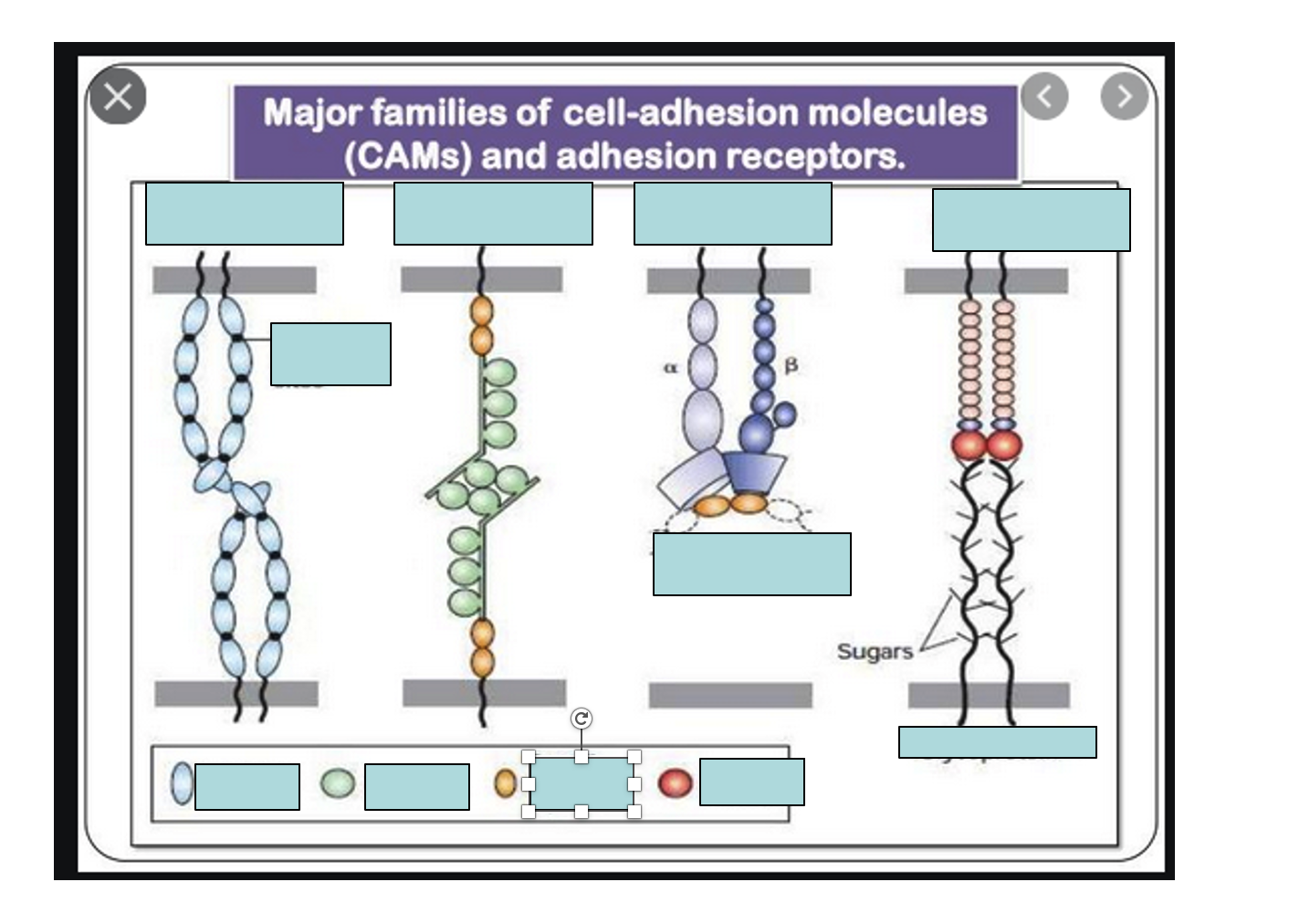
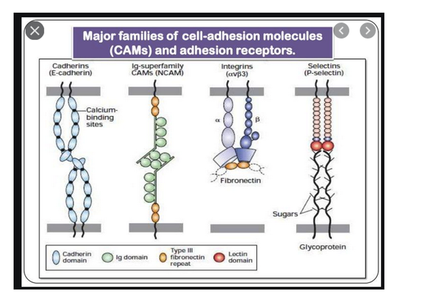
Label the four major families of cell-adhesion
This image illustrates the four major families of cell-adhesion molecules (CAMs) and adhesion receptors that mediate cell-to-cell and cell-to-extracellular matrix interactions. These include Cadherins (calcium-dependent, often involved in homophilic binding), Ig-superfamily CAMs like NCAM (typically homophilic and calcium-independent), Integrins(heterodimeric receptors that bind to extracellular matrix components like fibronectin), and Selectins (which bind to carbohydrate moieties on glycoproteins, involved in transient cell-cell adhesion). Together, these CAMs are crucial for tissue formation, cell migration, immune responses, and maintaining tissue integrity.
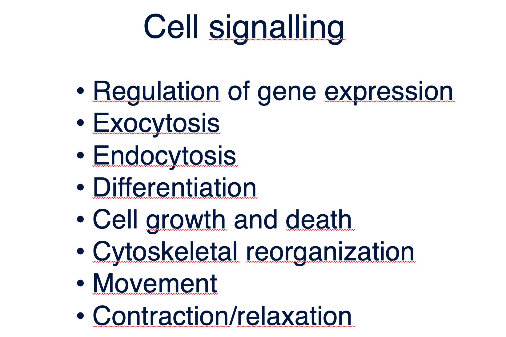
What is Cell signalling?
Cell signaling is the process by which extracellular stimuli are received and conveyed by the cell to regulate its own physiological responses. A single cell may receive many signals at the same time and it needs to integrate them into a unified action plan. Signaling process are involved in all processes listed.
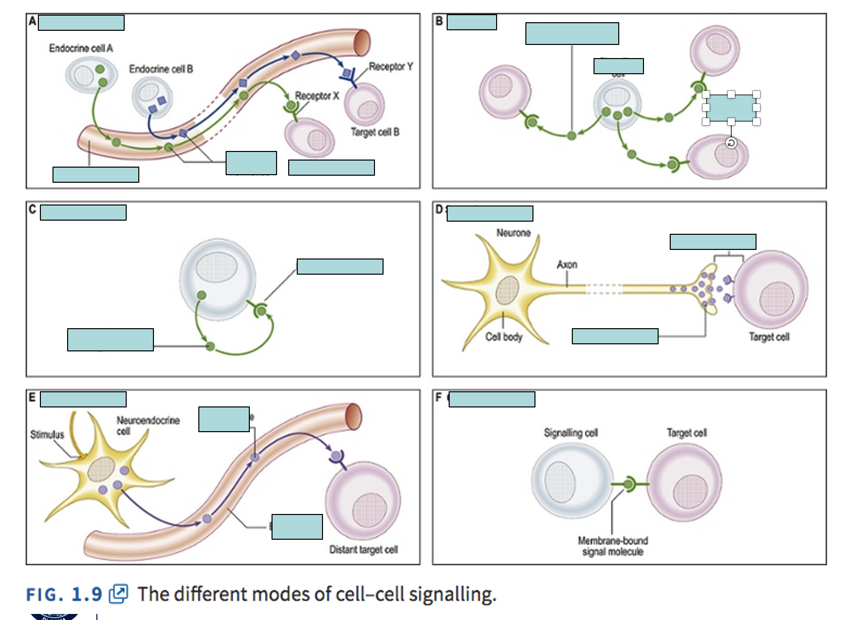
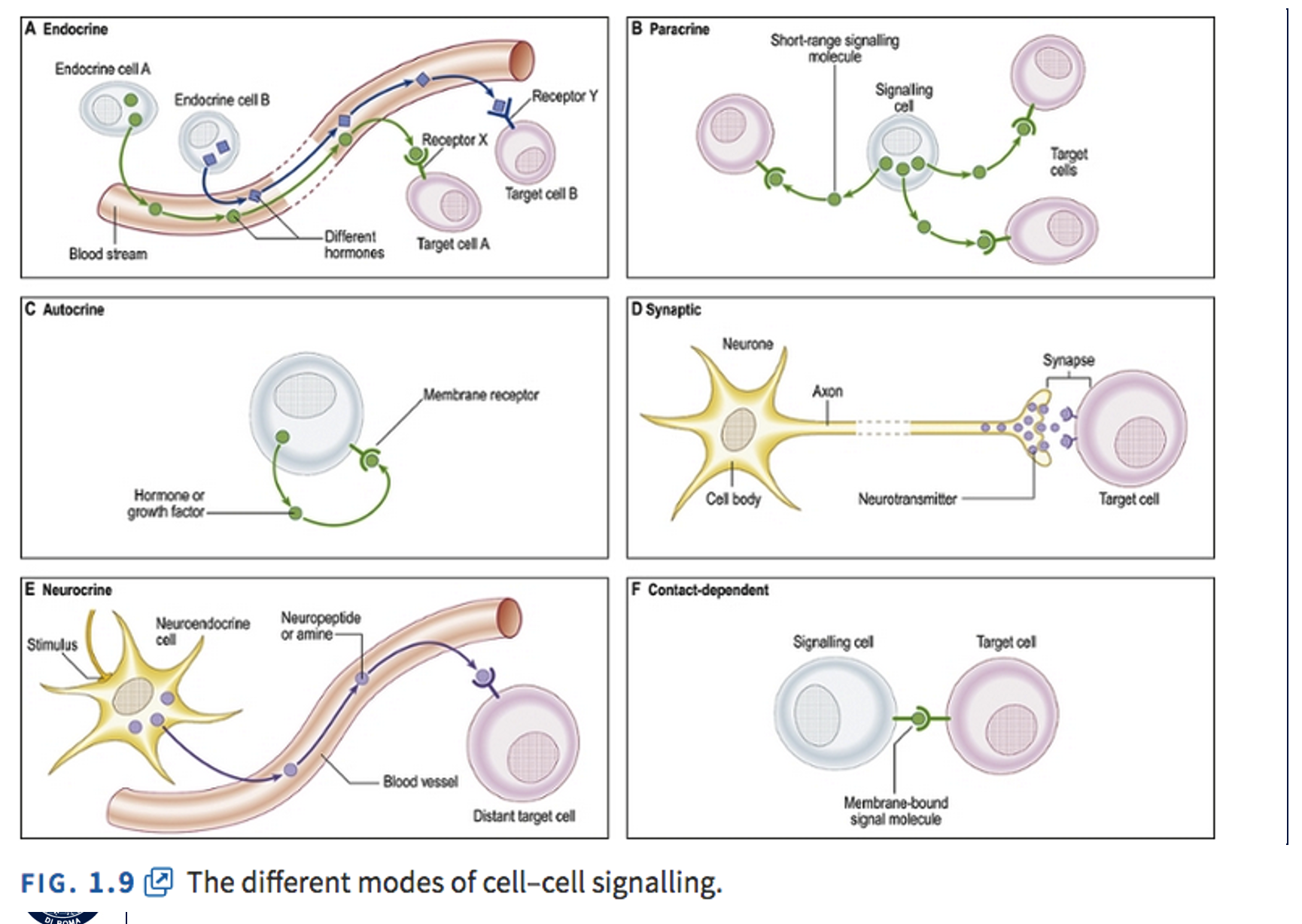
Label and explain signal transduction pathways
Cell signaling
process by which extracellular stimuli are received, processed and conveyed by the cell
Signal transduction pathways
Mechanisms by which cell respond to external environment
Signal transduction pathways are mechanisms by which cells respond to external environment, allow for amplification and modulation of the signal, are involved in biochemical and physiologic regulation. They are initiated by externalsignaling molecules. They can be soluble, act locally (autocrine or paracrine) or be transmitted by vascular system(endocrine signalling). They can also be insoluble, tethered to cell membranes or localized in extracellular matrix. Themajority of signaling pathways are initiated by the binding of primary messangers to specific receptors then are conveyedto target molecules inside the cells by the second messanger system
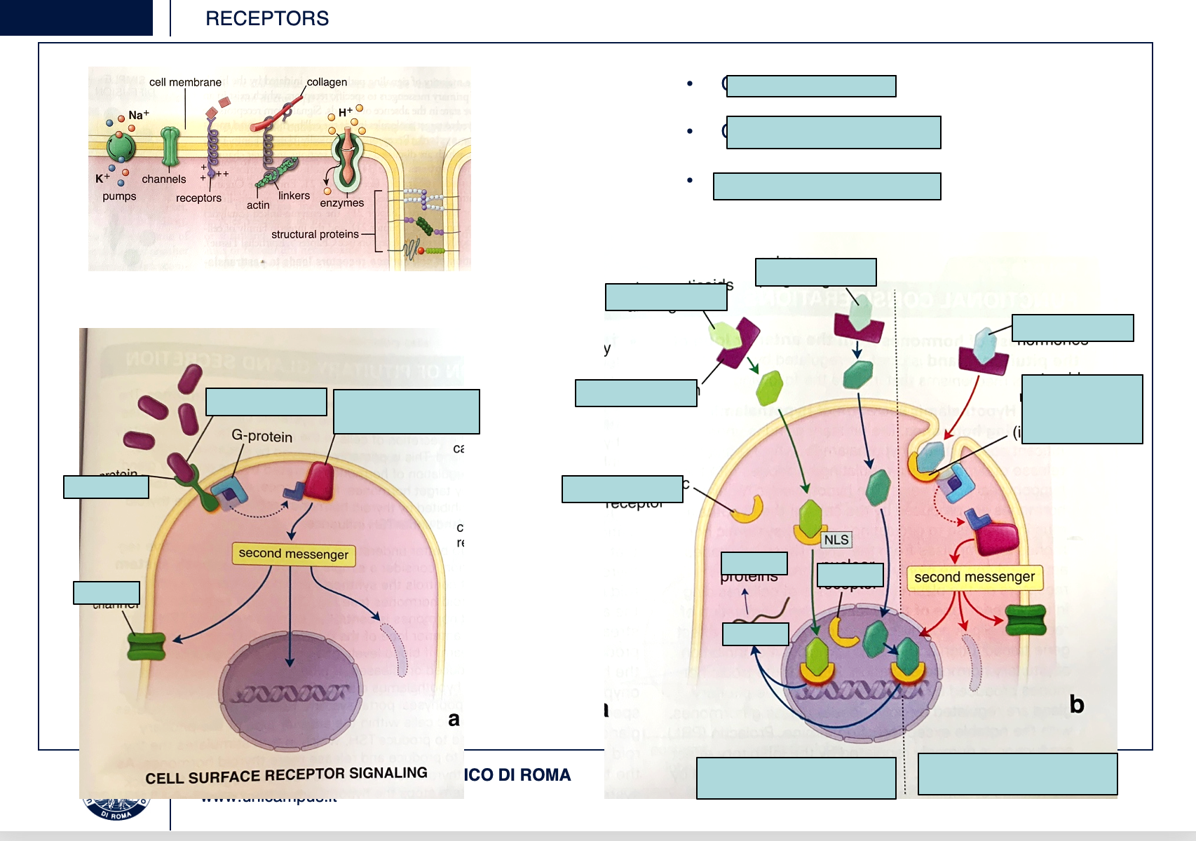
LABEL AND EXPLAIN THIS EXAMPLE OF INTRACELLULAR RECEPTORS
The first class of receptors is constituted by channel proteins receiving signals from extracellular environment or directly from other cells (i.e. synapses in nervous tissue or gap junctions present in many tissues). Binding of an hormone with surface receptor, initiate a process involving signaling reactions which may include
G proteins, so called because OF their ability to hydrolize GTP and various protein kinases resulting in the synthesis of second messanger molecules.
Examples of such systems include:
cyclic adenosine monophosphate cAMP,
cGMP guanosine,
tyrosine kinase system.
Examples of second messanger include
cAMP,
diacylglicerol,
inositol triphosphate or Ca.
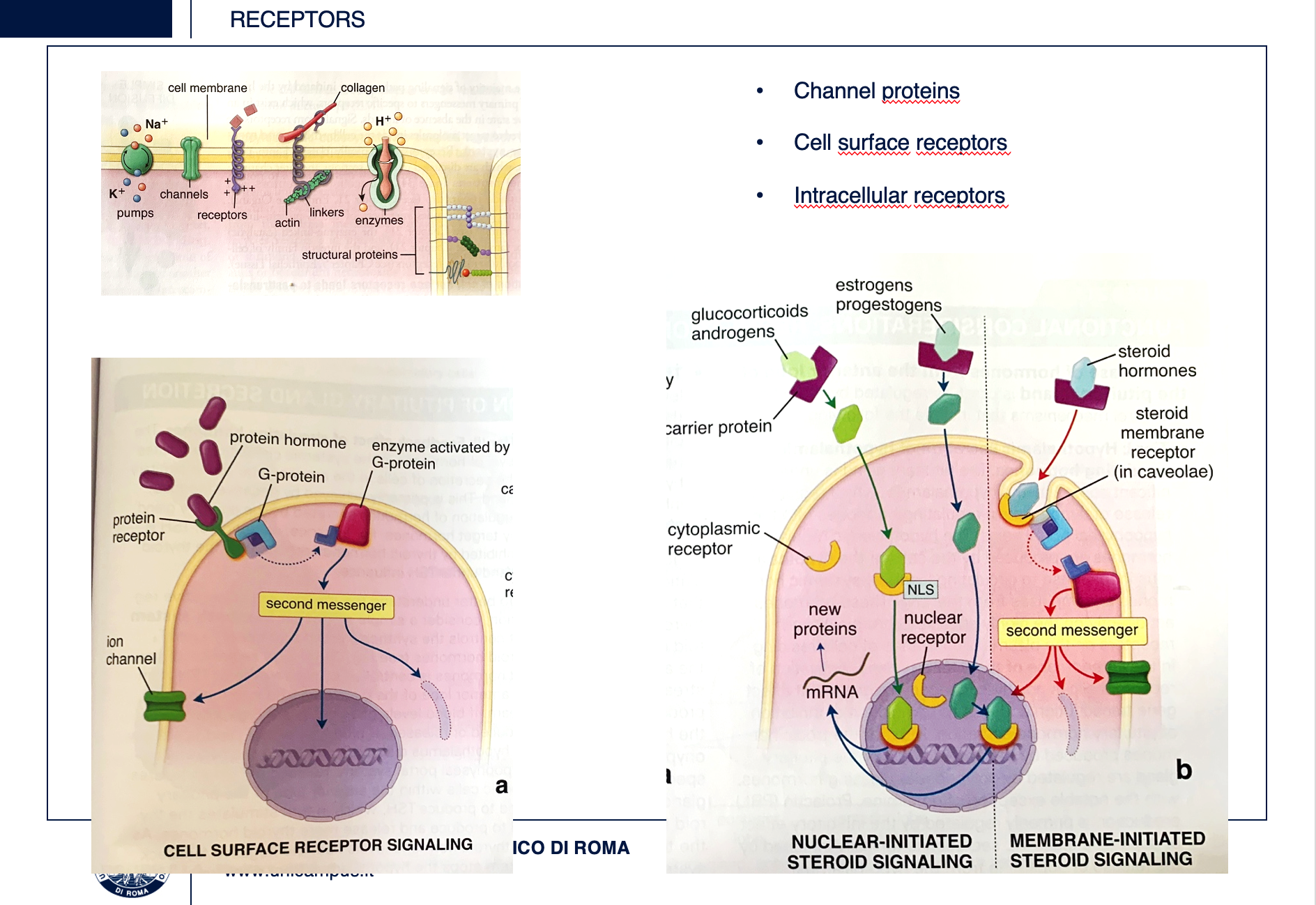
As example of intracellular receptors the action of steroid hormones is shown here (panel b). Some steroid hormones cross the membrane and bind to specific intracellulars receptors causing allosteric transformation of these molecules and the complex travel to nucleus, guided by the nucleus localizator signal where they binds DNA and promote or regulate transcription of specific genes. Other hormones go directly into the nucleus. The complex of hormone and nuclear receptor is the transcription factor.

EXPLAIN WHAT’S HAPPENING HERE
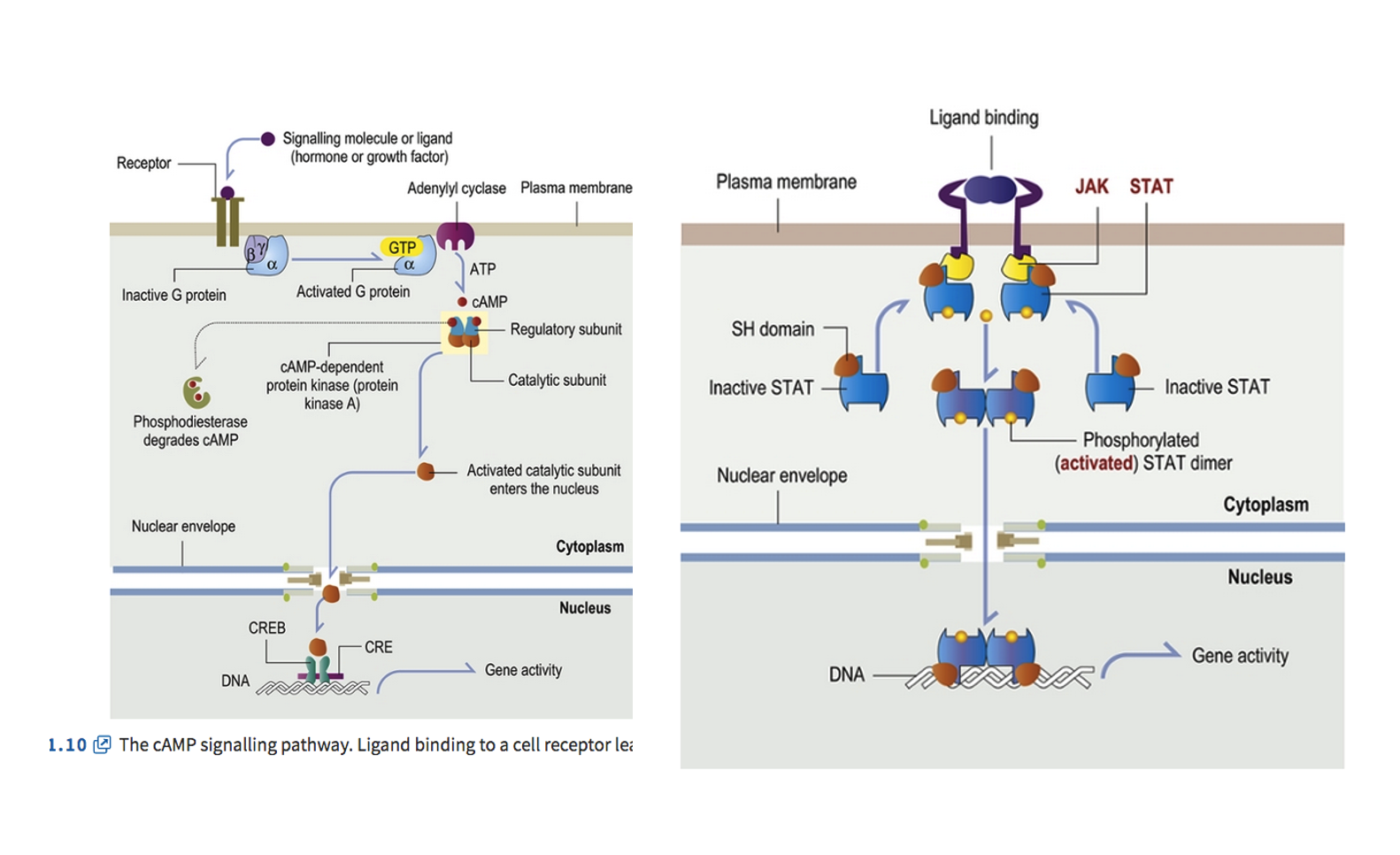
In essence, both diagrams illustrate different molecular mechanisms by which an external signal (ligand) is received at the cell surface, relayed through a series of intracellular steps (signal transduction), and ultimately leads to changes in gene expression in the nucleus, thereby altering cell behavior. The image displays two distinct signal transduction pathways that occur in cells, initiated by ligand binding to receptors on the plasma membrane and ultimately leading to changes in gene activity within the nucleus.
Left Panel: The cAMP Signalling Pathway
Ligand Binding: A signalling molecule (ligand), such as a hormone or growth factor, binds to a receptor on the plasma membrane.
G Protein Activation: This binding activates a G protein (specifically, the Gα subunit) by causing it to exchange GDP for GTP.
Adenylyl Cyclase Activation: The activated Gα subunit then activates Adenylyl cyclase, an enzyme located on the plasma membrane.
cAMP Production: Adenylyl cyclase converts ATP into cAMP (cyclic AMP). cAMP acts as a "second messenger."
PKA Activation: cAMP binds to and activates cAMP-dependent protein kinase (protein kinase A, PKA) by causing its regulatory subunits to dissociate from the catalytic subunits.
Nuclear Entry & Gene Regulation: The activated catalytic subunit of PKA enters the nucleus, where it phosphorylates CREB (cAMP response element-binding protein).
Gene Activation: Phosphorylated CREB then binds to CRE (cAMP response element) on the DNA, which leads to increased gene activity.
cAMP Degradation: Phosphodiesterase degrades cAMP, turning off the signal.
Right Panel: The JAK-STAT Signalling Pathway
Ligand Binding & Receptor Dimerization: A ligand binds to two separate receptors on the plasma membrane, causing them to come together and form a dimer.
JAK Kinase Activation: This receptor dimerization brings together and activates associated JAK (Janus kinase) proteins, which are tyrosine kinases.
Receptor Phosphorylation: Activated JAKs then phosphorylate specific tyrosine residues on the receptor tails.
STAT Recruitment & Phosphorylation: These phosphorylated tyrosines act as docking sites for STAT (Signal Transducer and Activator of Transcription) proteins. Once bound, STATs are also phosphorylated by JAKs.
STAT Dimerization: Phosphorylated STAT proteins detach from the receptor and form (activated) STAT dimers.
Nuclear Translocation: These activated STAT dimers then move through the nuclear envelope into the nucleus.
Gene Activation: Inside the nucleus, the STAT dimers bind directly to specific DNA sequences, leading to changes in gene activity.
In essence, both diagrams illustrate how extracellular signals are received at the cell surface and then transduced through a series of molecular events involving proteins and second messengers to ultimately alter gene expression, thereby controlling cellular responses.
Transmembrane receptors and second messanger
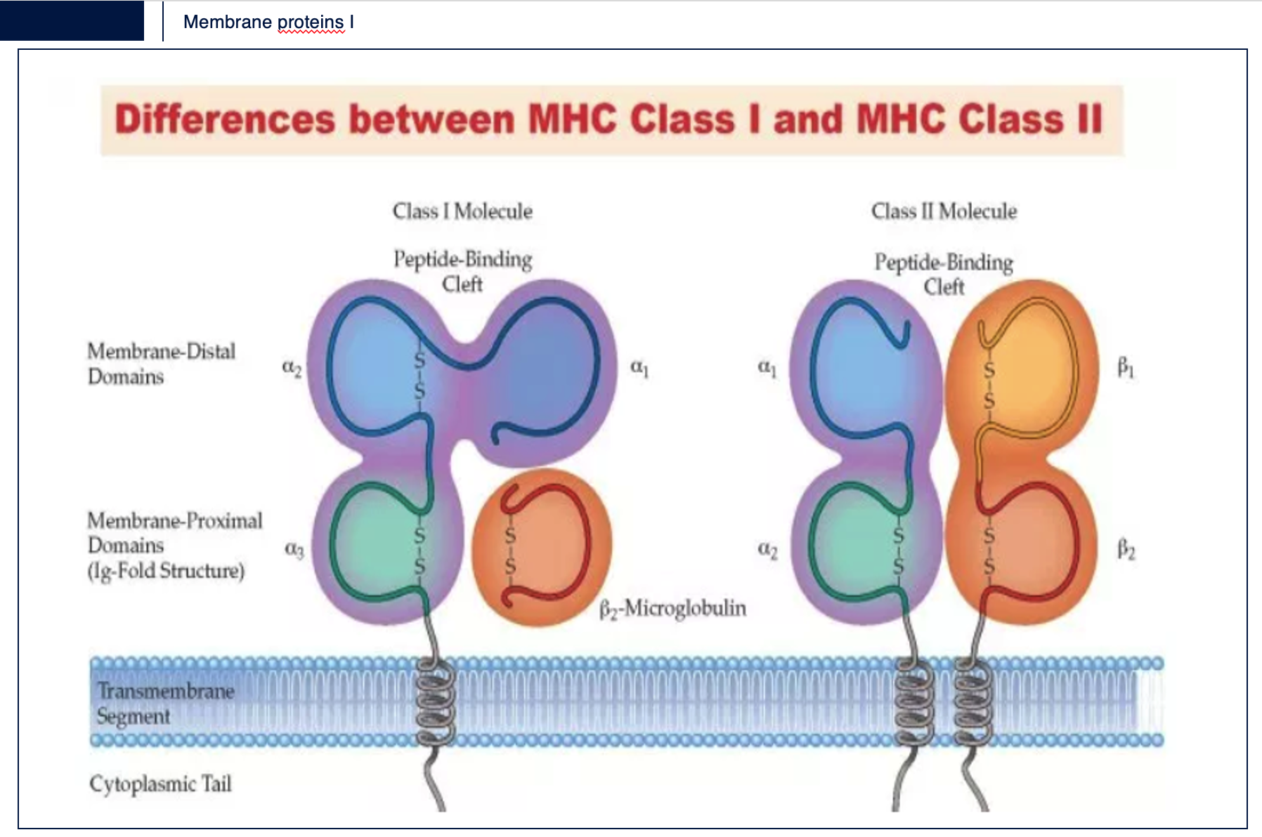
Explain the differences between MHC Class I and MCH Class II
The MHC II molecules are normally found only on professional antigen-presenting cells (APC) such as dendritic cells,mononuclear phagocytes, some endothelial cells, thymic epithelial cells, and B cells. These cells are important in initiating immune responses. The antigens presented by class II peptides are derived from extracellular proteins (not cytosolic as in MHC class I).
Interferon-y (IFN-y) is also responsible for converting monocytes which are MHC class II negative cells into functional APCs that express MHC class II on their surfaces.
The MHC II molecules present foreign peptides to helper CD4+ T lymphocytes
Label and describe the process of antigen presentation, highlighting the role of MHC I in presenting endogenous peptides to CD8+ T cells, which are crucial for the immune response against intracellular pathogens.
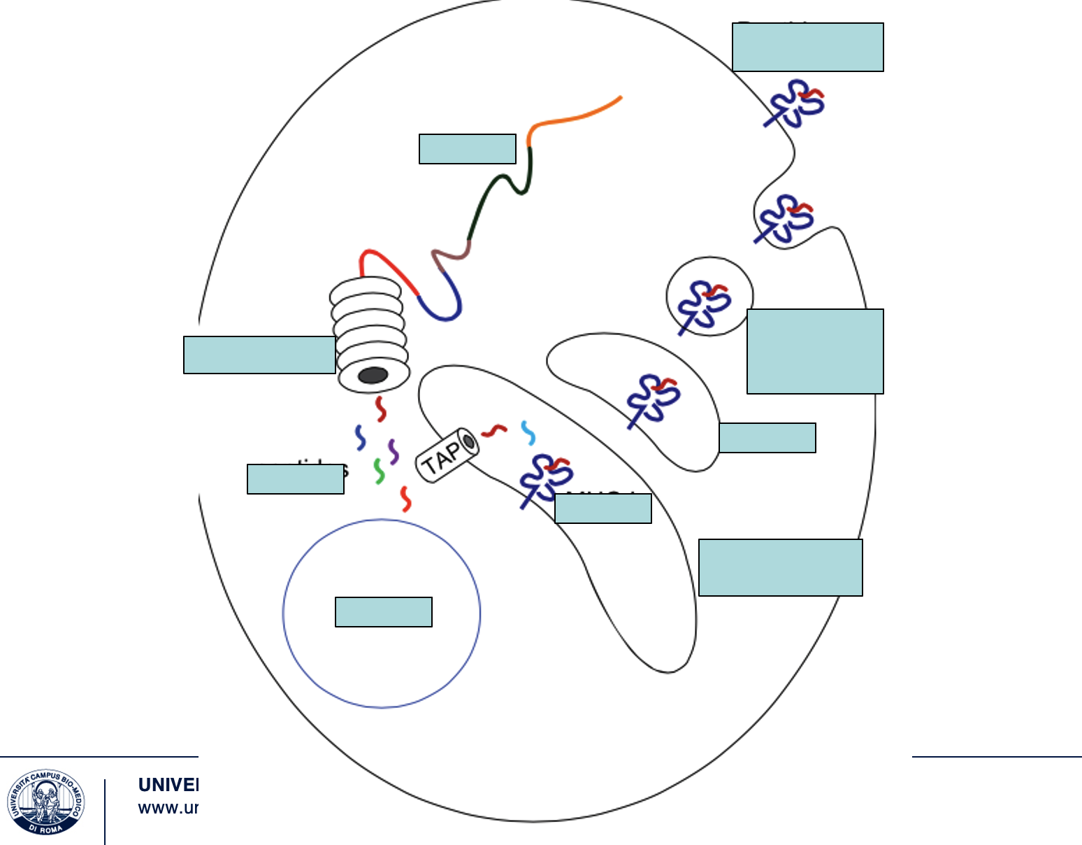
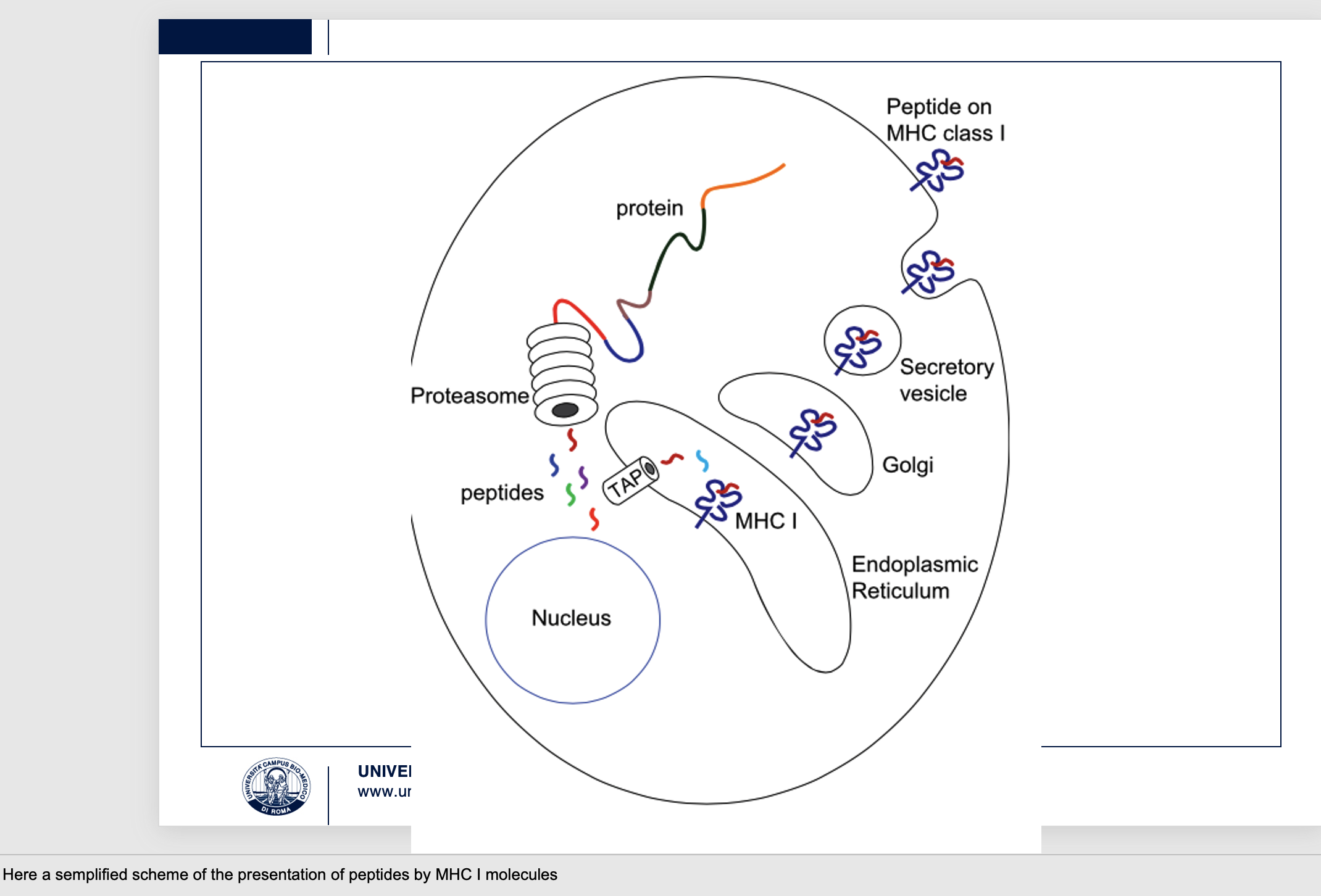
Here a semplified scheme of the presentation of peptides by MHC I molecules
The image depicts the MHC Class I antigen presentation pathway.
Protein Degradation: Endogenous proteins (e.g., viral proteins, abnormal self-proteins) are ubiquitinated and degraded into peptides by the proteasome in the cytoplasm.
Peptide Transport: These peptides are then transported into the Endoplasmic Reticulum (ER) by the TAP(Transporter Associated with Antigen Processing) complex.
MHC I Loading: Inside the ER, newly synthesized MHC Class I molecules bind to these peptides.
Surface Presentation: The peptide-loaded MHC Class I complex then transits through the Golgi apparatus and is transported via a secretory vesicle to the cell surface, where the peptide on MHC Class I is displayed.
This process allows the cell to present internal "self" or "non-self" (e.g., viral) peptides to CD8+ T cells. CD8+ T cells recognize these complexes and, if the peptide is foreign, can trigger an immune response to eliminate infected or abnormal cells, which is crucial for combating intracellular pathogens.
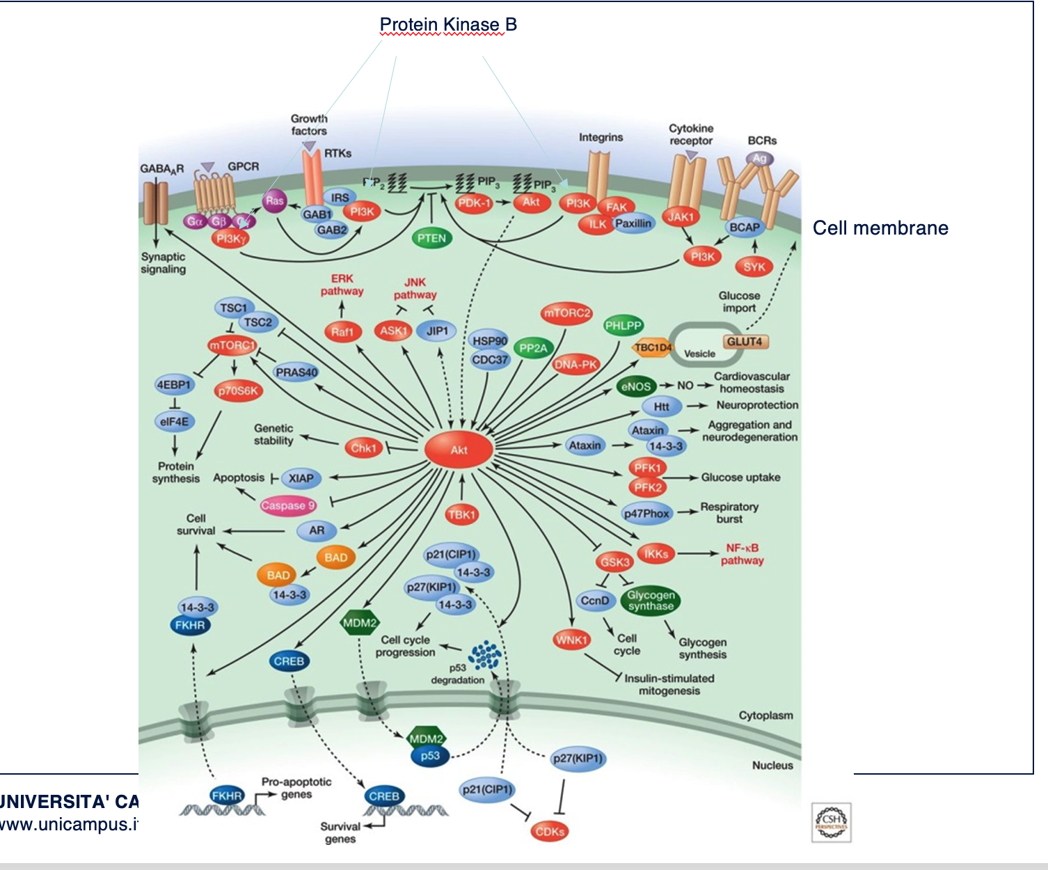
Describe what you see
This is a scheme of activity of protein kinase B .
The pathway begins with fosphorilation of PI3K (phosphatitylinositol 3 Kinase) then fosphorilation of phosphotydilinositol#,4,5-triphosphate (PIP3) the second messanger.
Downstream response(by Akt) include cell survival, growth, proliferation,cell migration, and angiogenesis.Impairment is linked to cancer and type II diabetes.
The key information is that the first signalling event (binding of molecules to surface receptors) is followed by a cascade of events mediated by second messengers able to amplify (promoting or repressing) the response. Knolewdge of the complex network of interactions between molecules help to use and deisgn drugs to impairepathological processes.

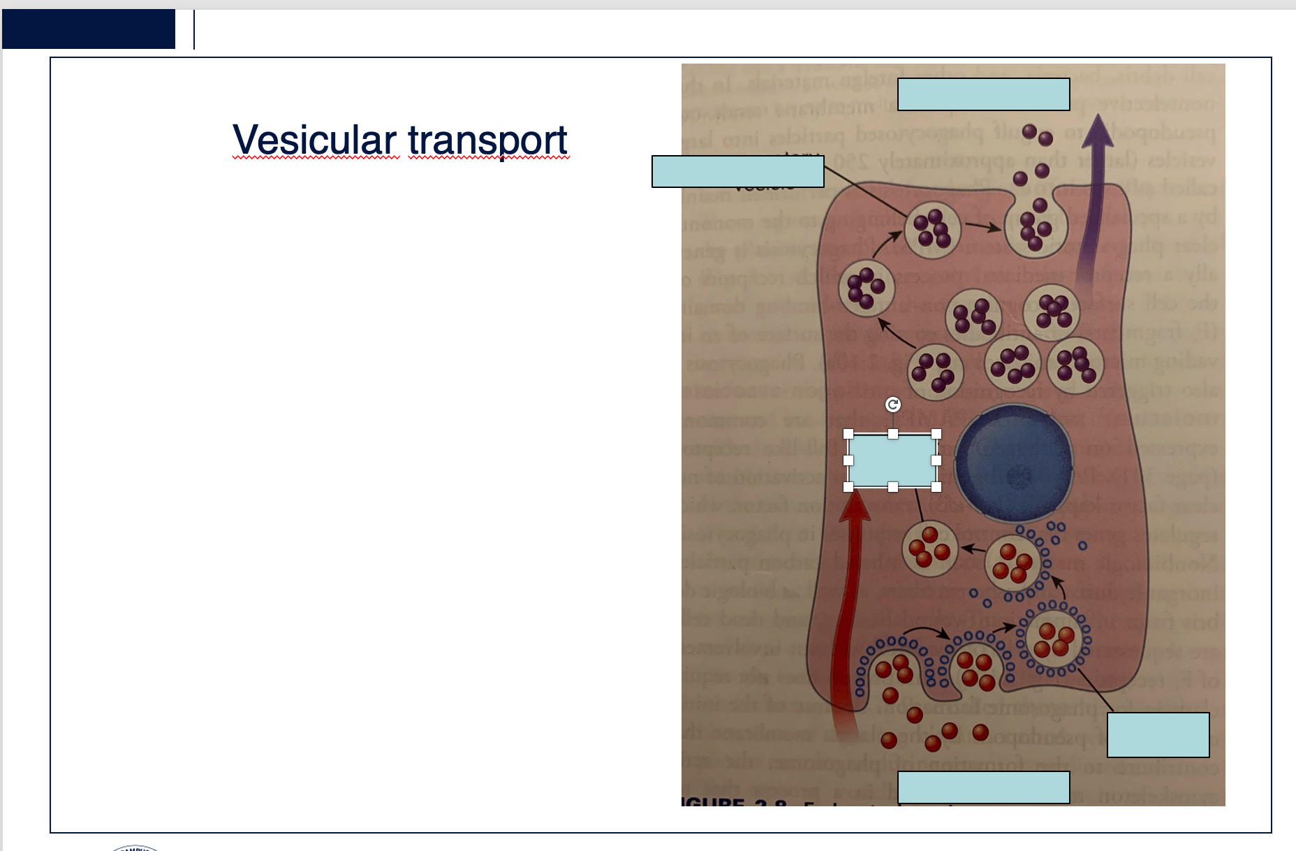
Label and describe
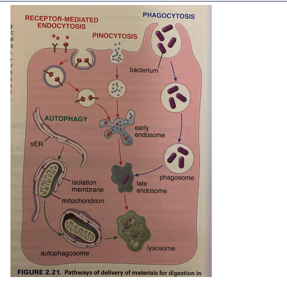
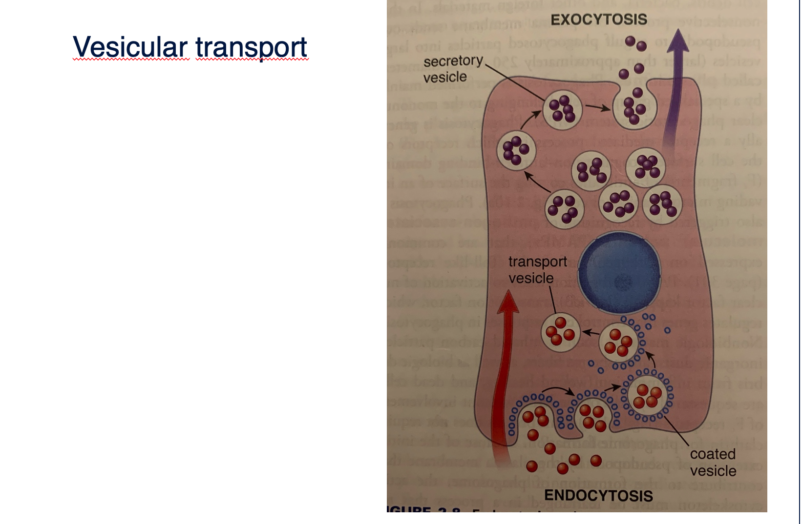
These two activities are coupled, if one is blocked also the second doesn’t work. Endocytosis is a general term for processes of vesicular transport in which substances enter the cell. It permit the uptake of membrane proteins, fluids , nutrients, lipids, singnalling molecules. Exocytosis is the general term for processes of vesicular transport in whichsubstances (cargo molecules) leave the cell. Exocytosis is also the process by which all cells deliver the intracellularplasma membrane to the cell surface.

Explain
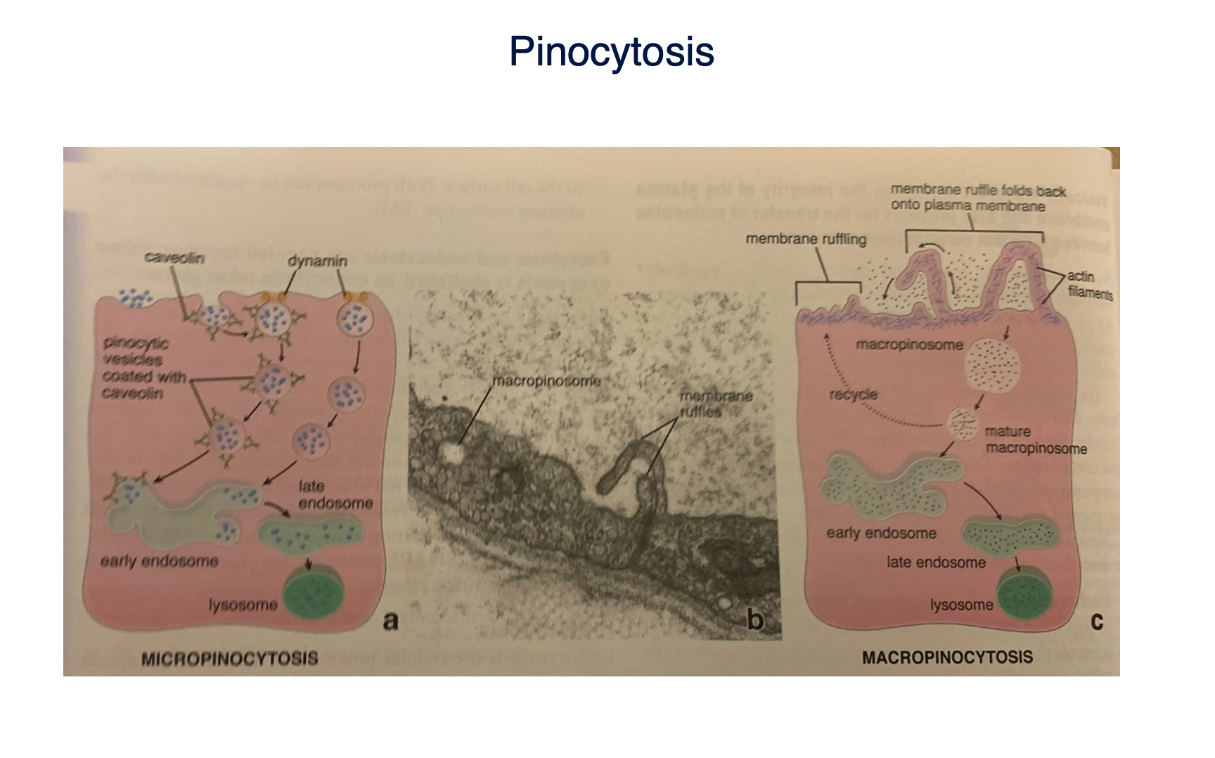
Micropinocytosis is the non specific ingestion of fluid and small protein via small vesicles (<150 nm). Is present in everycell and is constitutive. Vesicle formation is usually associated with the presence of caveolin and flotillin.Macropinocytosis involved a rearrangement of the plasma membrane and underlying actin cytoskeleton.
Receptor-mediated
endocytosis
Explain
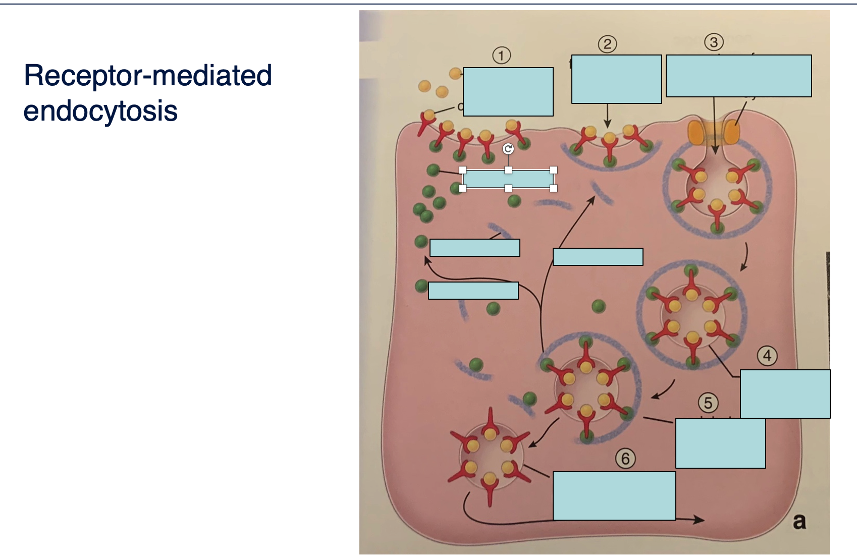
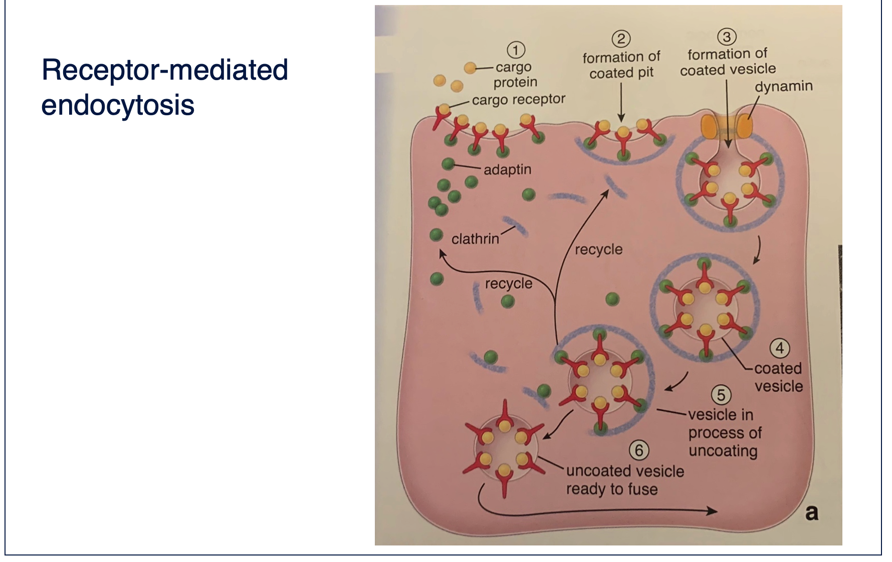
This is a special endocytic pathway initiated from the binding of extracellular moleculaes to surface receptors. Severalreceptors, in a close space. After binding there is the formation of the so called coated pits mediated by clathrin, dynaminpinch and coated vesicle is formed
Label
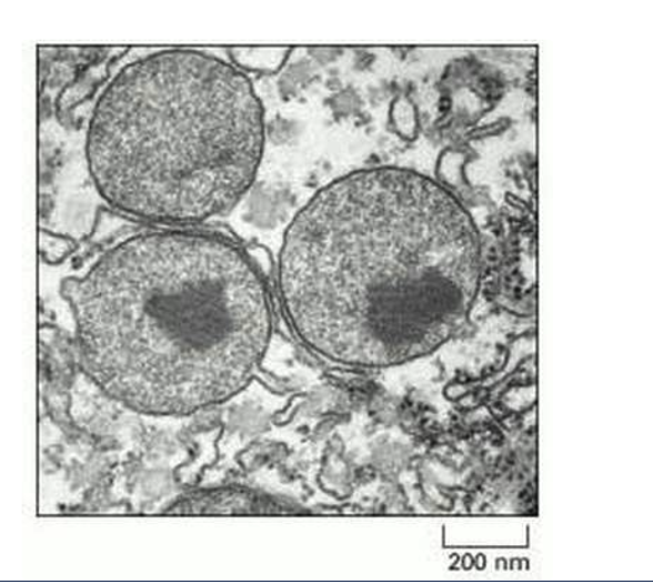
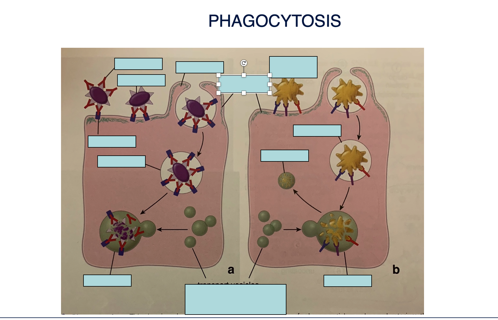
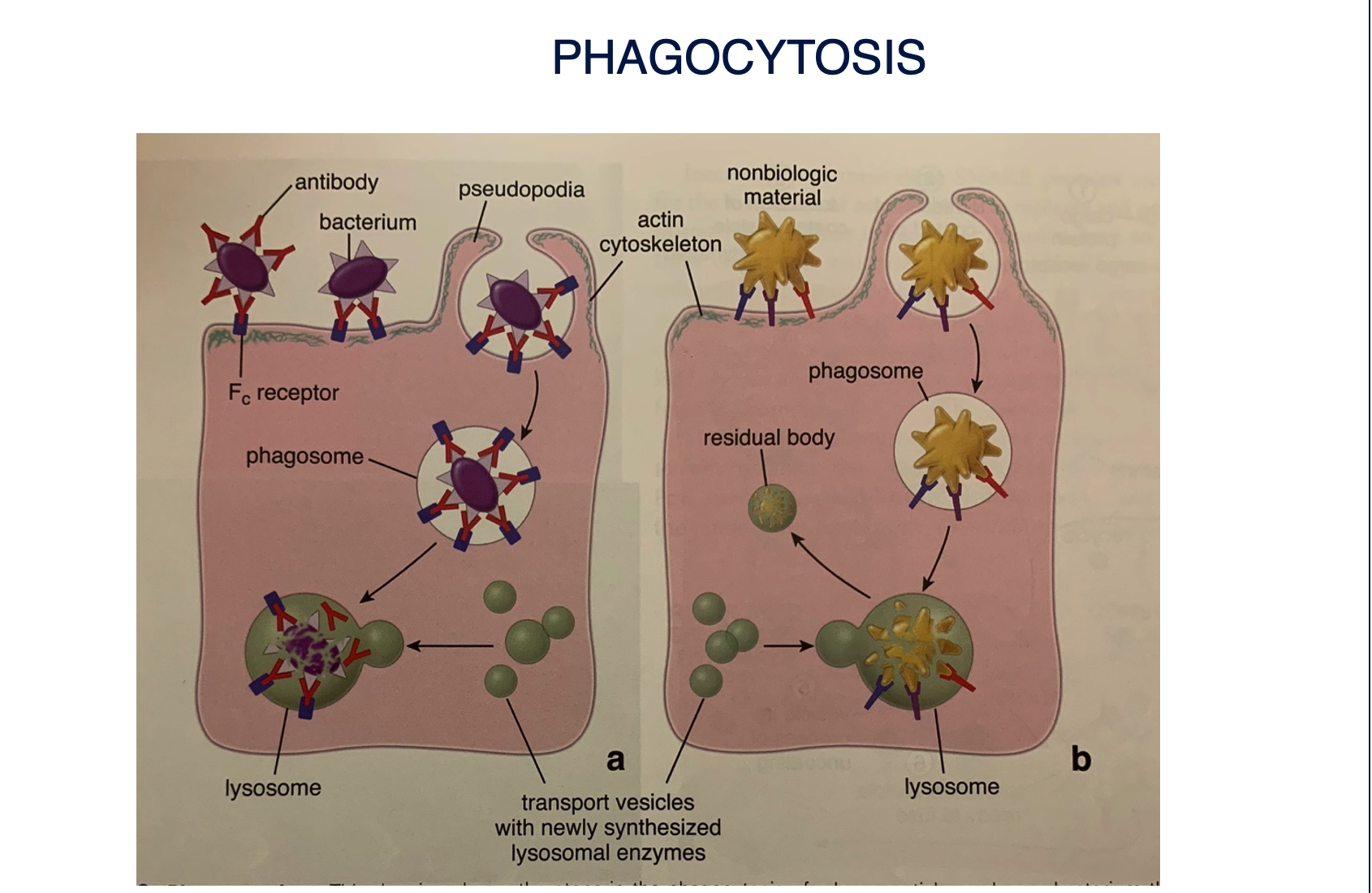
Phagocyt. Is a special receptor mediated endocytosis. Is performed by a specialized group of cells belonging to the Mononuclear phagocytic system and occurs when we have to neutralize big bad things(from a cell POV)

Article
what’s this
cytoplasm
Describe feature
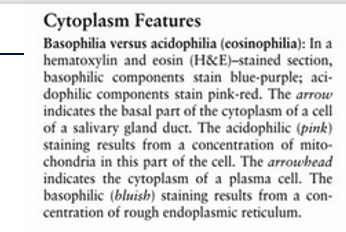
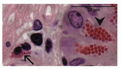
Describe feature
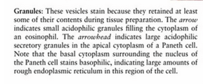
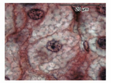
Describe feature
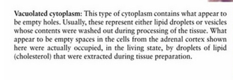
Describe feature
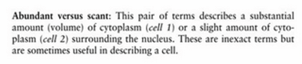
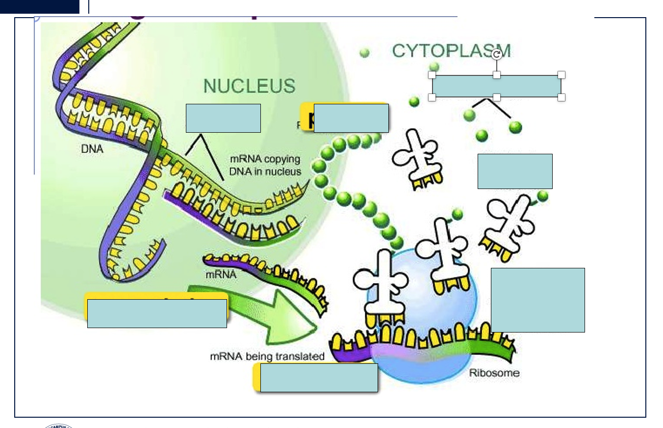
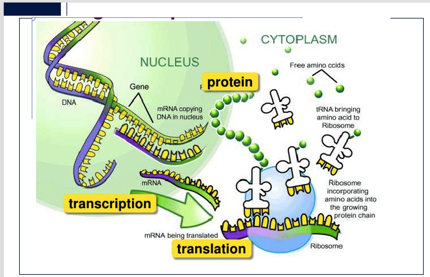
describes the flow of genetic information within a biological system.
This slide illustrates the central dogma of molecular biology)
: transcription in the nucleus, where a DNA gene is copied into mRNA, followed by translation in the cytoplasm on a ribosome, where mRNA's genetic code is used to assemble free amino acids (brought by tRNA) into a protein chain.
Describe
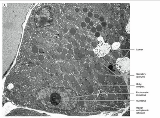
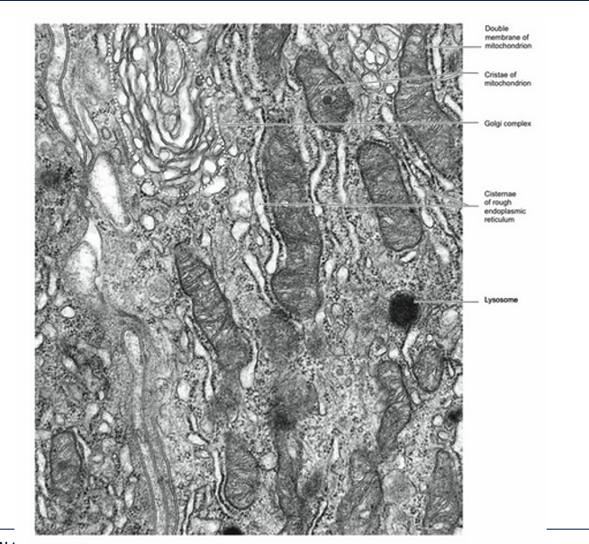
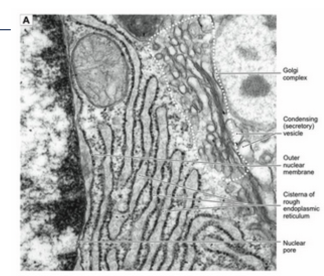
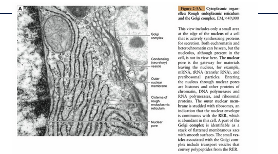
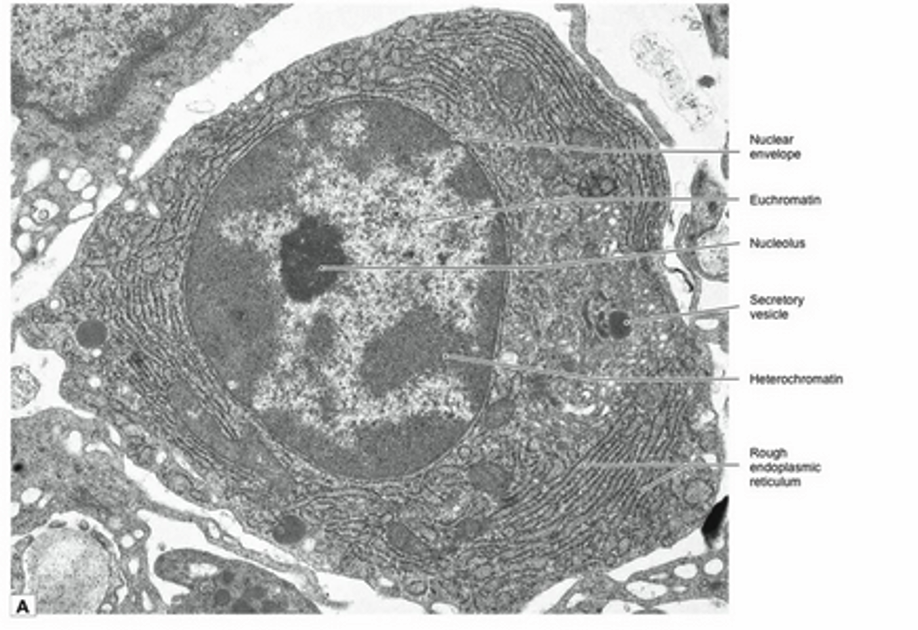
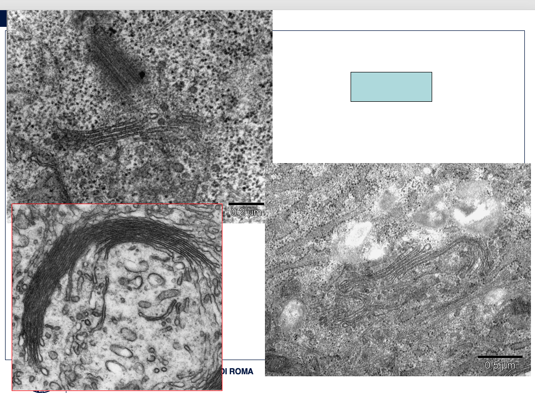
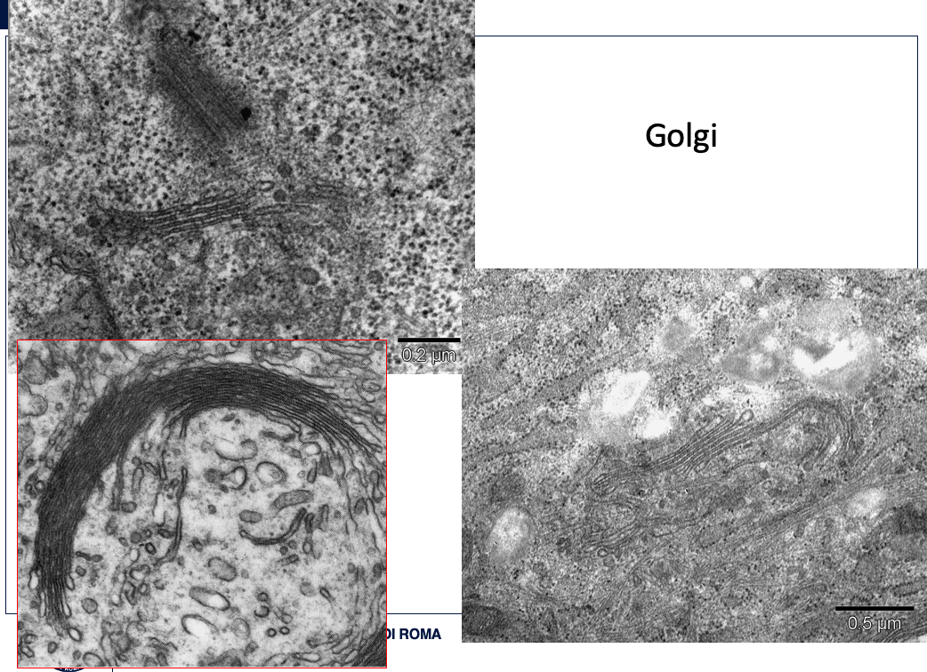
1.This view includes only a small area at the edge of the nucleus of a cell that is actively synthesizing proteins for secretion. Both euchromatin and heterochromatin can be seen, but the nucleolus, although present in the cell, is not in view here. The nuclear pore is the gateway for materials leaving the nucleus, for example, mRNA, tRNA (transfer RNA), and preribosomal particles. Entering the nucleus through nuclear pores are histones and other proteins of chromatin, DNA polymerases and RNA polymerases, and ribosomal proteins. The outer nuclear membrane is studded with ribosomes, an indication that the nuclear envelope is continuous with the RER, which is abundant in this cell. A part of the Golgi complex is identifiable as a stack of flattened membranous sacs with smooth surfaces. The small vesicles associated with the Golgi complex include transport vesicles that convey polypeptides from the RER.
Overall View:
Context: A small area at the edge of the nucleus of a cell actively synthesizing proteins for secretion.
Magnification: Electron Micrograph (EM), x49,000.
Nucleus:
Components Visible: Euchromatin and heterochromatin.
Components Not Visible (but present in cell): Nucleolus.
Nuclear Pore:
Structure: Gateway.
Function (Materials Leaving Nucleus): mRNA, tRNA (transfer RNA), preribosomal particles.
Function (Materials Entering Nucleus): Histones and other proteins of chromatin, DNA polymerases, RNA polymerases, ribosomal proteins.
Outer Nuclear Membrane:
Structure: Studded with ribosomes.
Association: Continuous with the Rough Endoplasmic Reticulum (RER).
Rough Endoplasmic Reticulum (RER):
Abundance: Abundant in this cell.
Structure: Indicated by ribosomes on the outer nuclear membrane and its continuity with the nuclear envelope.
Function: Involved in active synthesis of proteins for secretion.
Golgi Complex:
Structure: Identifiable as a stack of flattened membranous sacs with smooth surfaces.
Associated Structures: Small vesicles.
Function of Associated Vesicles: Transport polypeptides from the RER.
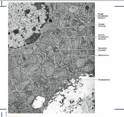
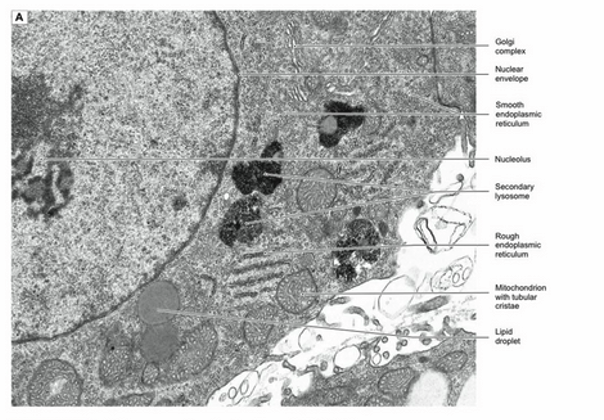
Describe what you see
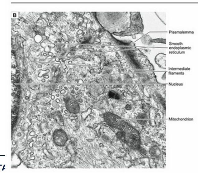
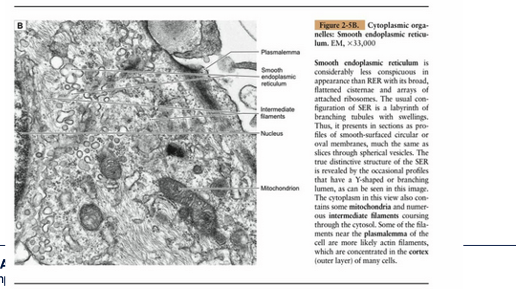
Smooth Endoplasmic Reticulum Characteristics: Smooth endoplasmic reticulum is considerably less conspicuous in appearance than RER with its broad, flattened cisternae and arrays of attached ribosomes. The usual configuration of SER is a labyrinth of branching tubules with swellings. Thus, it presents in sections as profiles of smooth-surfaced circular or oval membranes, much the same as slices through spherical vesicles. The true distinctive structure of the SER is revealed by the occasional profiles that have a Y-shaped or branching lumen, as can be seen in this image.
Cytoplasmic Contents: The cytoplasm in this view also contains some mitochondria and numerous intermediate filaments coursing through the cytosol. Some of the filaments near the plasmalemma of the cell are more likely actin filaments, which are concentrated in the cortex (outer layer) of many cells.
Describe what you see
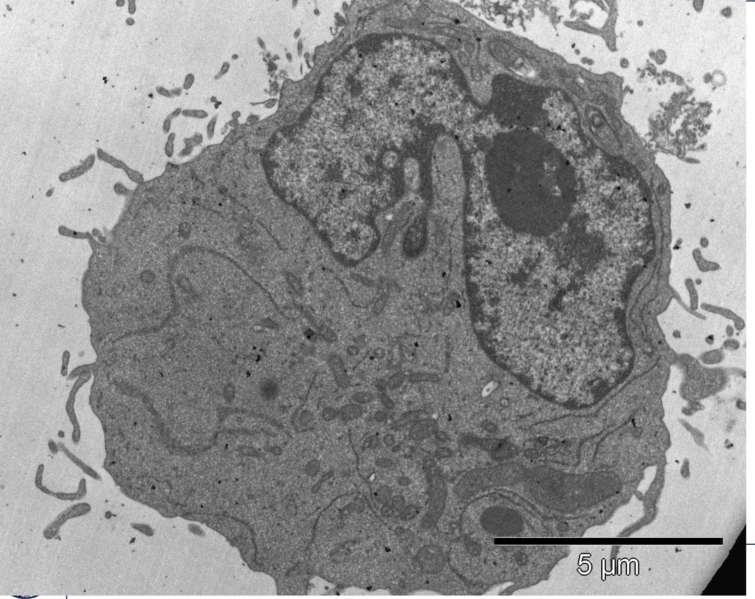
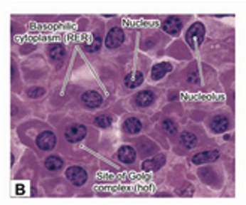
Plasma cells function to synthesize and secrete immunoglobulin, a glycoprotein. Once these cells have differentiated from stimulated B lymphocytes, they secrete the antibodies as fast as they are generated for 1 to 2 weeks before they die. The major structures required for this process are RER, the Golgi complex, and secretory vesicles. The RER is the site of synthesis and sequestering of the polypeptides of the antibody. Posttranslational modification and packaging occur in the Golgi complex, and the secretory vesicles convey the product to the cell surface. Because plasma cells do not store the immunoglobulin, few, if any, secretory vesicles are seen in the cytoplasm. Rather, the cytoplasm is packed with RER, and there is a large Golgi complex (not visible in this section) located adjacent to the nucleus. The nucleus has one or more well-developed nucleoli, but there is a considerable amount of heterochromatin, considering that this is an active protein-secreting cell. The explanation may be that only one protein, an antibody molecule, is secreted, and the cell is terminally differentiated, so it will never divide. The immunoglobulin is secreted into the surrounding interstitial compartment, from which it can enter circulation through walls of small blood or lymph vessels. Even though the cell is not sharply polarized, the nucleus tends to occupy an eccentric position, with the Golgi complex near the center of the cell.


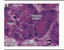
describe what you see
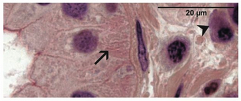
The appearance of plasma cells in light microscopy is consistent with the ultrastructure of these antibody-secreting cells. The nucleus has a mixture of heterochromatin and euchromatin in an arrangement that is variously described as a "clock face" or checkerboard pattern. A large nucleolus or two occupy the center of the nucleus. The cytoplasm is basophilic as a result of the ribonucleic acid associated with the extensive RER. Depending upon the orientation of the cell in the section, a pale area, called the hof, can be seen adjacent to the nucleus. This unstained area is the location of the Golgi complex.
describe
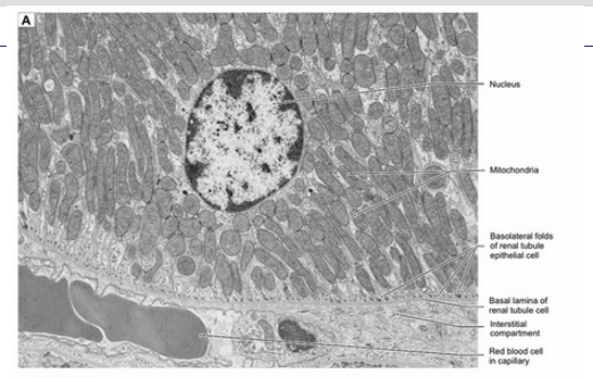
Steroid hormine-secreting cells, adrenal cortex. EM,x 21.000
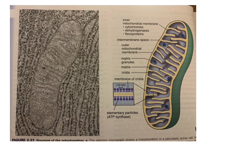
Adrenal cortical cells, testicular interstitial cells, and ovarian follicular cells are examples of cell types that function to synthesize and secrete steroid hormones. Each of these cells have in common the equipment needed for synthesis of steroid hormones. The necessary components include lipid droplets containing cholesterol esters as substrates, SER, and mitochondria that function together in the synthesis of hormones. The mitochondria characteristically have cristae that are tubular rather than shelflike.


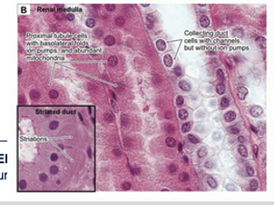
describe
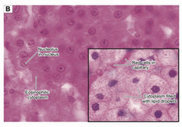
Light microscopic appearance of steroid hormone-secreting cells. H&E, x800 main panel and inset
Light microscopic views of steroid hormone-synthesizing cells reveal only a hint of the distinctive set of structures that characterize the cytoplasm of these cells. The eosinophilia (acidophilia) of the cytoplasm is largely attributable to the abundance of mitochondria. Steroid hormone-secreting cells vary in the amount of cholesterol stored in the form of lipid droplets, which appear in most preparations as empty vacuoles because their contents are extracted during specimen preparation. The cells in the main panel are in the zona reticularis and have few droplets, similar to the cell in the electron micrograph (Fig. 2-10A). The cells in the inset are from the zona fasciculata and have numerous lipid droplets.
what are these?
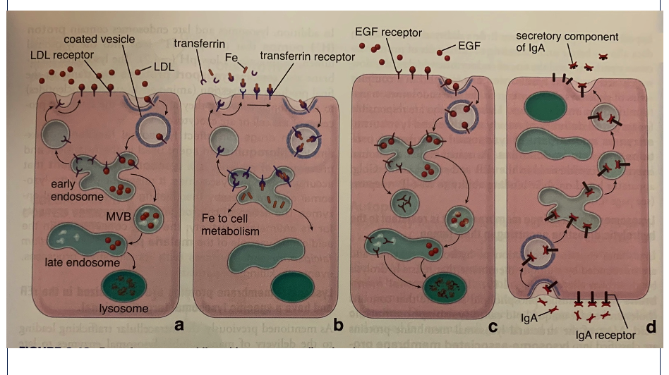
describe the major pathways of protein and lipid trafficking within a eukaryotic cell
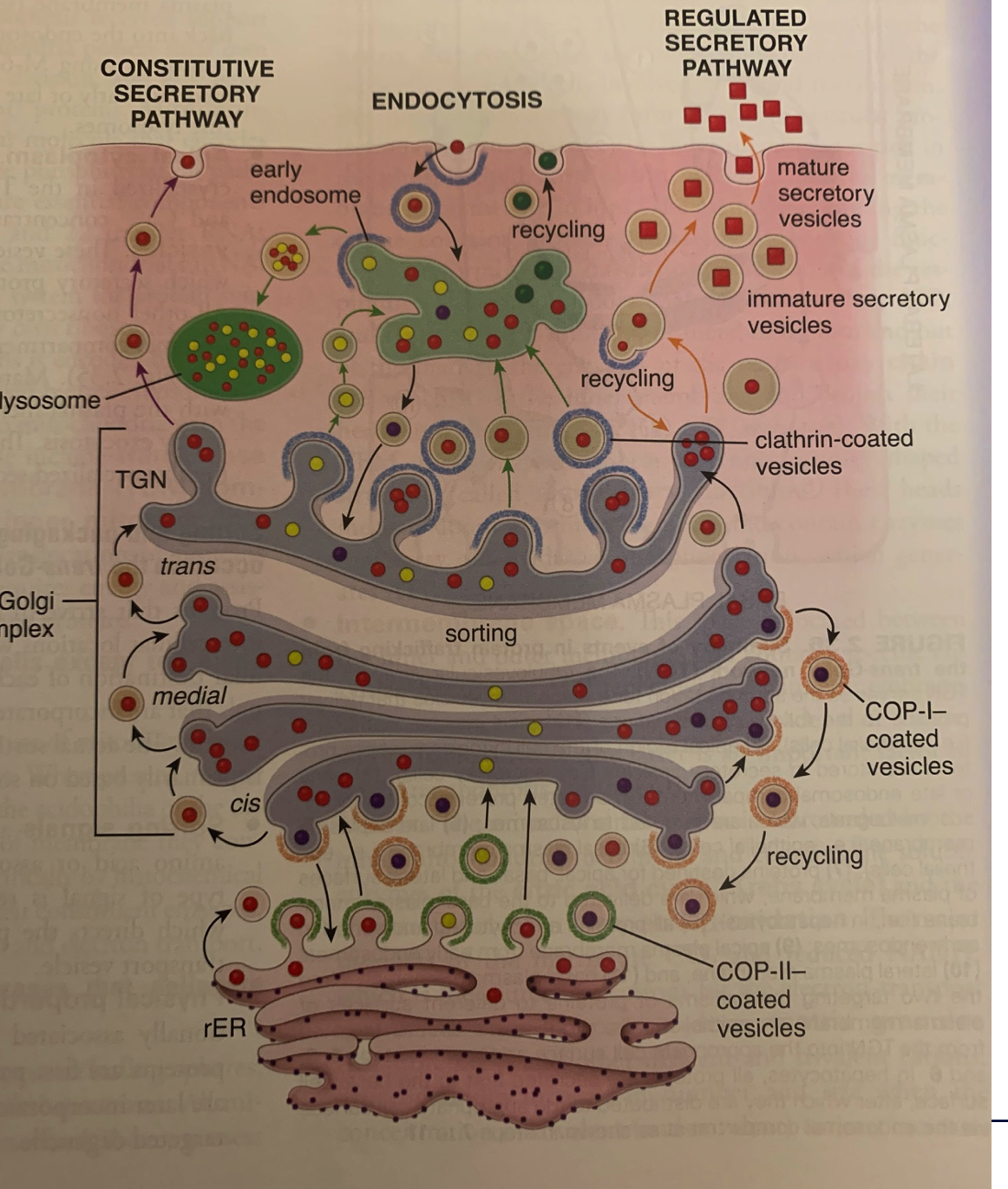
This image illustrates the major pathways of protein and lipid trafficking within a eukaryotic cell: the Constitutive Secretory Pathway, the Regulated Secretory Pathway, and Endocytosis.
Let's break down each part:
1. Rough Endoplasmic Reticulum (rER):
Starting Point: The rER is where newly synthesized proteins, especially those destined for secretion, insertion into membranes, or delivery to organelles like the Golgi, lysosomes, and endosomes, begin their journey.
COP-II-coated vesicles: Proteins bud off from the rER in COP-II-coated vesicles (light green vesicles with yellow contents). These vesicles move towards the Golgi apparatus.
Recycling (light green vesicles with dark green contents): Some vesicles (light green with dark green contents) move back to the rER, indicating recycling of components or retrieval of ER resident proteins.
2. Golgi Complex:
Structure: The Golgi is a series of flattened sacs called cisternae, typically divided into cis, medial, and transcompartments.
Cis-Golgi Network (CGN or cis): Vesicles from the rER fuse with the cis face of the Golgi.
Medial-Golgi: Proteins move through the medial cisternae.
Trans-Golgi Network (TGN): This is the final sorting station of the Golgi. Proteins are sorted into different pathways here.
COP-I-coated vesicles: These vesicles (light red with dark red contents) mediate retrograde transport within the Golgi (from trans to cis) and also from the Golgi back to the rER, ensuring recycling of Golgi resident proteins and retrieval of ER proteins that might have escaped.
3. Constitutive Secretory Pathway (Left Side):
Continuous Secretion: This pathway operates continuously in all eukaryotic cells.
Process: Proteins and lipids exit the TGN in vesicles that move directly to the plasma membrane, where they fuse, releasing their contents outside the cell (secretion) or integrating membrane proteins into the plasma membrane.
Examples: Components of the extracellular matrix, plasma membrane proteins, and lipids are secreted or integrated this way.
4. Regulated Secretory Pathway (Right Side):
Specific Stimulus Required: This pathway is found in specialized cells (e.g., endocrine cells, nerve cells) that store secretory products and release them only in response to a specific signal or stimulus.
Immature Secretory Vesicles: At the TGN, proteins destined for regulated secretion are concentrated into immature secretory vesicles (larger, irregularly shaped vesicles with dark red contents).
Maturation: These immature vesicles undergo maturation, involving further condensation of cargo and removal of excess membrane, becoming mature secretory vesicles (smaller, dense-core vesicles with dark red contents).
Storage and Release: Mature secretory vesicles are stored near the plasma membrane and fuse with it only when a specific signal is received, releasing their contents (e.g., hormones, neurotransmitters) in a burst.
Clathrin-coated vesicles: Excess membrane and some receptors from immature secretory vesicles are recycled back to the TGN via clathrin-coated vesicles (light blue with light red contents).
5. Endocytosis (Top Middle):
Uptake from Outside: This process involves the internalization of substances from the extracellular environment into the cell.
Early Endosome: Endocytic vesicles (e.g., clathrin-coated vesicles from the plasma membrane) fuse with early endosomes.
Sorting within Early Endosome: From the early endosome, internalized molecules can take several routes:
Recycling (Back to Plasma Membrane): Some receptors and membrane components are recycled back to the plasma membrane (light green vesicles with dark green contents).
To Lysosome (Degradation): Other cargo, particularly those destined for degradation, are sorted into vesicles that mature into or fuse with late endosomes and eventually lysosomes.
Lysosome: Lysosomes are organelles containing hydrolytic enzymes that break down waste materials, cellular debris, and foreign substances.
In summary, the image provides a comprehensive overview of the dynamic trafficking pathways that allow eukaryotic cells to synthesize, process, sort, transport, and secrete proteins and lipids, as well as internalize substances from their environment.

Describe