head and neck anatomy pin test
1/125
There's no tags or description
Looks like no tags are added yet.
Name | Mastery | Learn | Test | Matching | Spaced |
|---|
No study sessions yet.
126 Terms
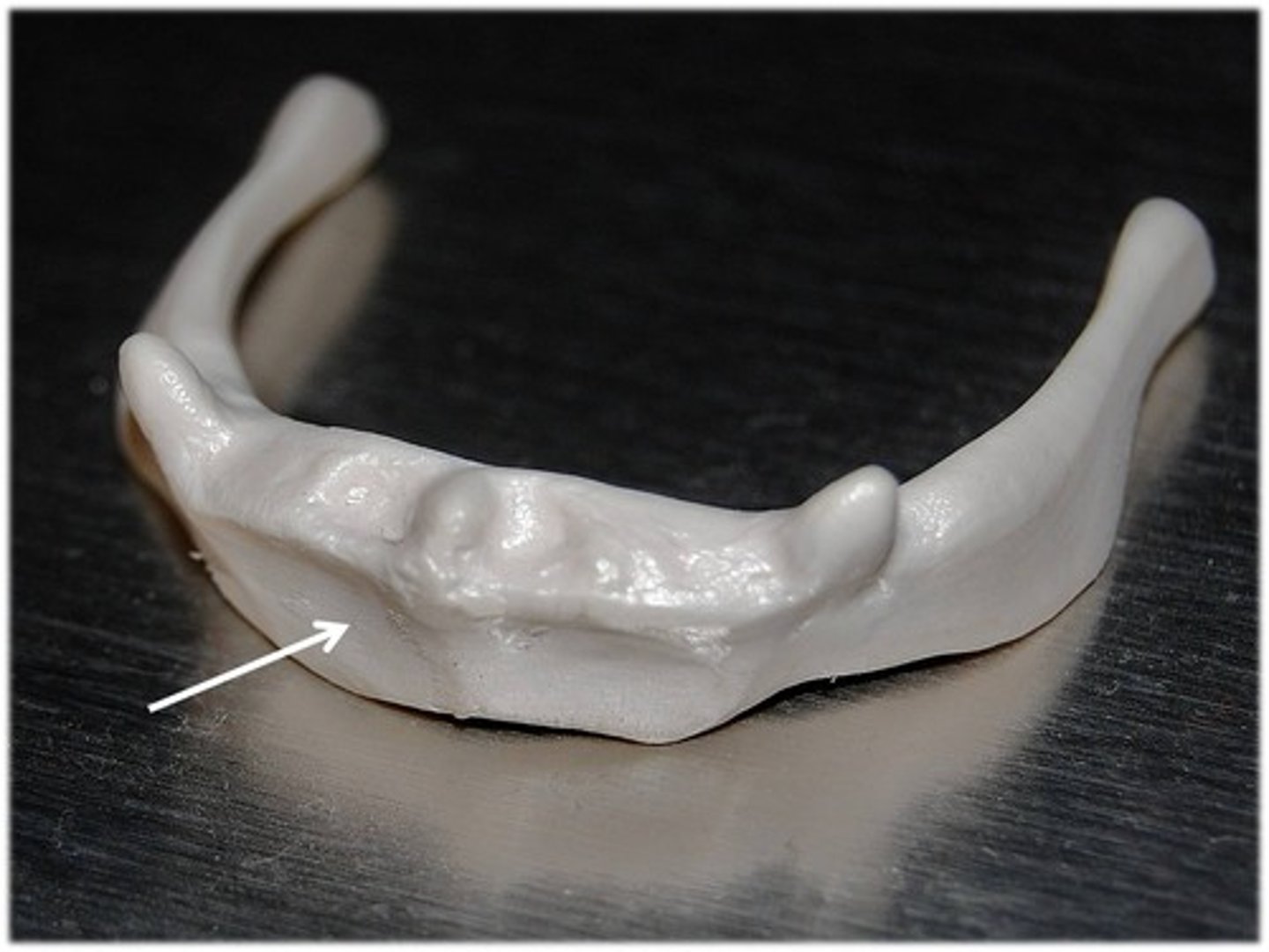
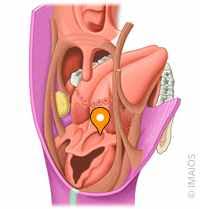
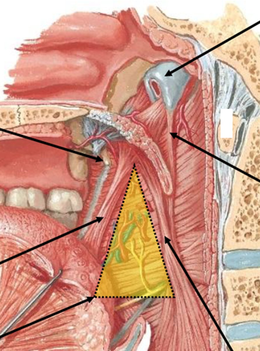
body of hyoid
what is this structure
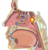
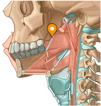
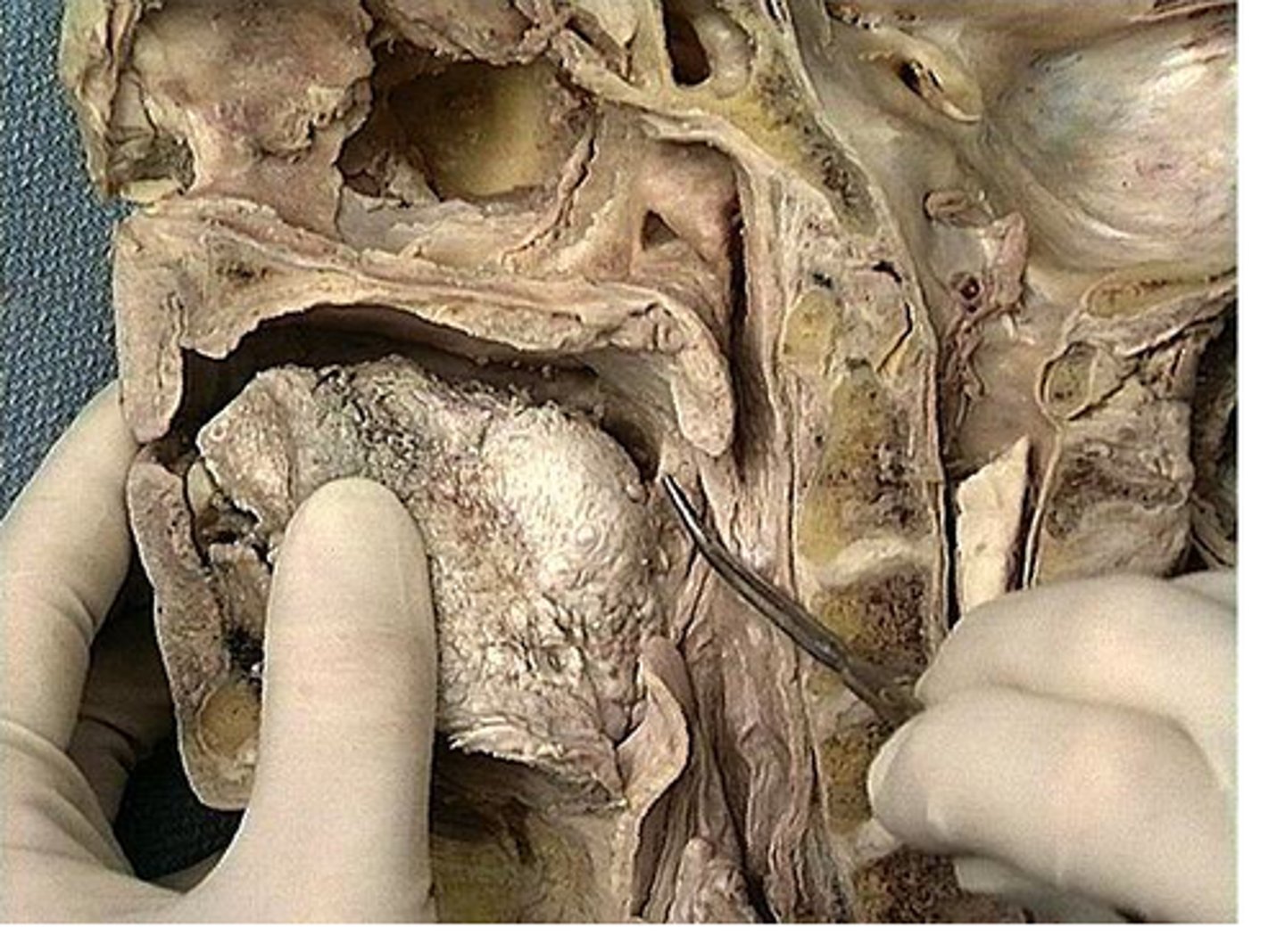
pharyngotympanic tube (eustachian tube)
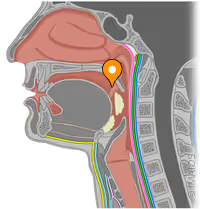
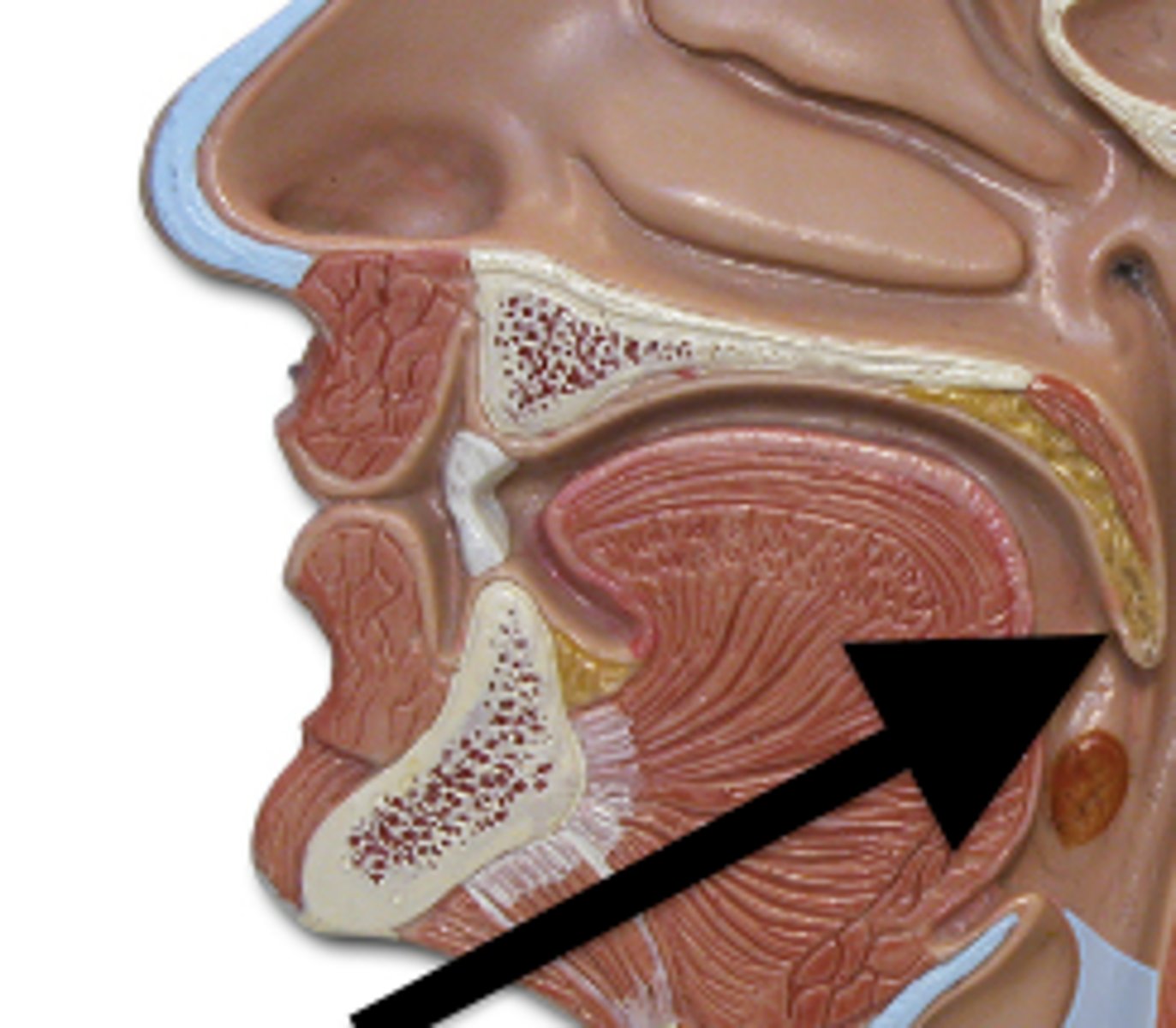
isthmus of oral fauces
what is this opening in the back of the oral cavity
piriform apeture
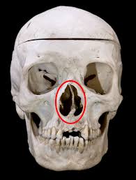
internal laryngeal nerve, superior laryngeal artery
what is the innervation and blood supply ABOVE the vocal cords
recurrent laryngeal nerve and inferior laryngeal artery
innervation and blood supply BELOW the vocal cords
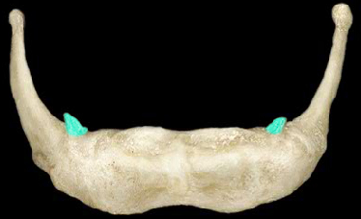
lesser horn of hyoid, stylohyoid ligament
structure and what attaches here
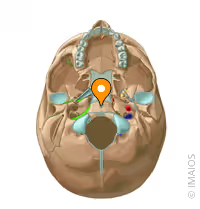
pharyngeal tubercle, superior constrictor muscle
structure, what attaches here
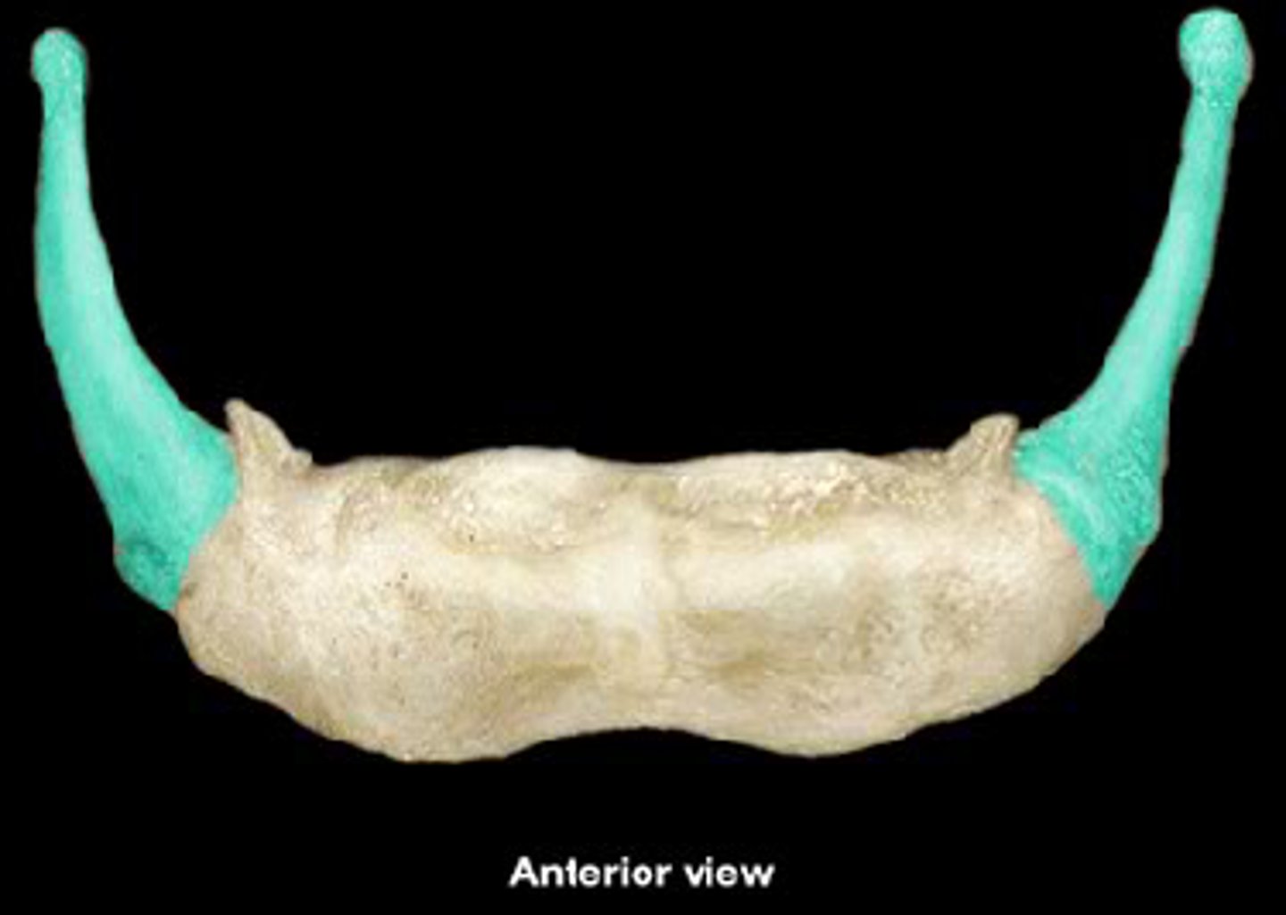
greater horns of hyoid, middle constrictor muscle
structure and what attaches heree
thyroid cartilage, inferior constrictor
structure and what attaches here
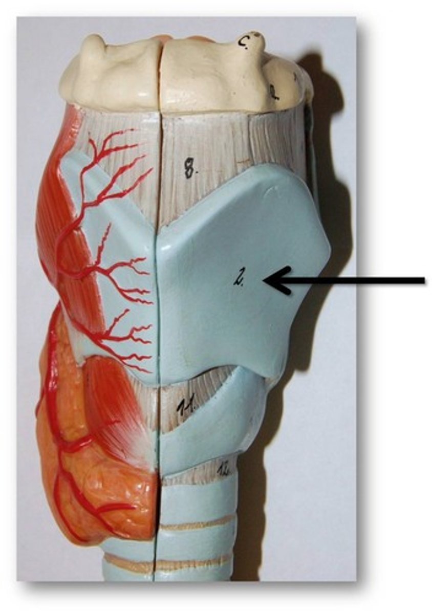
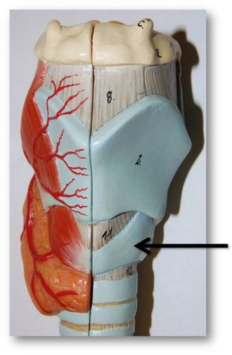
cricoid cartilage
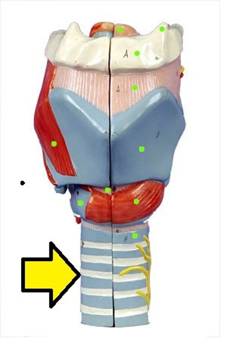
tracheal rings
pharyngeal raphe
Posterior midline attachment for the constrictors

stylohyoid ligament
holds the hyoid bone in place.
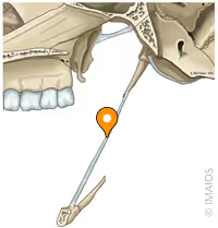
pterygomandibular raphe, superior constrictor
structure and what attaches here
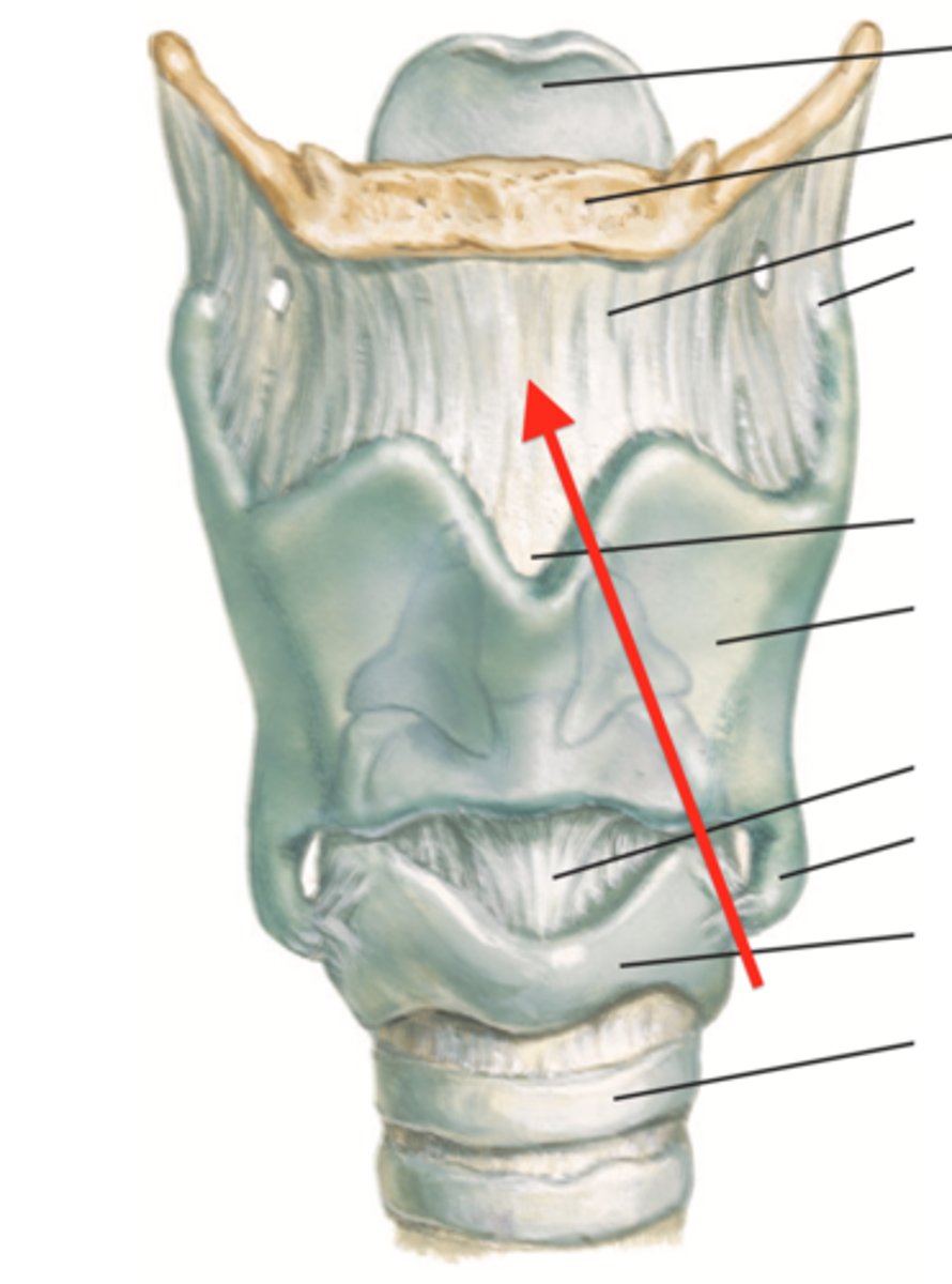
thyrohyoid membrane, internal laryngeal nerve and superior laryngeal artery
structure and what passes through it
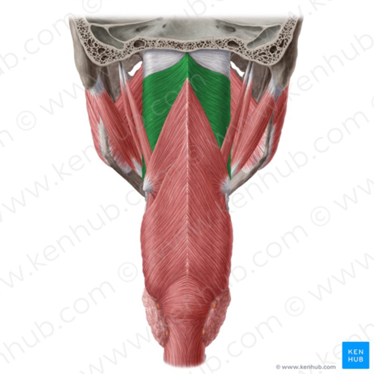
superior constrictor (CN X)
middle constrictor (CN X)
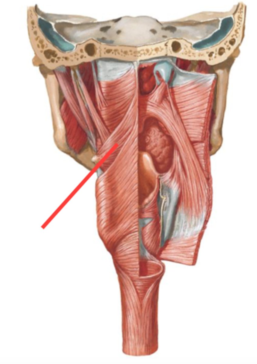
inferior constrictor (CN X)
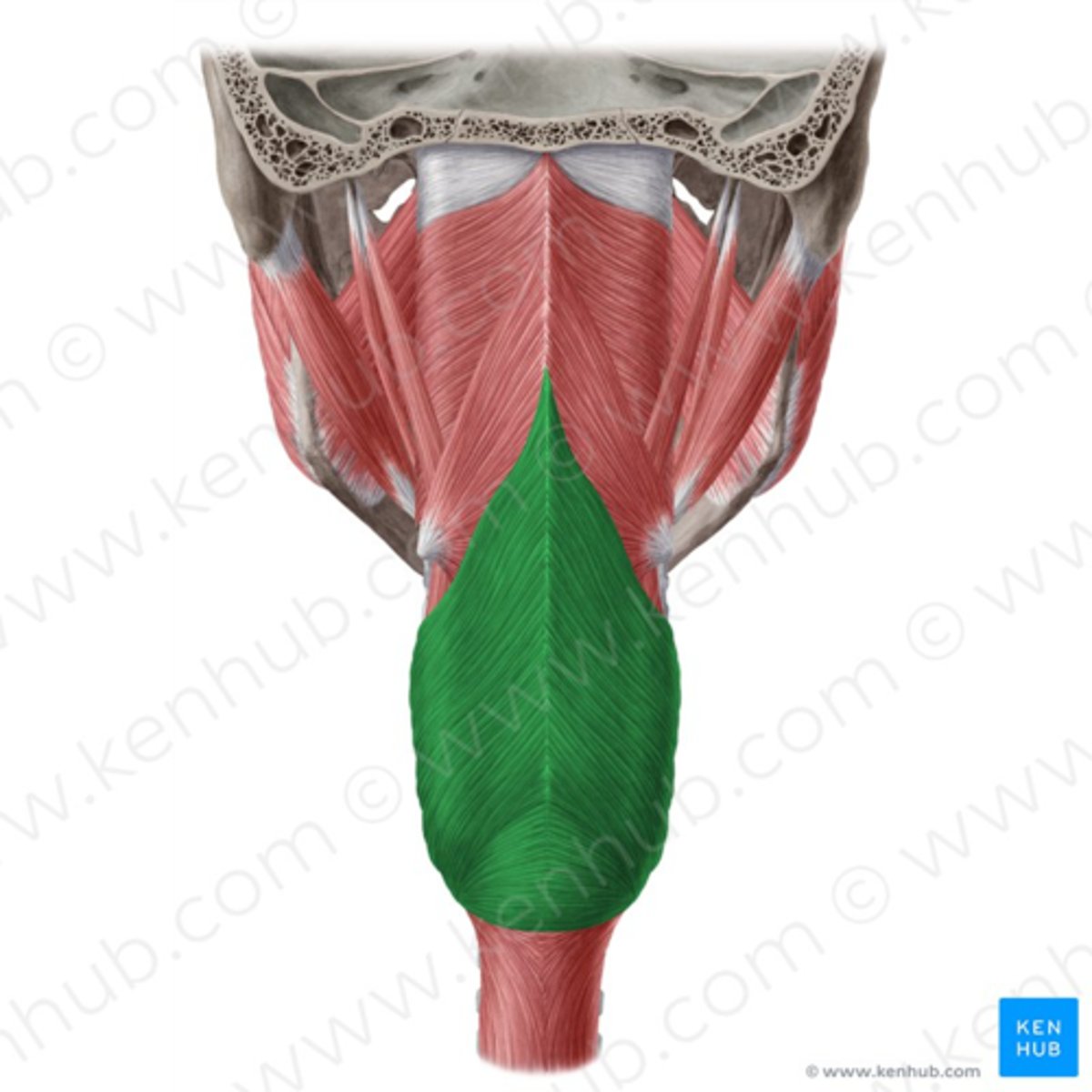
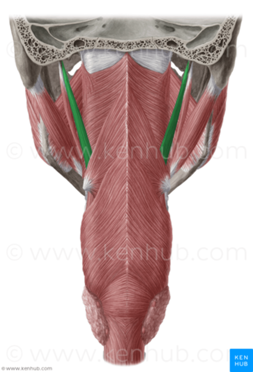
Stylopharyngeus (CN IX)
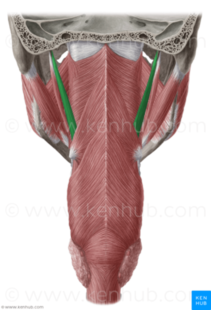
CN IX (glossopharyngeal)
what innervates this muscle?
internal laryngeal nerve
SENSORY innervation to larynx ABOVE vocal cords
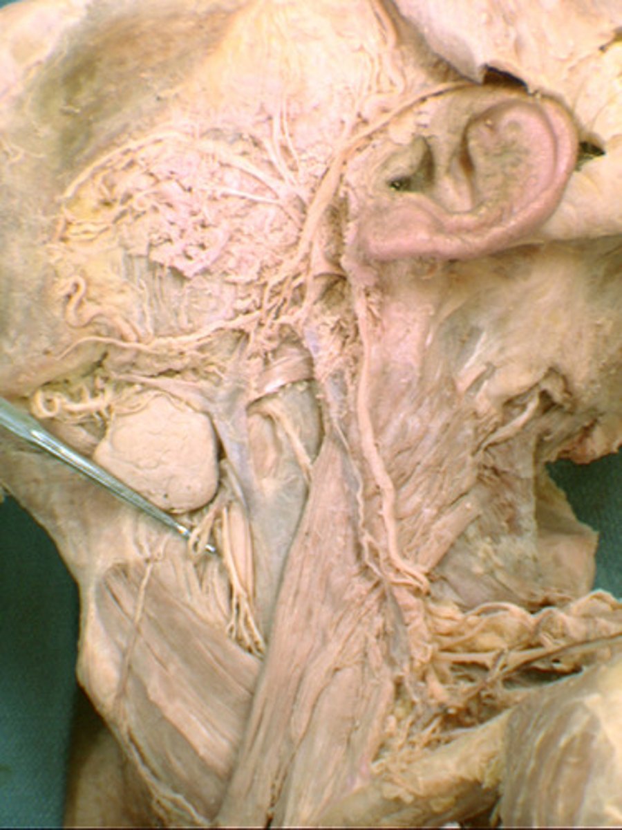
recurrent laryngeal
SENSORY nerve to larynx BELOW to vocal cords
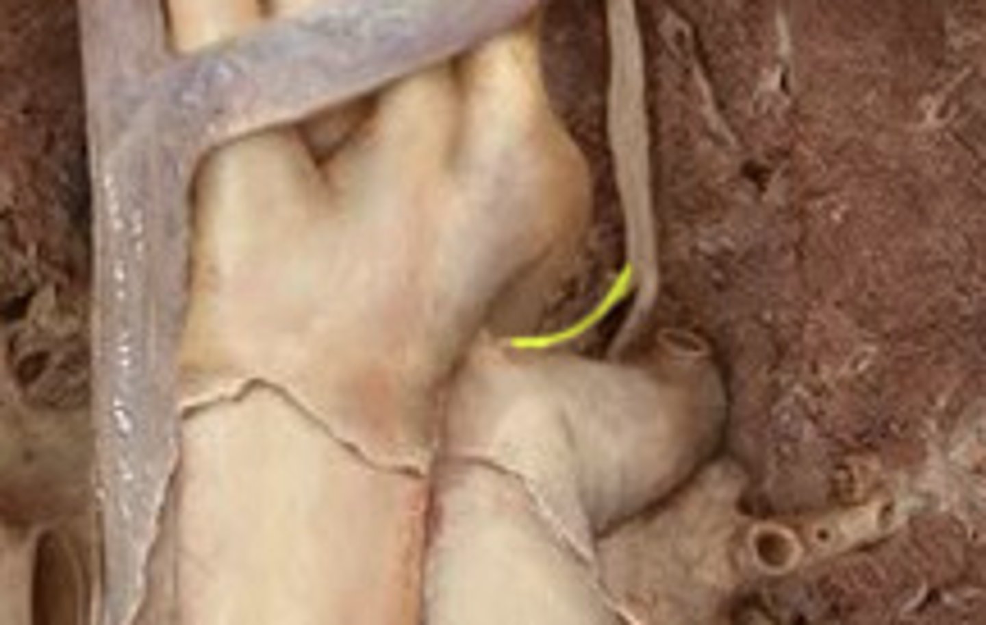
sympathetic trunk and superior cervical sympathetic ganglion
supplies postganglionic sympathetic innervation to the smooth muscle and glands in the head
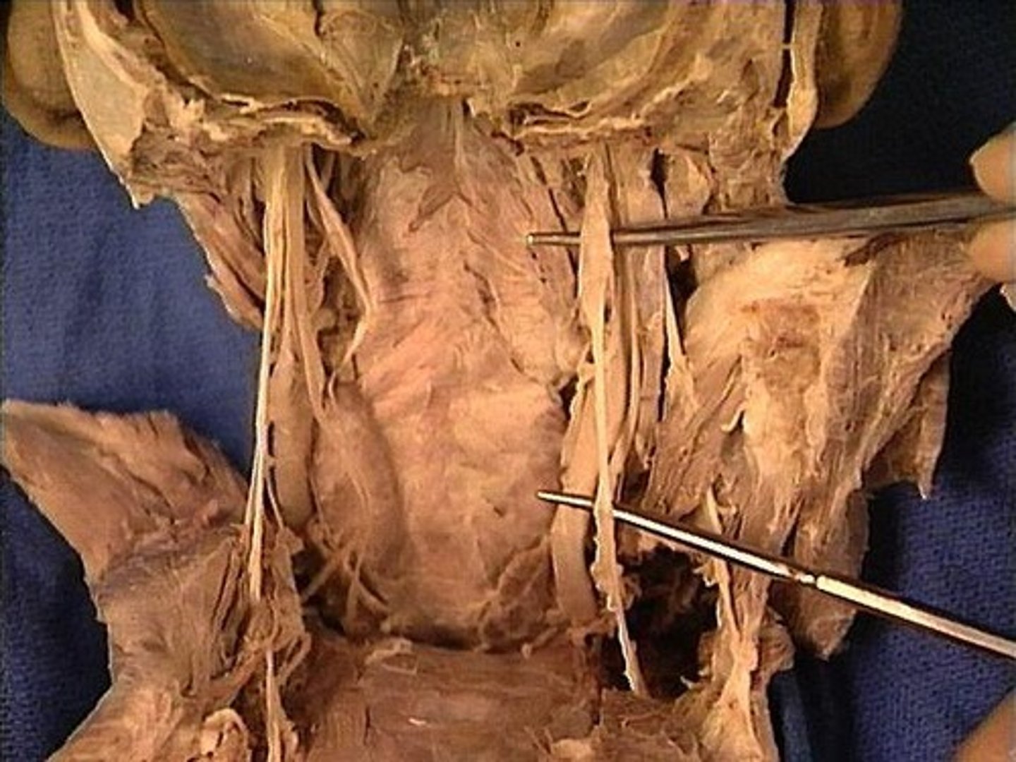
superior thyroid artery
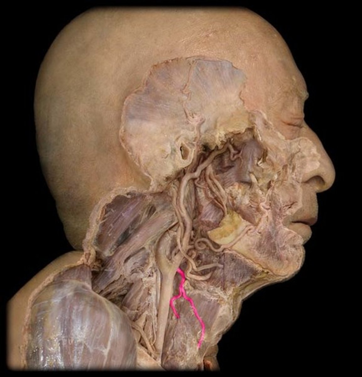
superior laryngeal artery
Travels with internal laryngeal nerve through the thyrohyoid membrane
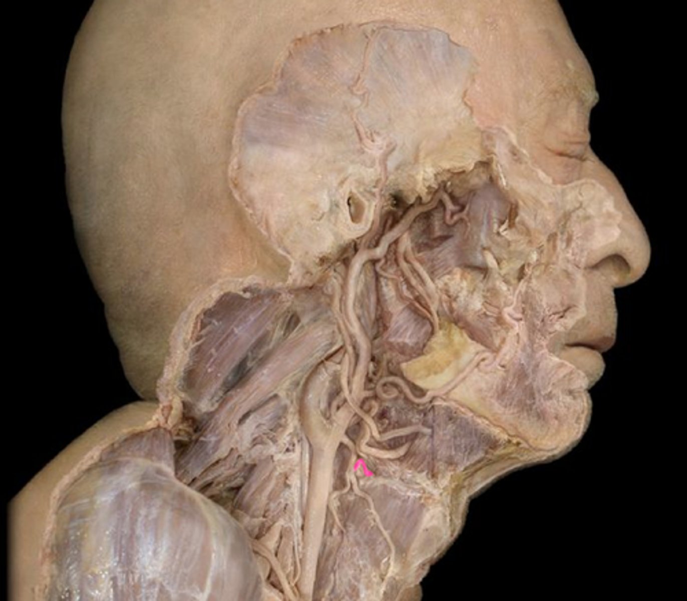
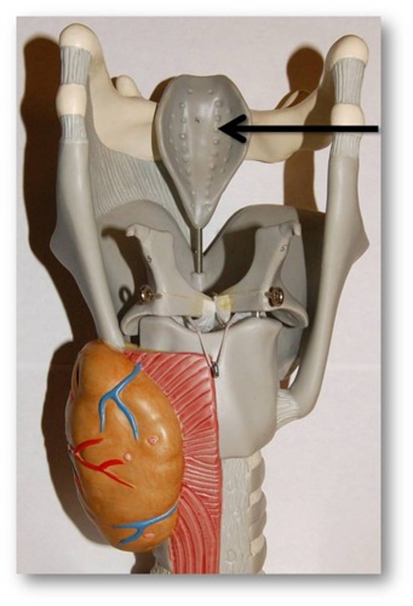
epiglottis
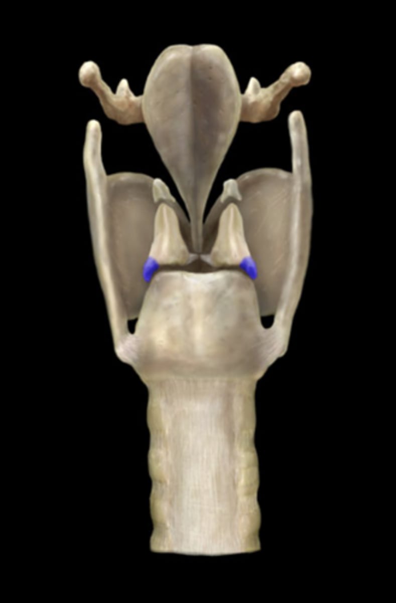
muscular process of arytenoid
Attaches to posterior and lateral cricoarytenoid muscles
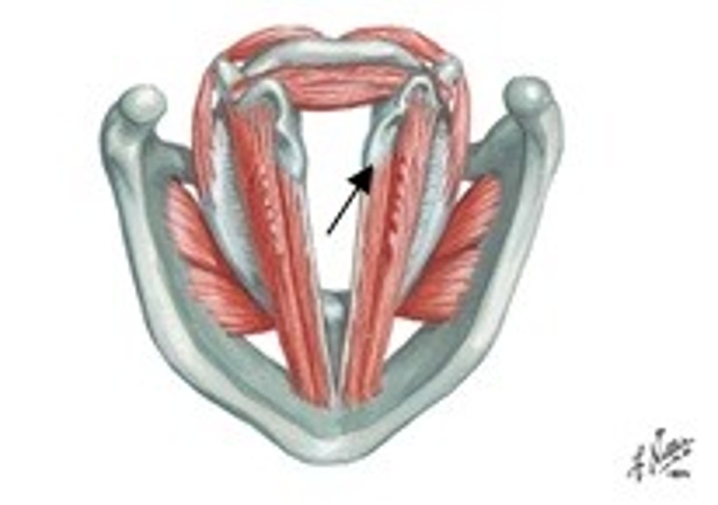
vocal process of arytenoid
Attached to vocal ligament
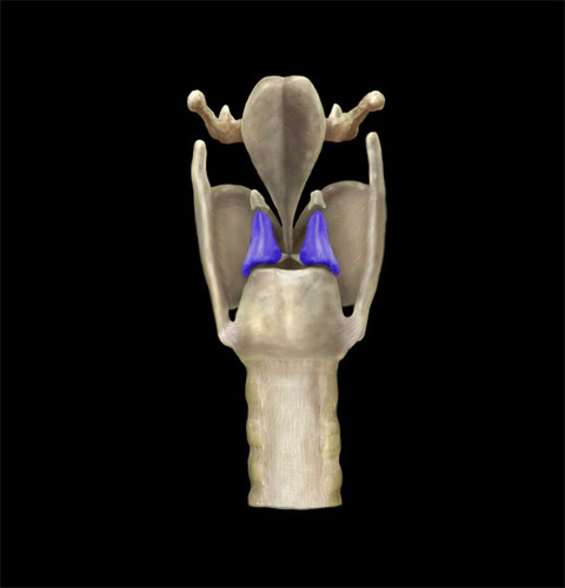
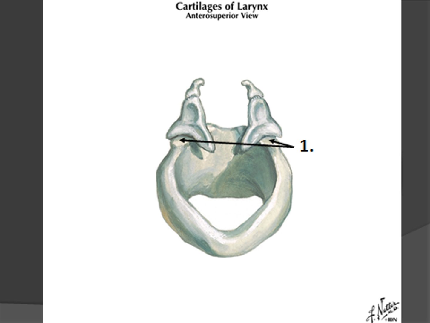
arytenoid cartilage
cricoarytenoid joint
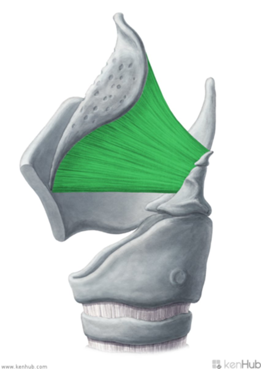
quadrangular membrane
lateral side of the epiglottis all the way to the ventricular fold
aryepiglottic fold/ligament
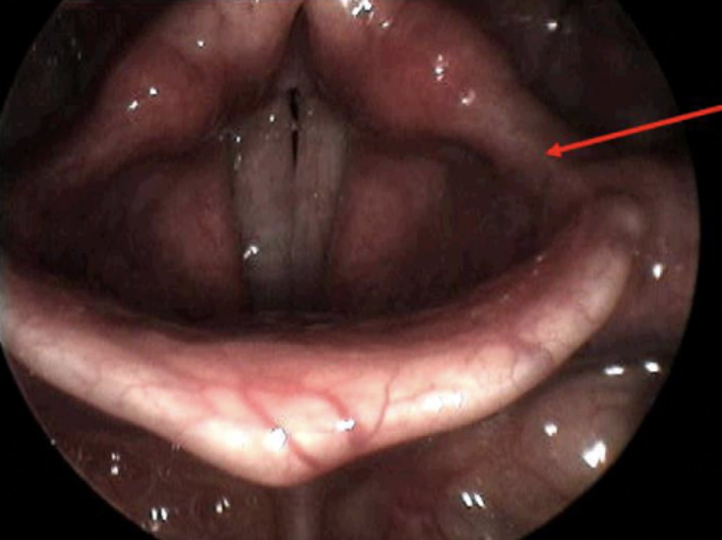
vestibular fold/ligament
false vocal cord
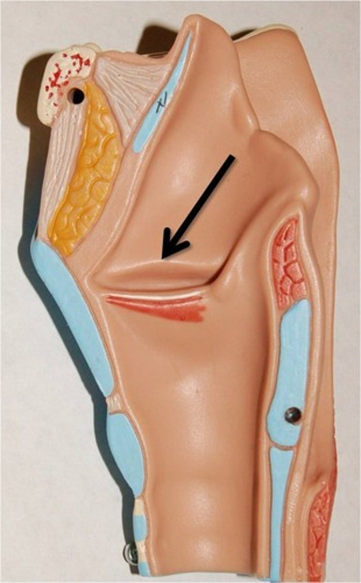
vocal fold/ligament
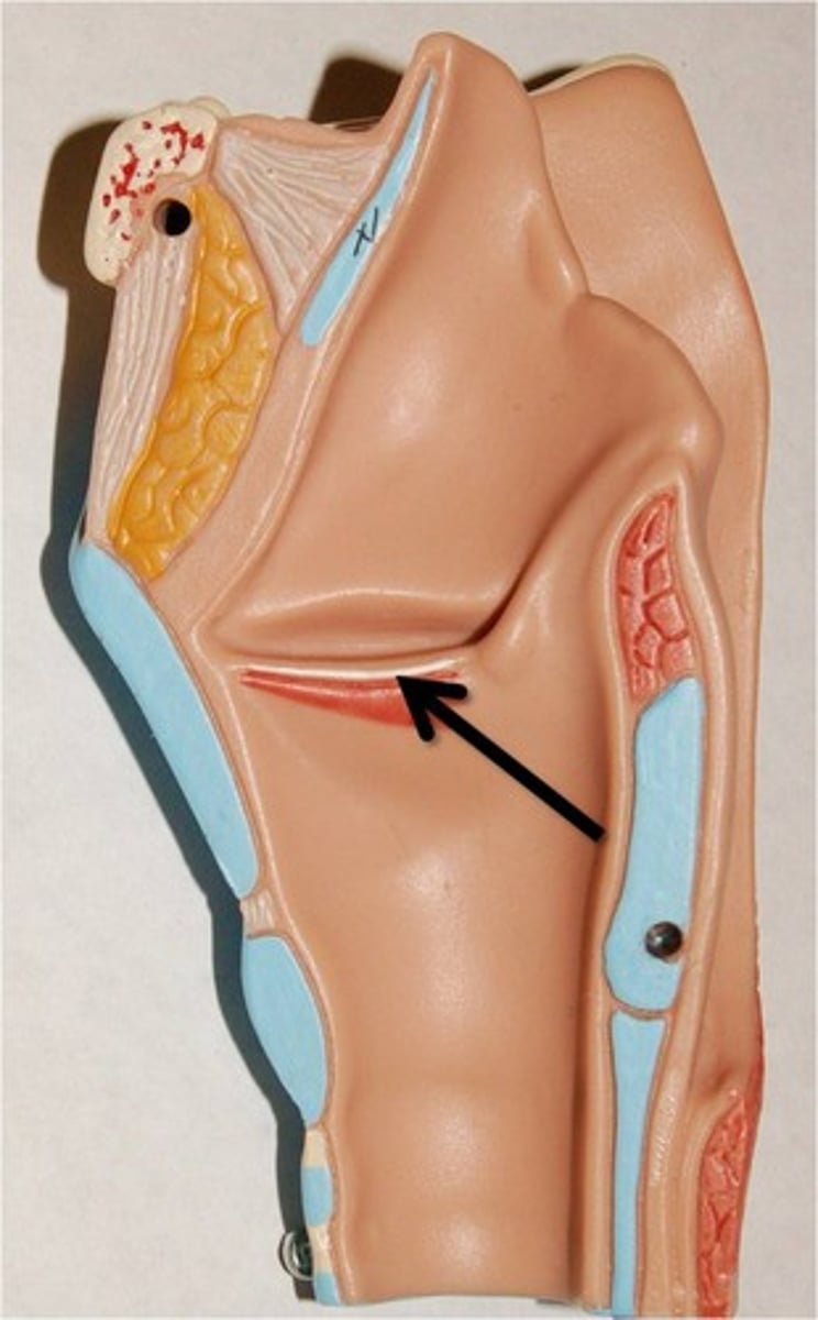
cricothyroid membrane
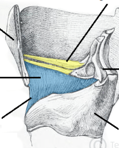
laryngeal inlet
opening that connects the pharynx and larynx
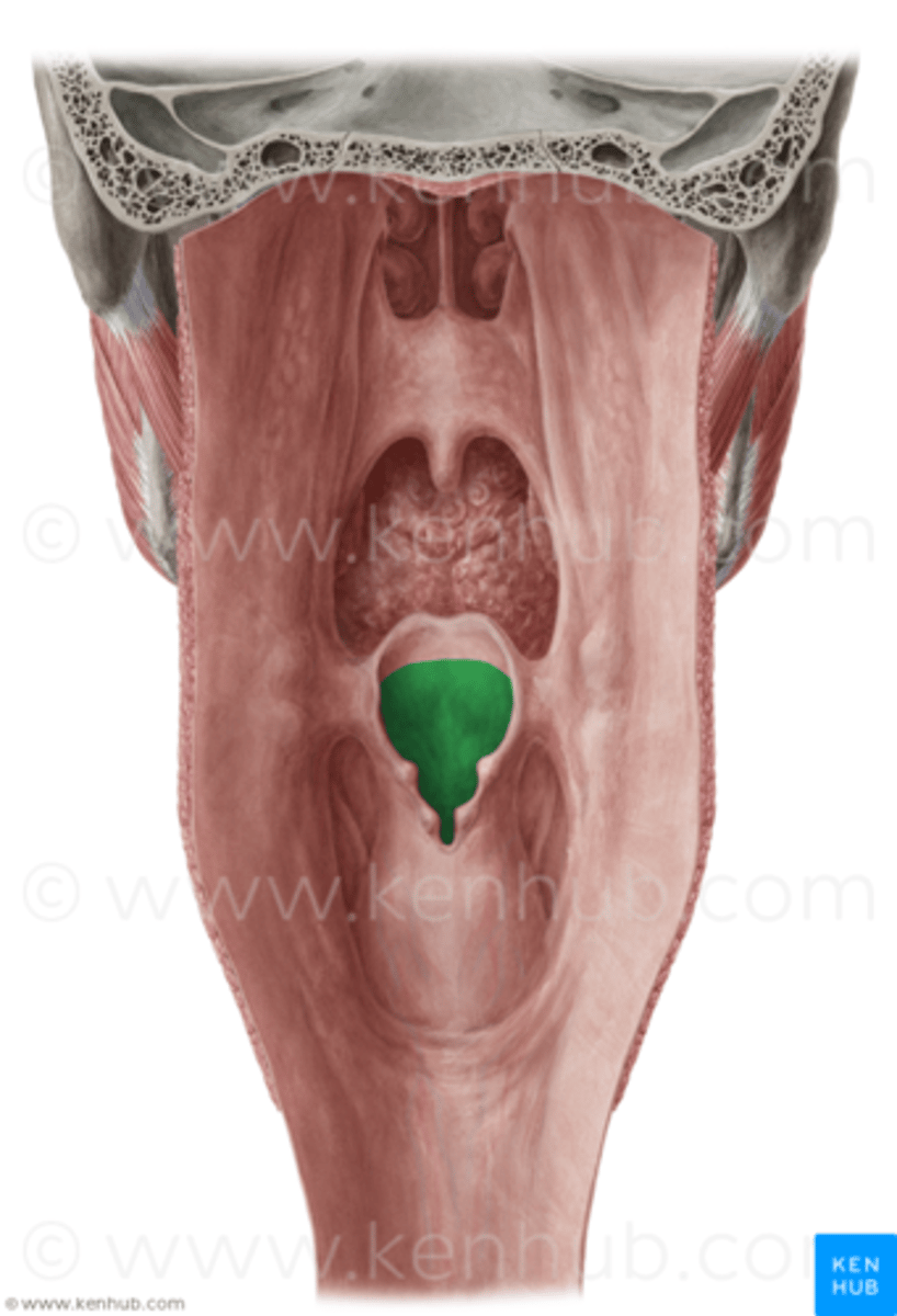
valleculae
space between tongue and epiglottis
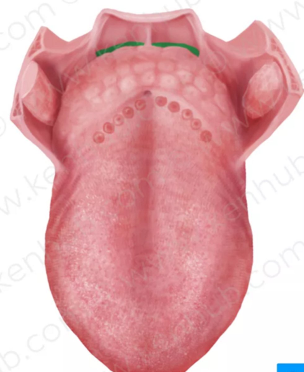
piriform fossa
hollow pockets on the lateral sides of the glottic opening
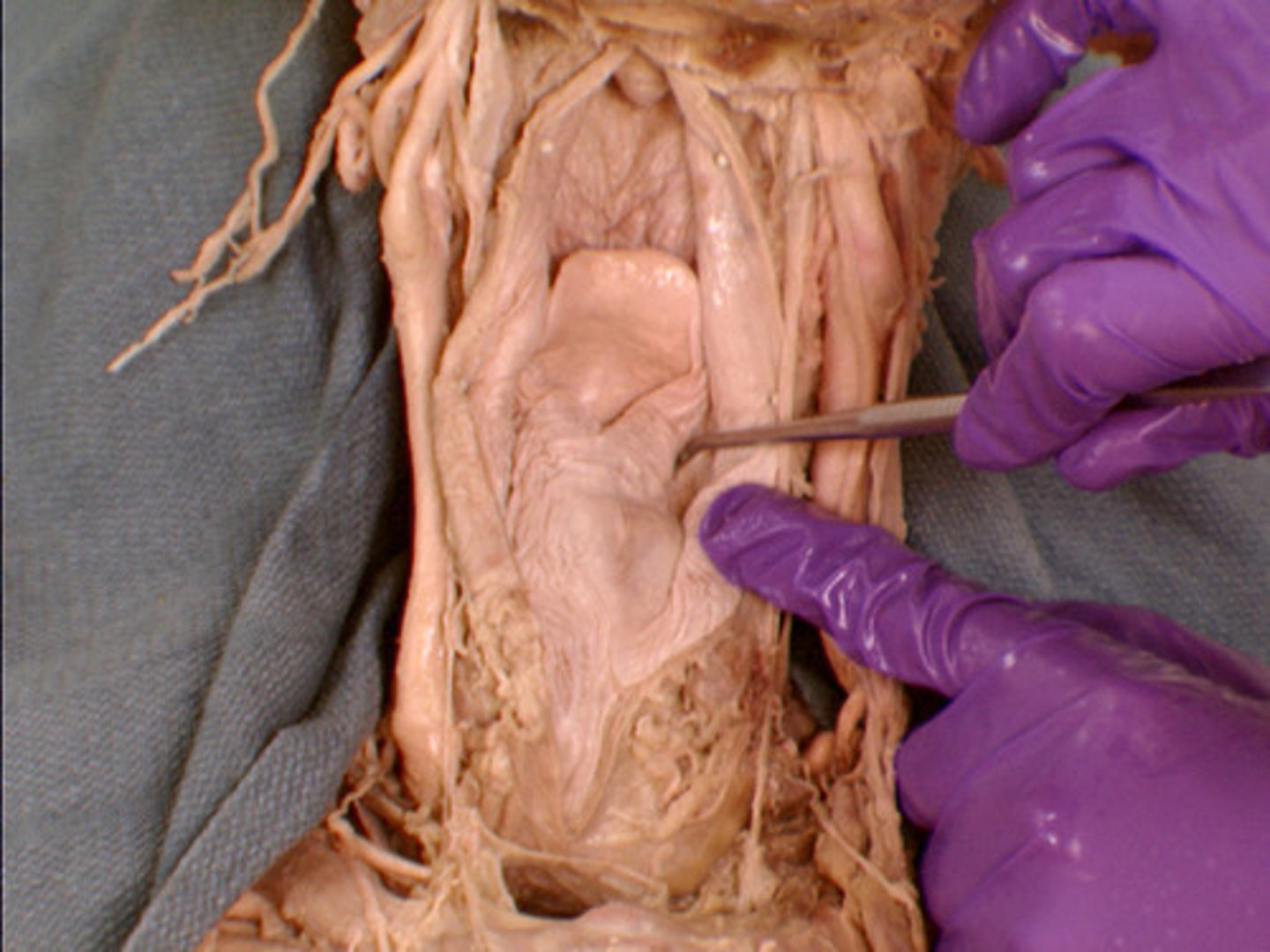
laryngeal vestibule/ supraglottic space
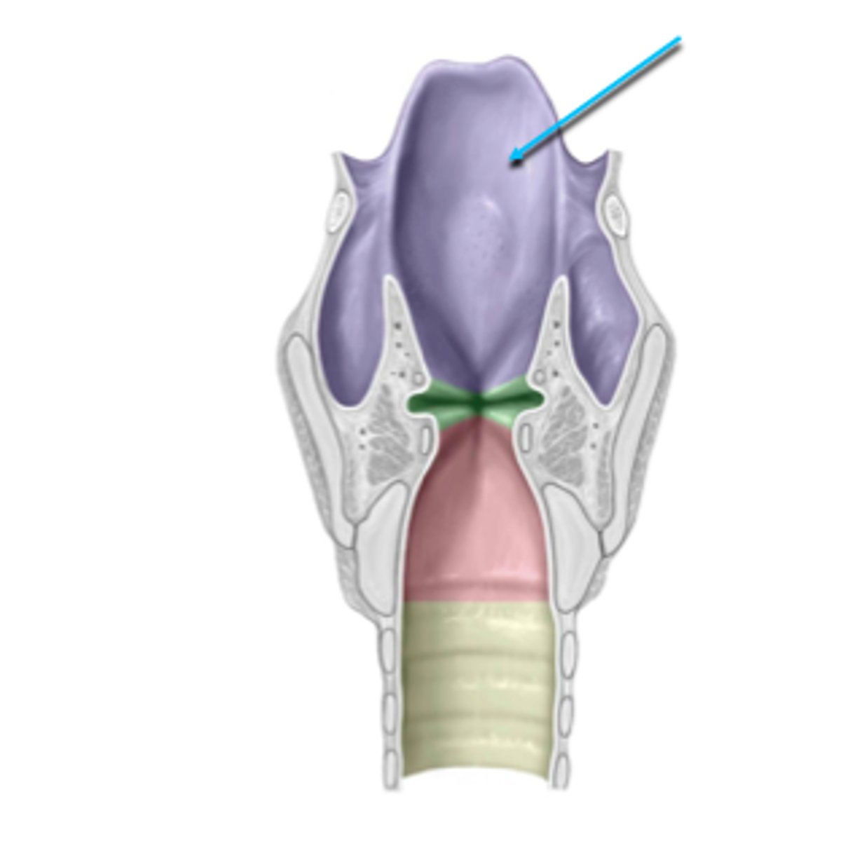
laryngeal ventricle
space between true and false vocal folds
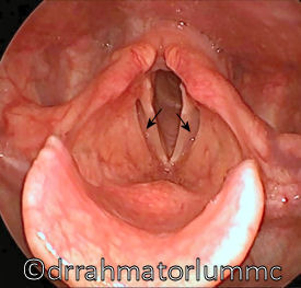
infraglottic space
area inferior to the vocal folds
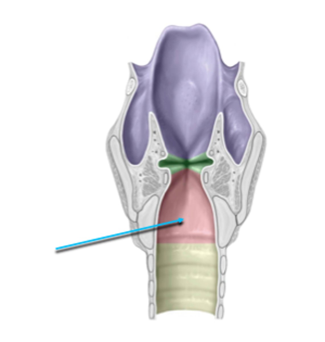
rima glottidis
opening between vocal folds
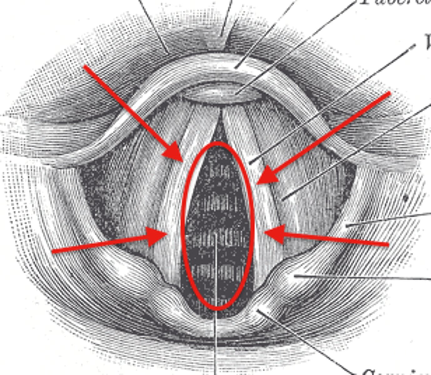
cricothyroid muscle, lengthen vocal cords, high pitch
structure, function, action
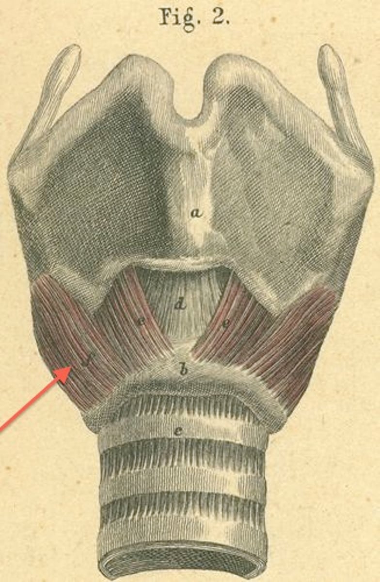
aryepiglottic muscle
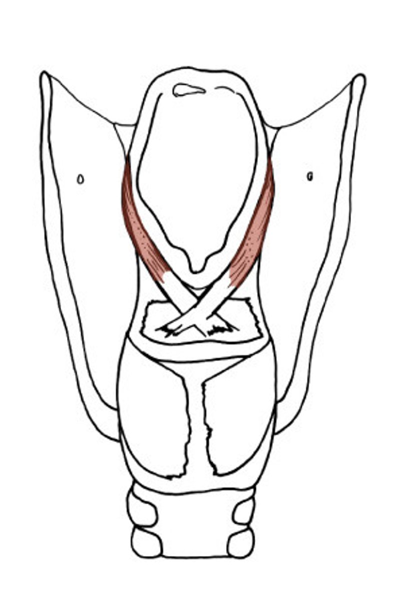
posterior cricoarytenoid, abducts vocal folds, forced respiration
name, function, action?
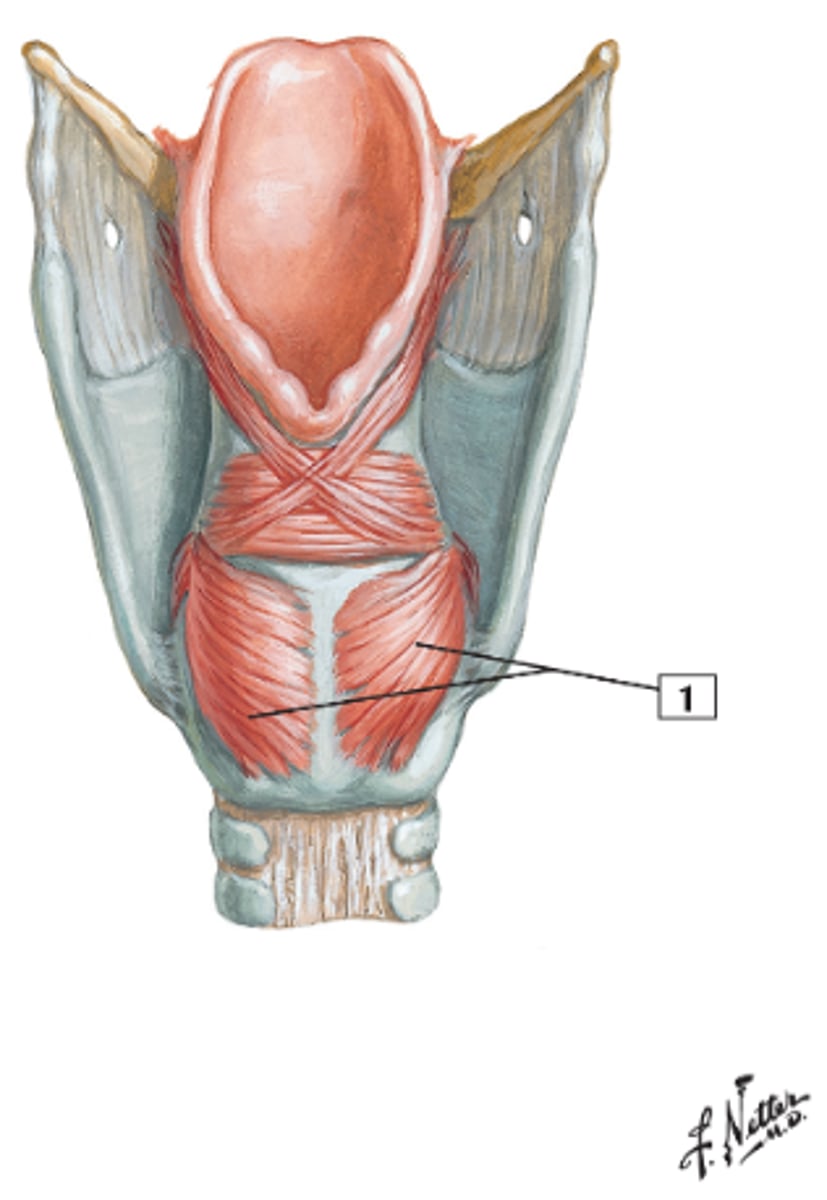
arytenoid muscle (transverse and oblique) , adducts vocal folds, bear down
name,function, action?
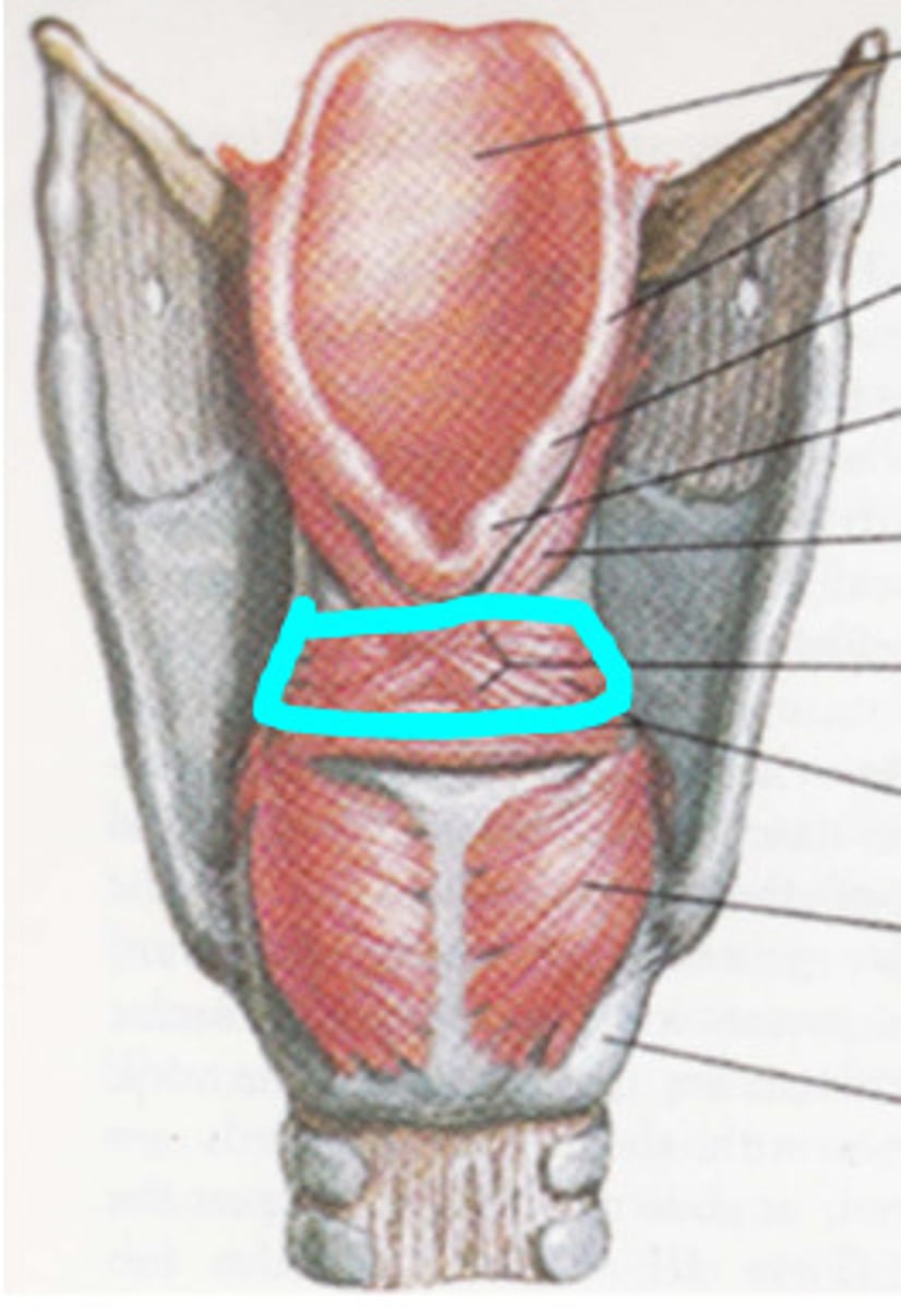
lateral cricoarytenoid, adducts vocal cords, bear down
name,function, action?
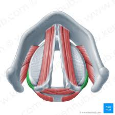
thyroarytenoid muscle, shorten vocal cords, low pitch
name, function, action
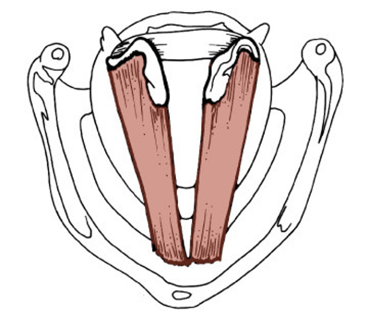
vocalis, fine tune pitch
structure and function
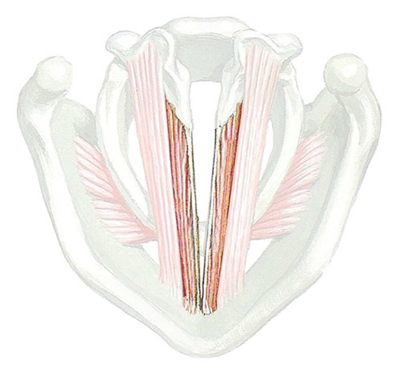
sensation of mucosa below vocal folds, larnygeal muscles (except cricothyroid)
what does the recurrent laryngeal innervate
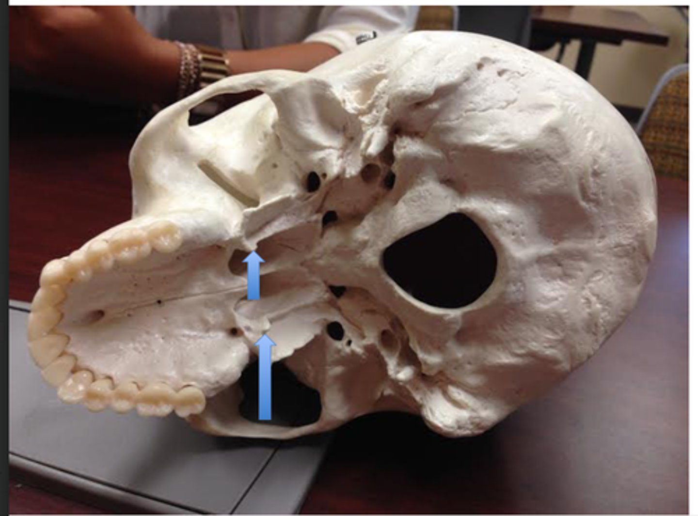
sphenopetrosal fissure
leads to bony potrion of pharyngotympanic tube
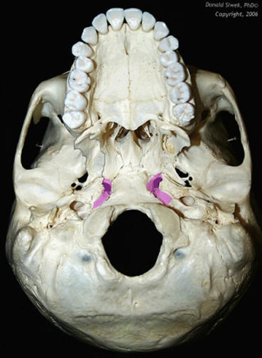
petrous part of temporal bone
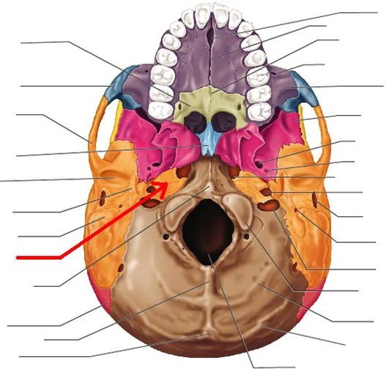
scaphoid fossa, tensor veli palatini
structure, what attaches here
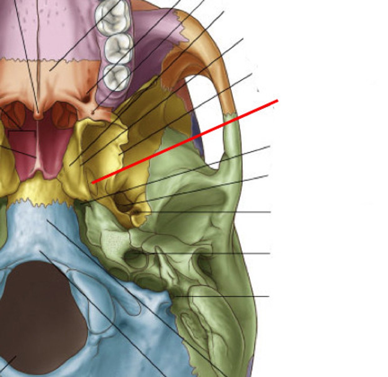
medial pterygoid plate
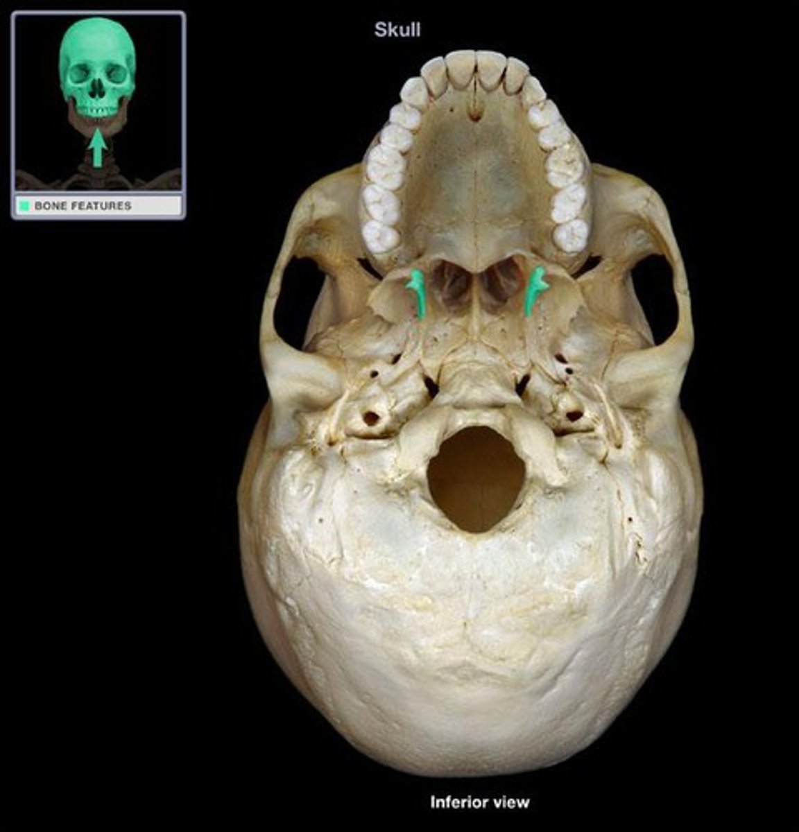
hamulus
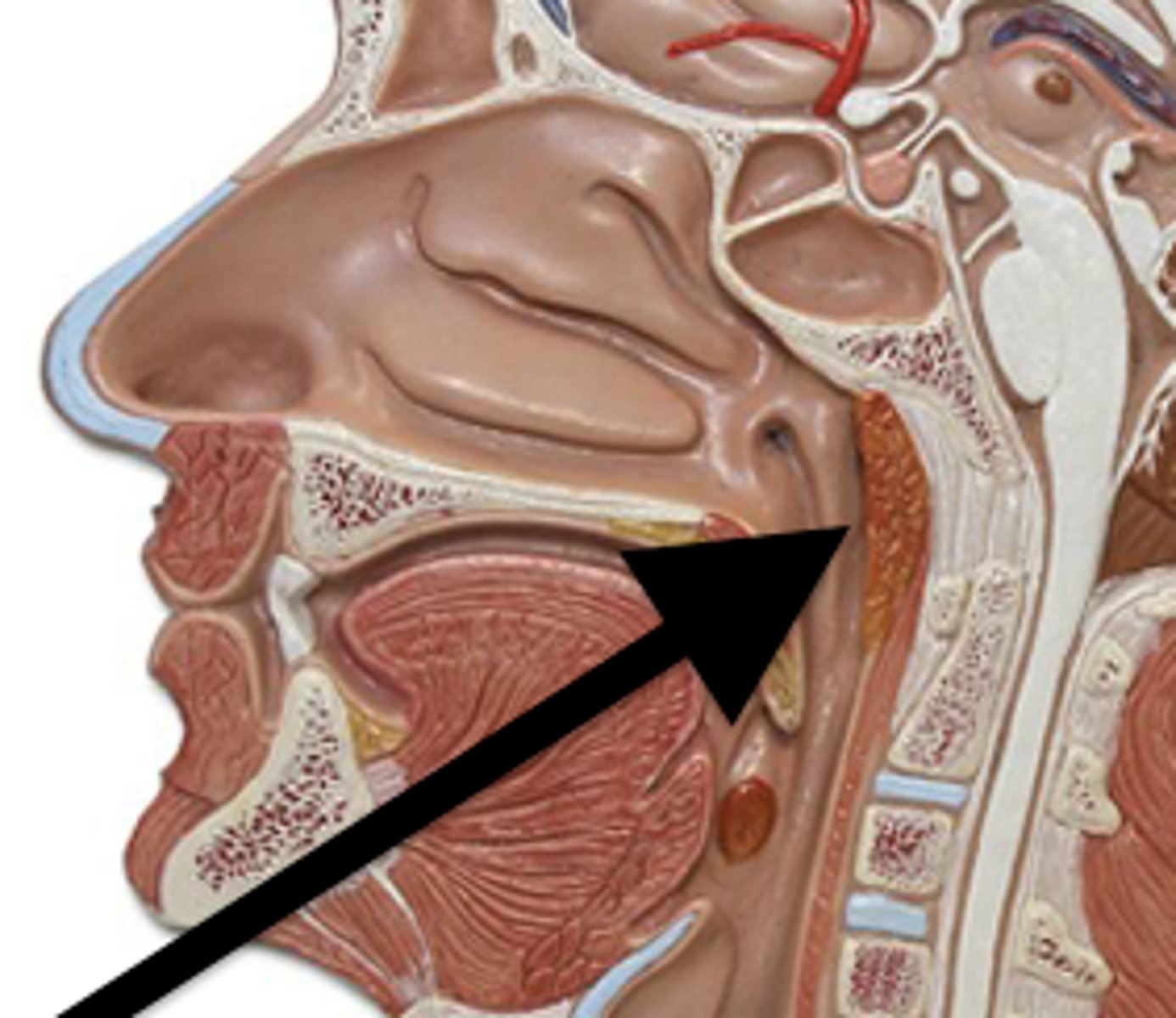
pharyngeal tonsils (adenoids)
what tonsilar tissue is found here
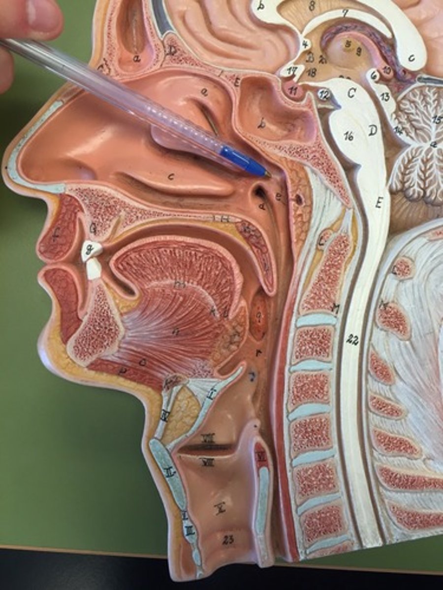
tubal tonsils
what tosilar tissue is found here
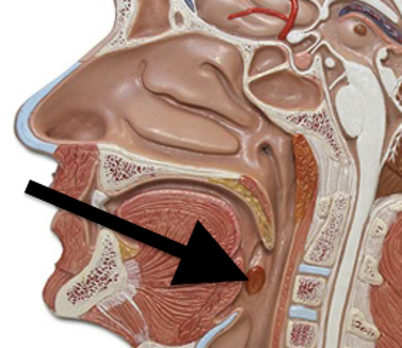
palatine tonsils
lingual tonsils
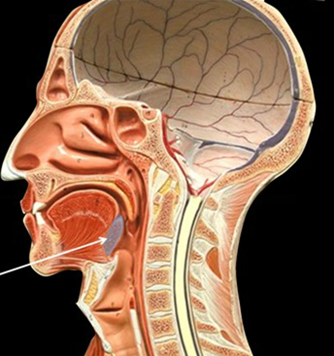
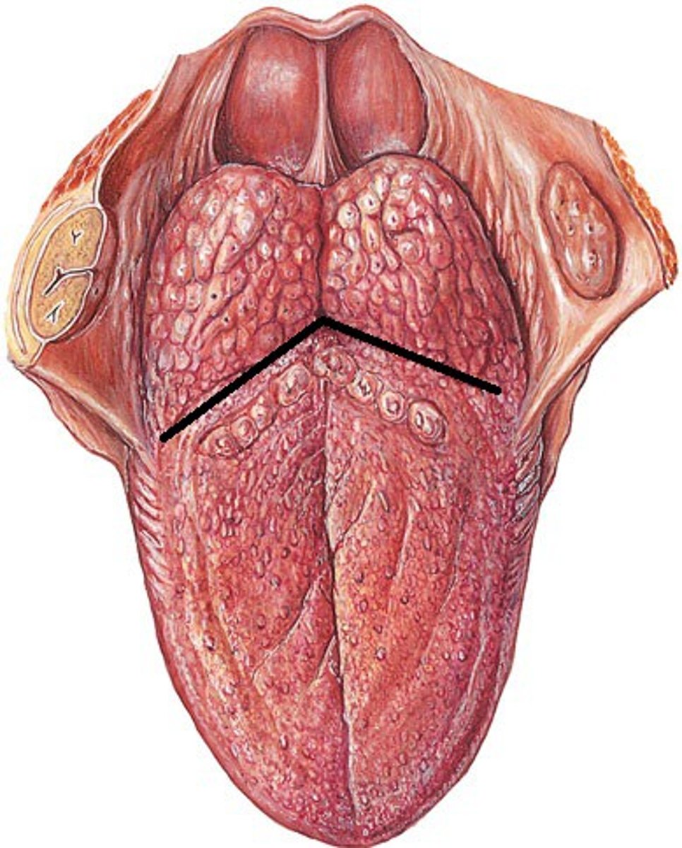
sulcus terminalis
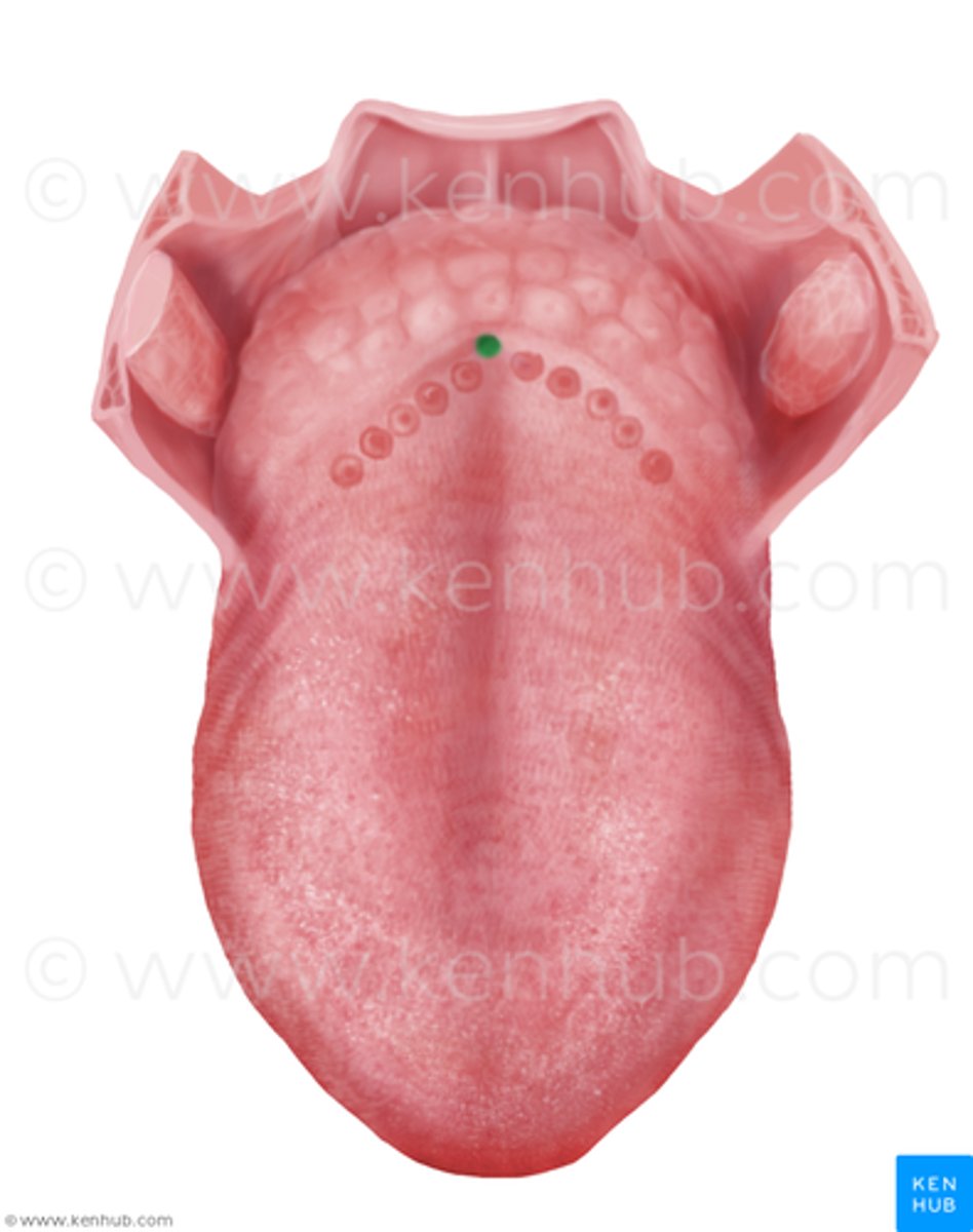
foramen cecum
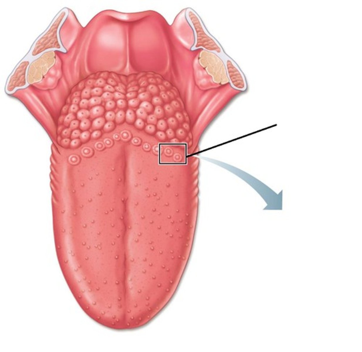
vallate papillae
valleculae
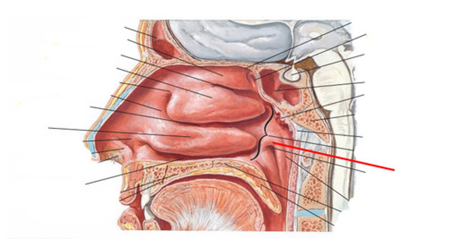
torus tubarius
elevation of cartilage caused by the auditory tube
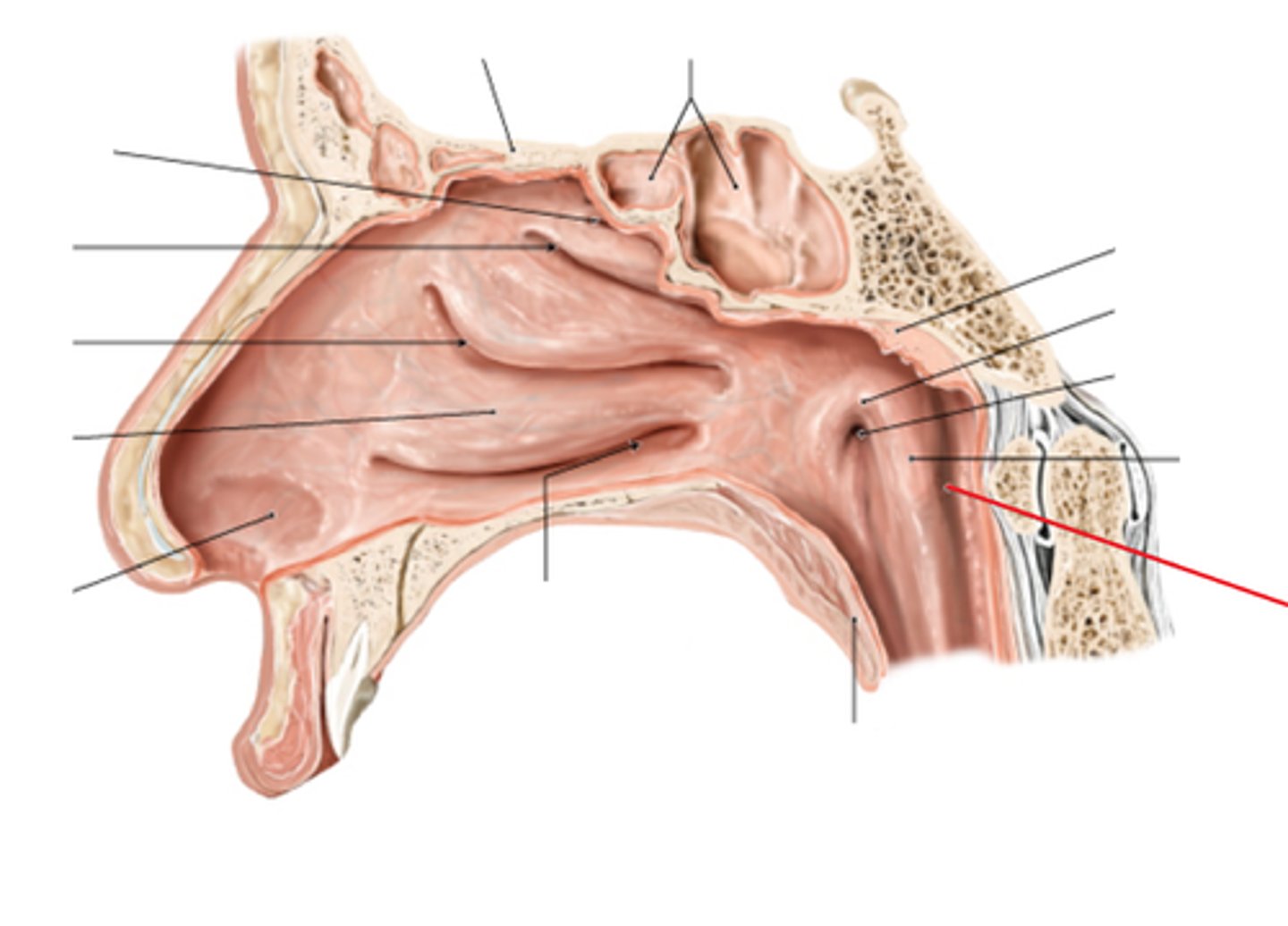
salpingopharyngeal fold and muscle (CN X)
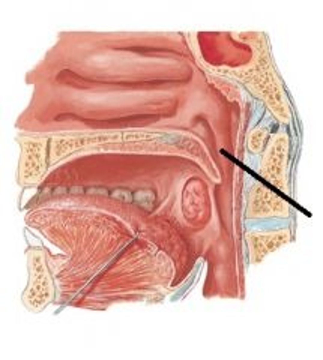
pharyngeal recess
a groove between salpingopharyngeal fold and
posterior wall of nasopharynx
tonsillar fossa
the space between the palatoglossal arch and the palatopharyngeal arch
uvula
small projection hanging from the back middle edge of the soft palate
musculus uvulae (CN X)
what muscle controls this structure
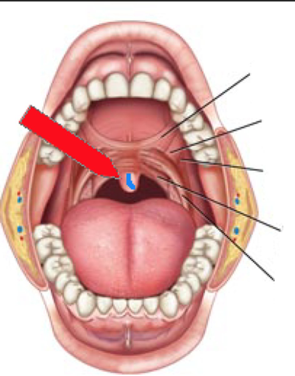
palatoglossal arch/muscle (Cn X)
palatopharyngeal arch/muscle (CN X)
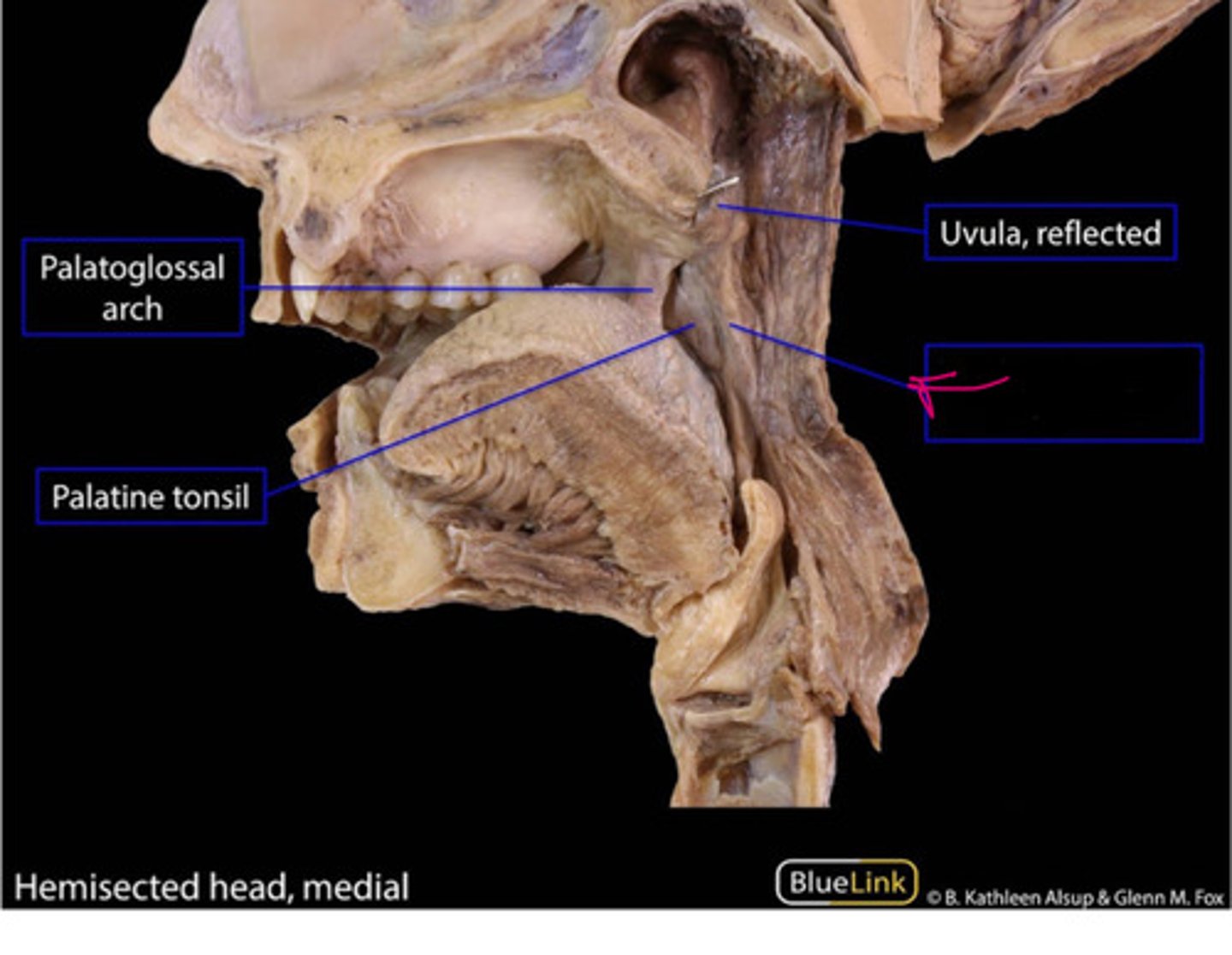
levator veli palatini muscle, elevate soft palate (CN X)
name and function
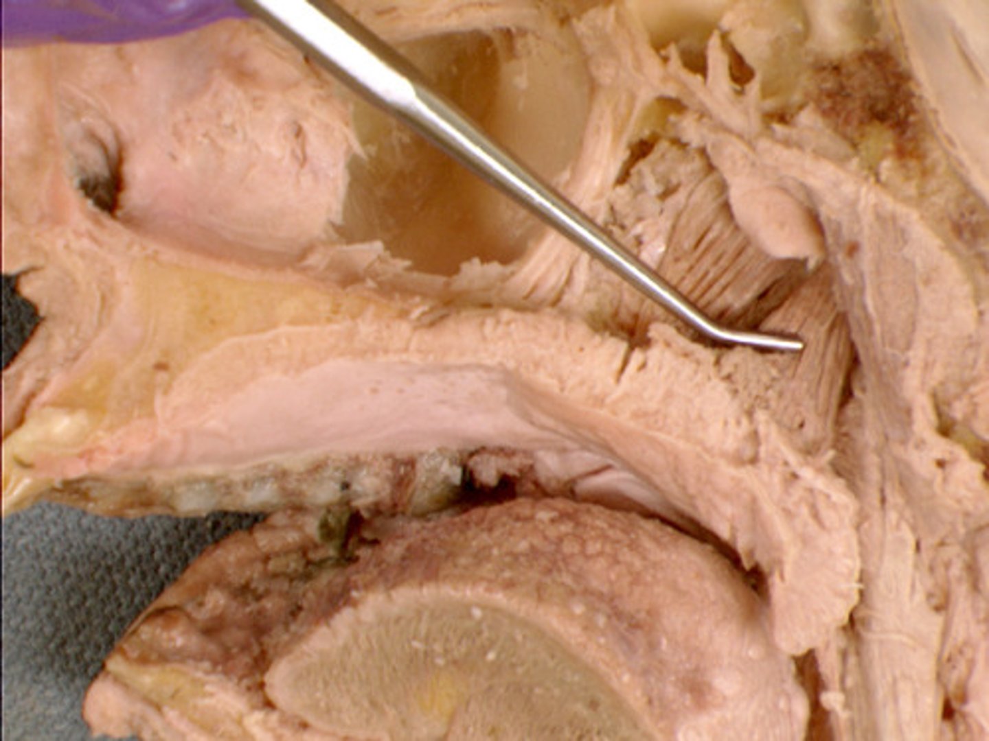
tensor veli palatini muscle, tenses soft palate (CN V3)
name and function
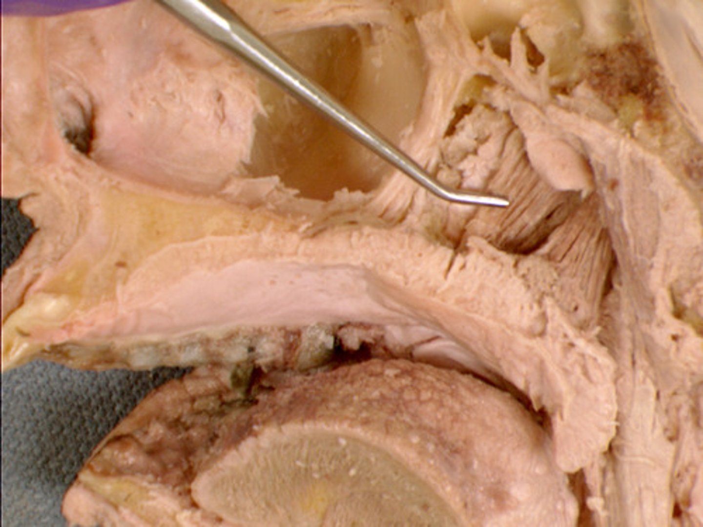
CN V3
what innervates this muscle?
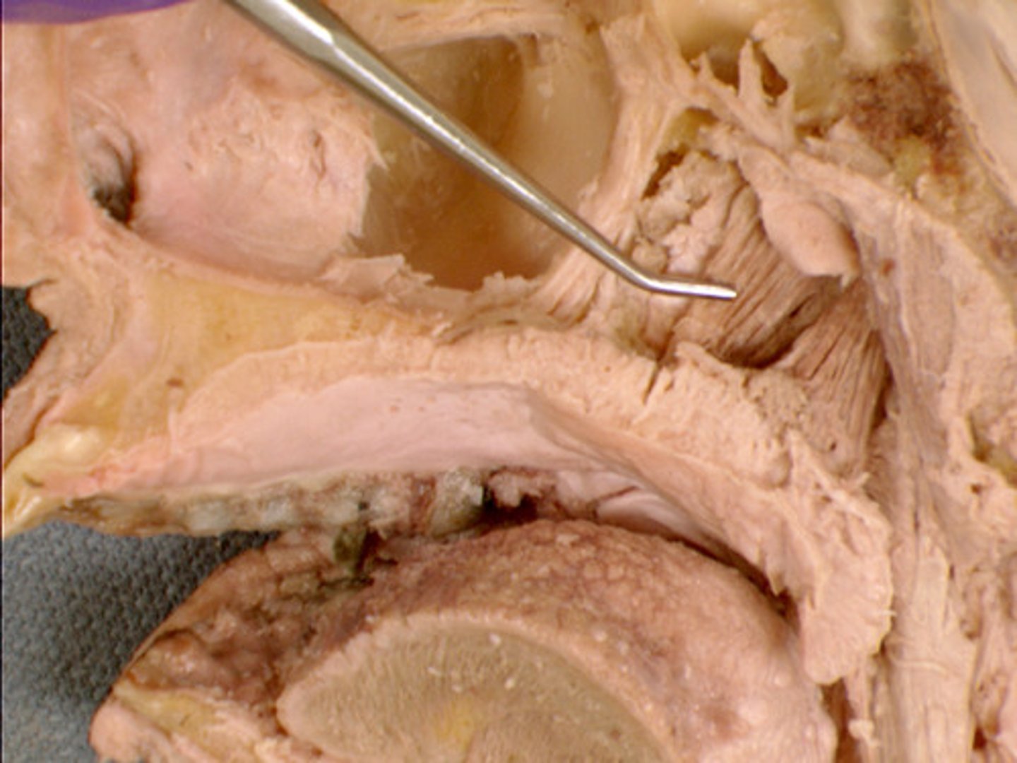
lacrimal fossa
depression where lacrimal sac sits
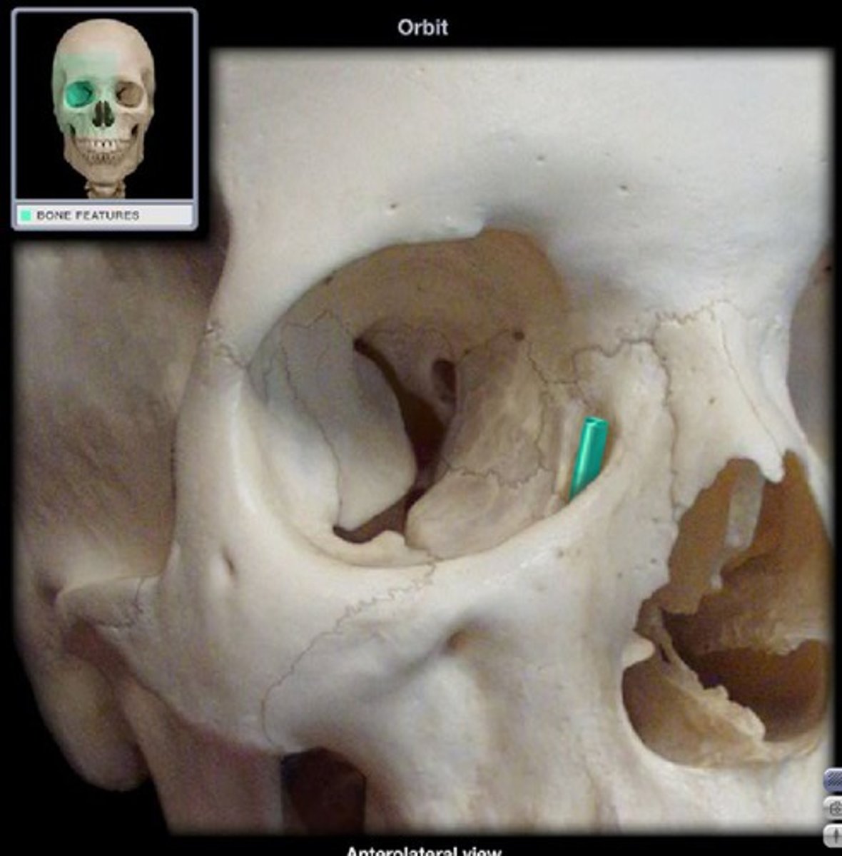
nasolacrimal canal/duct
empties lacrimal fluid into the nasal cavity
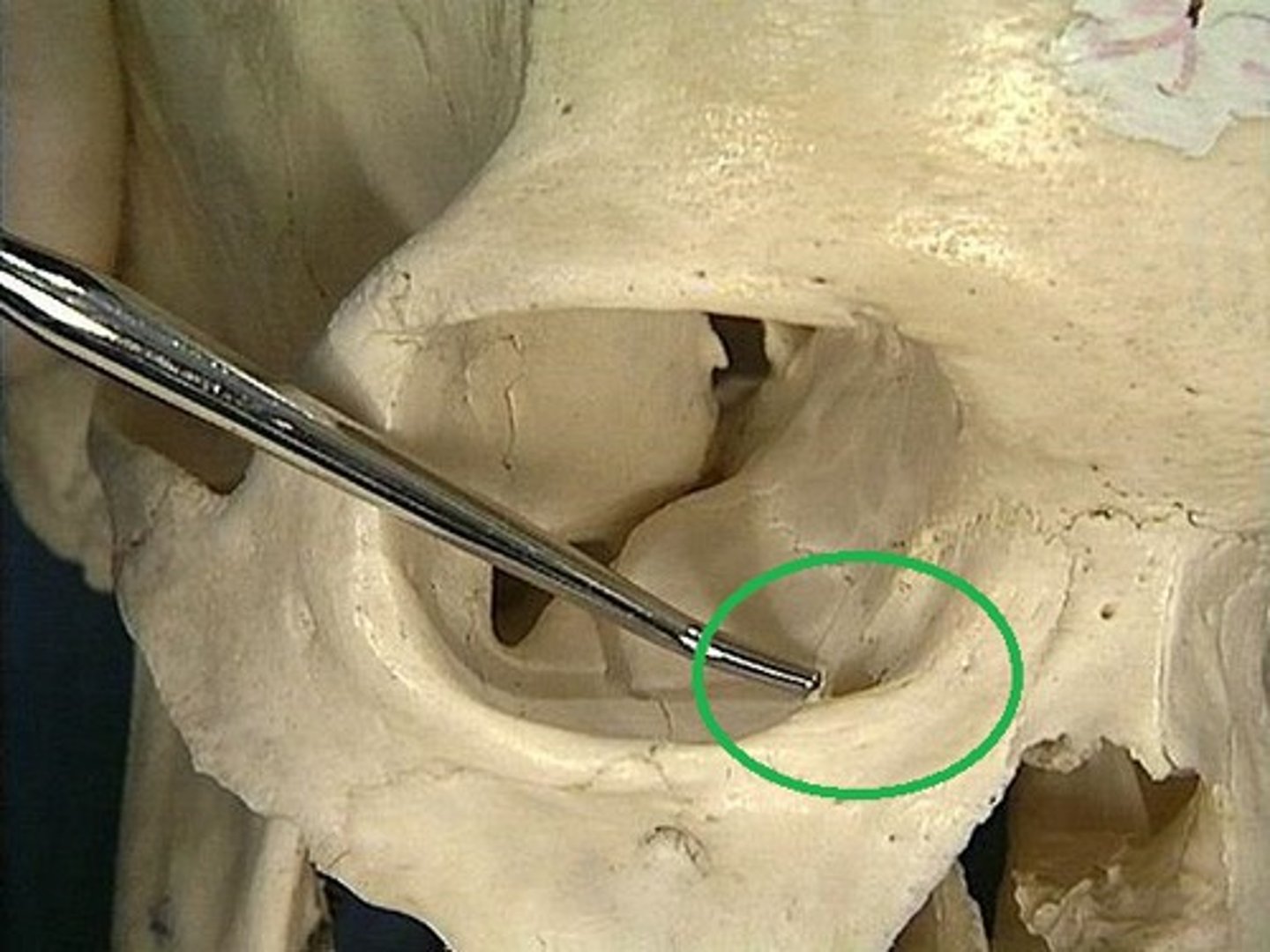
inferior meatus
where does the nasolacrimal duct drain into
superior concha
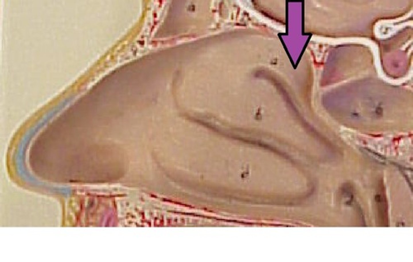
middle concha
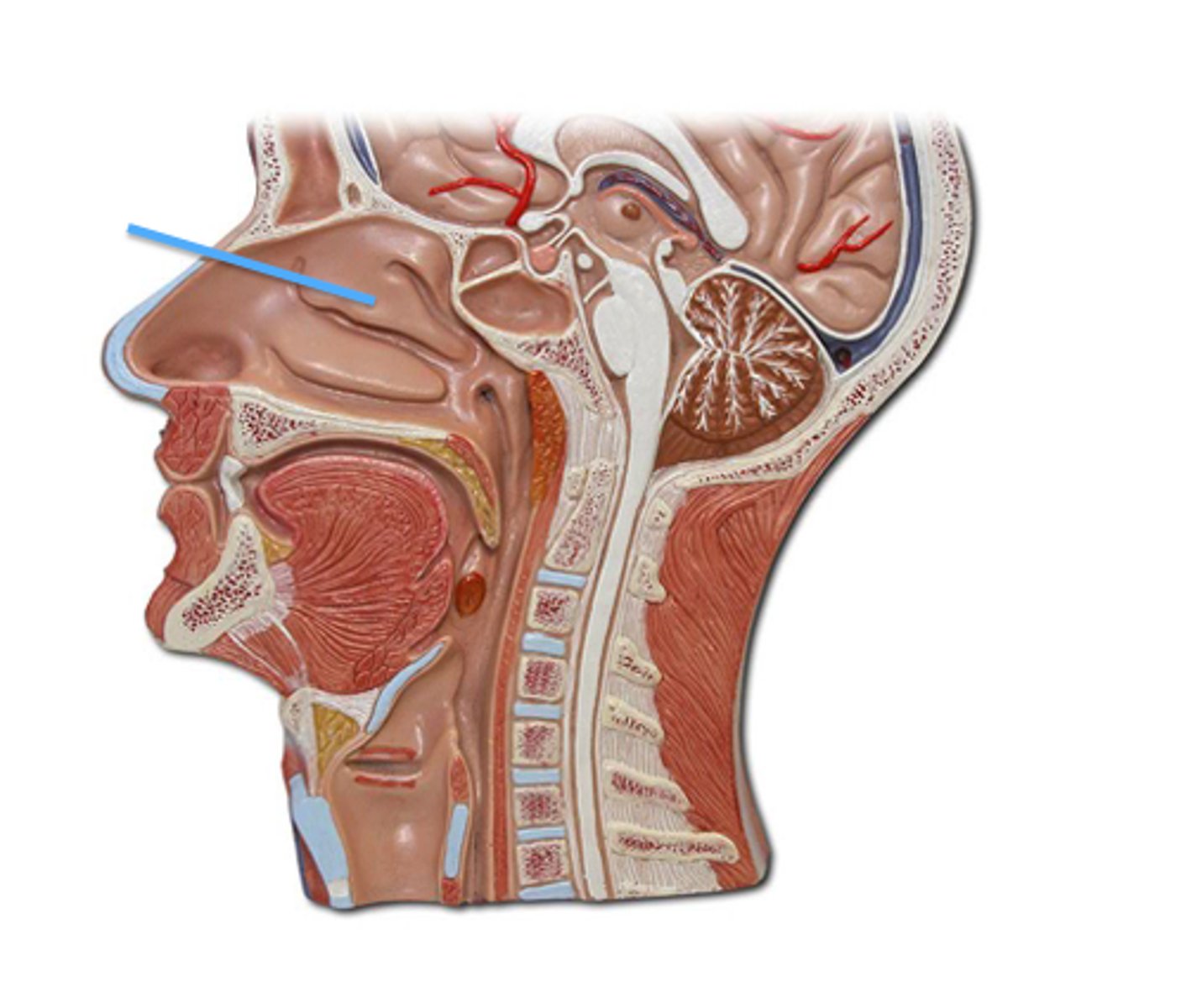
inferior concha
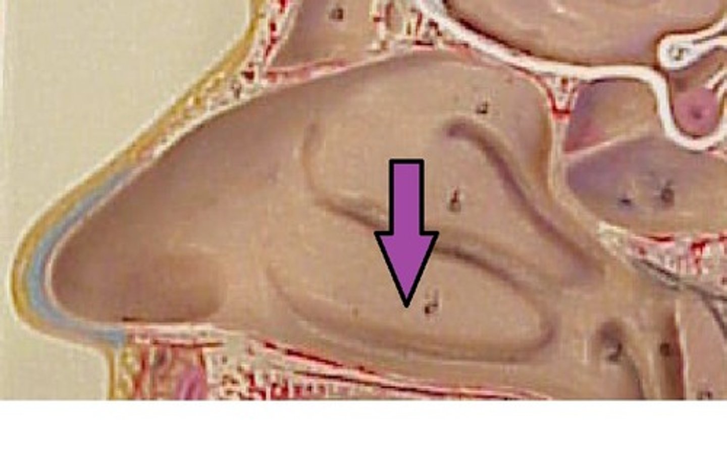
superior nasal meatus
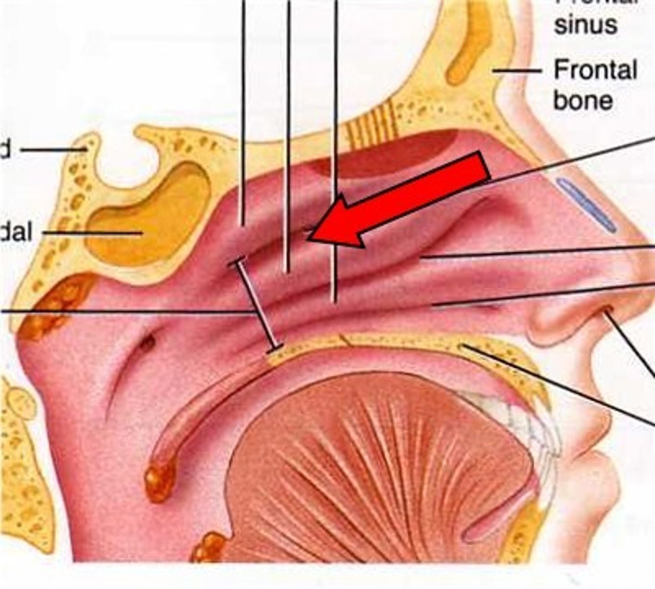
middle nasal meatus
where frontal, maxillary, sinuses and ethmoid air cells drain
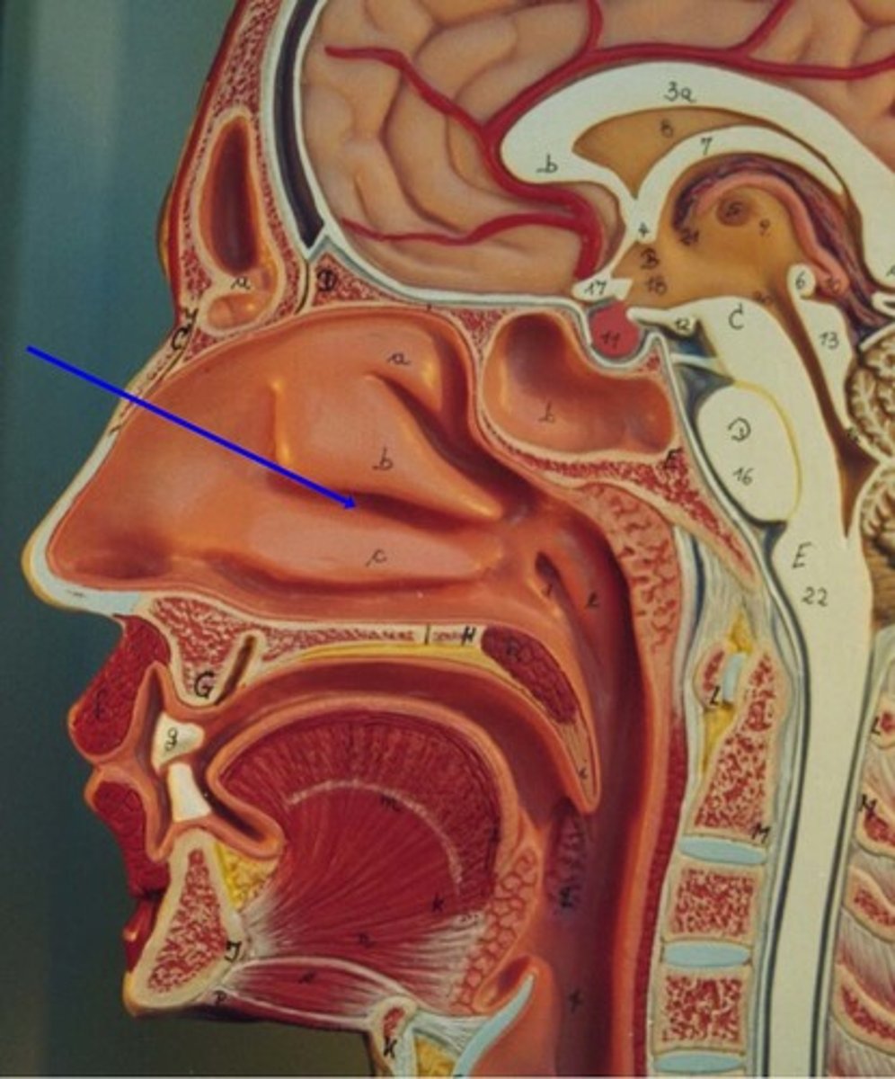
inferior nasal meatus
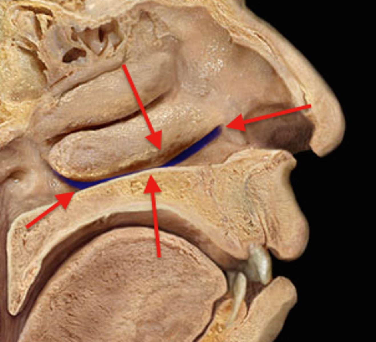
sphenoethmoidal recess
sphenoid sinus drains into
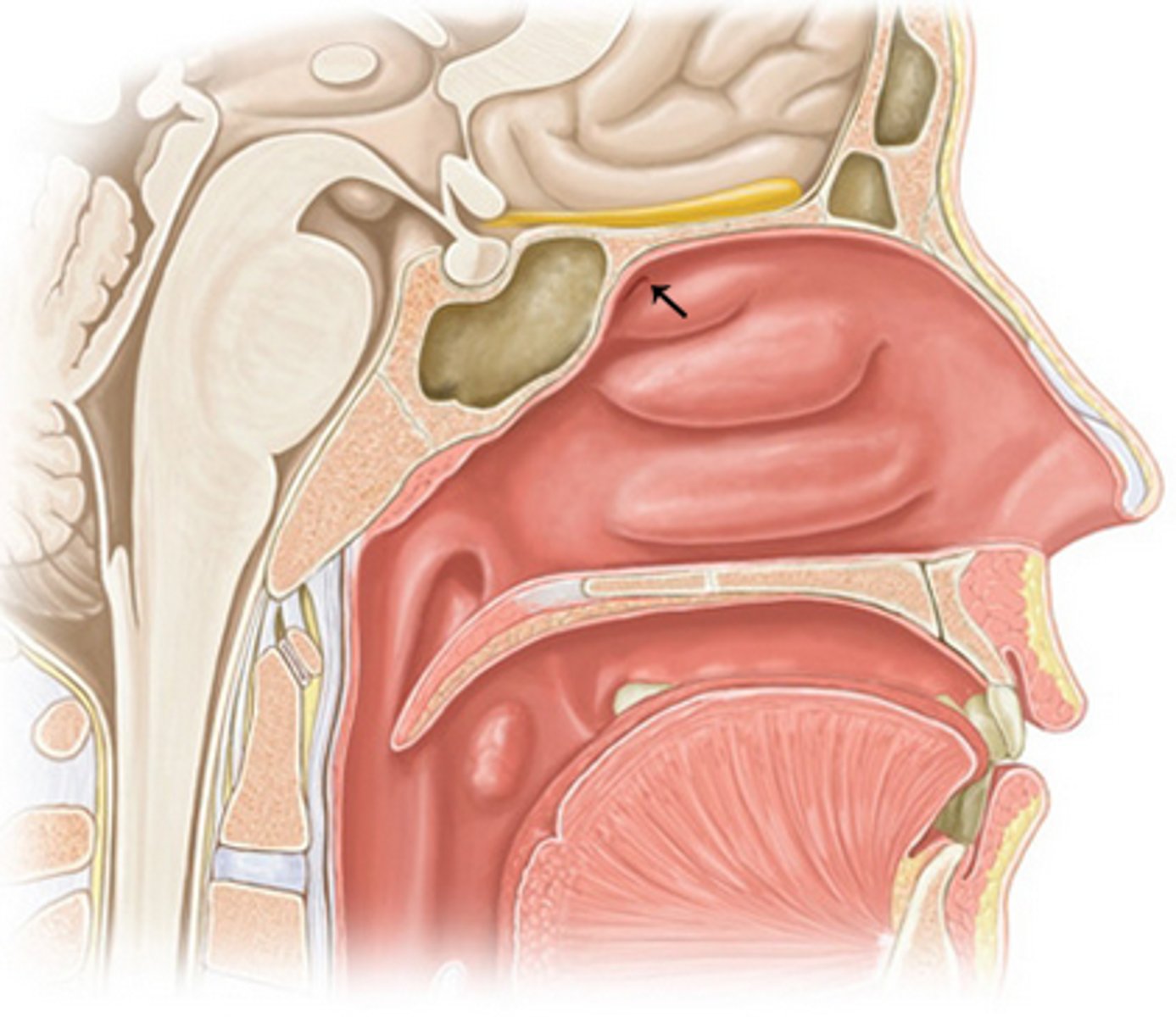
ethmoid bulla
locateed in middle meatus, where middle ethmoidal air cells drain
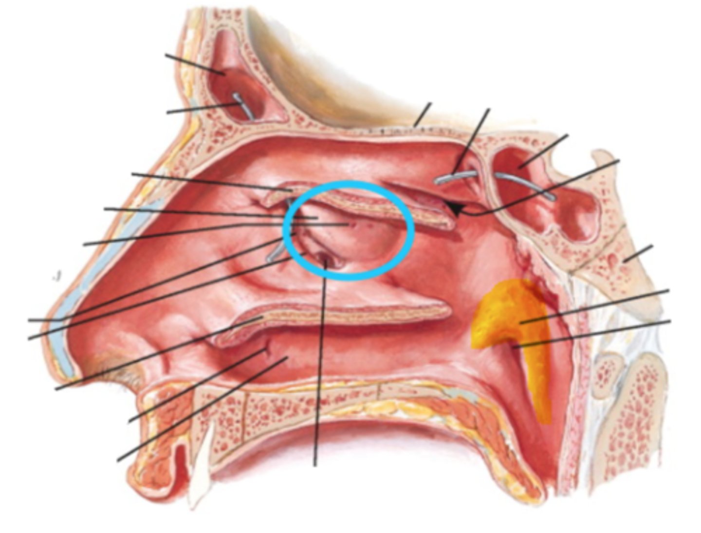
hiatus semilunaris
opening separating middle meatus and infundibulum
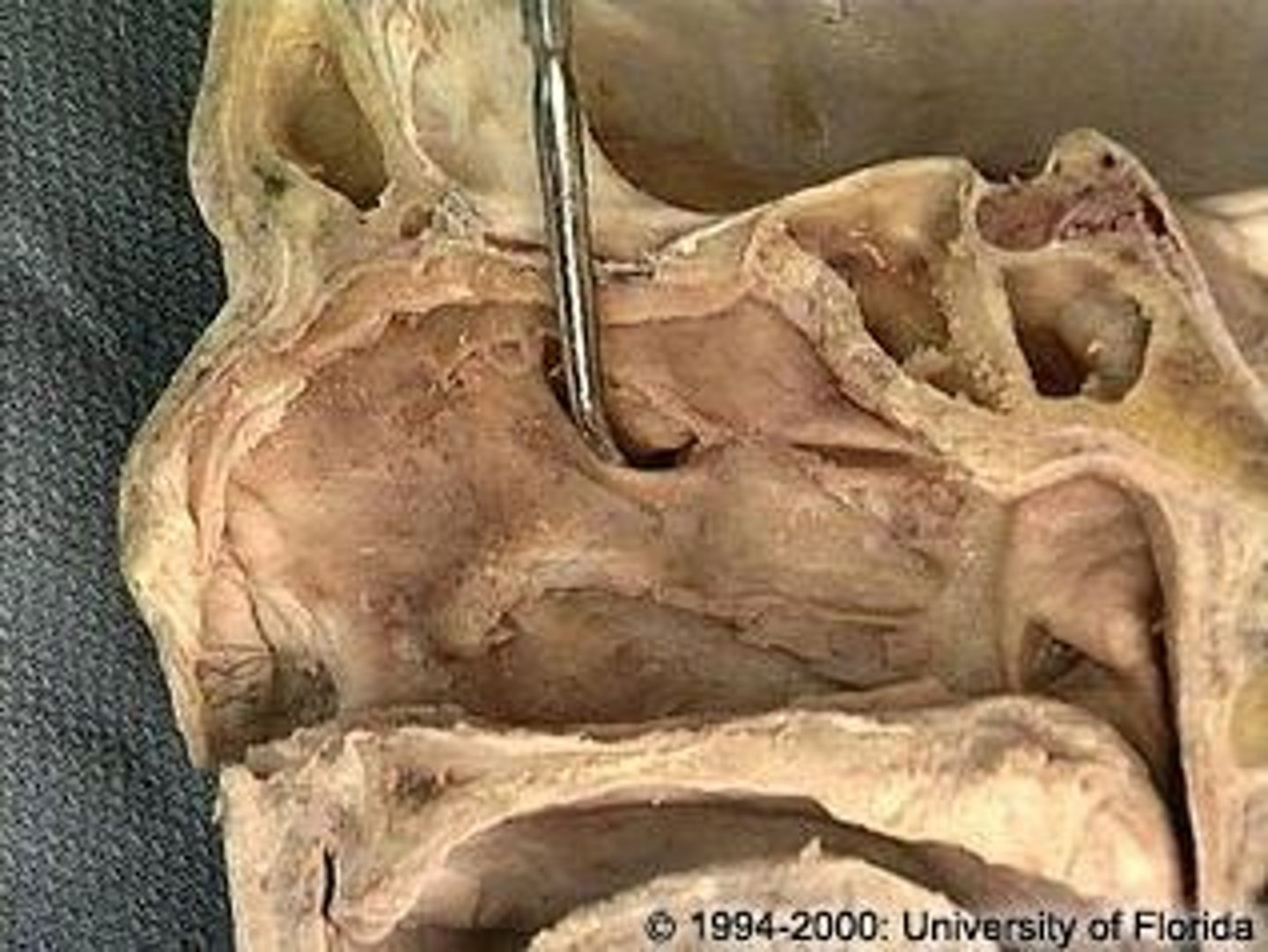
sphenoid sinus, sphenoethmoidal recess
structure and where it drains
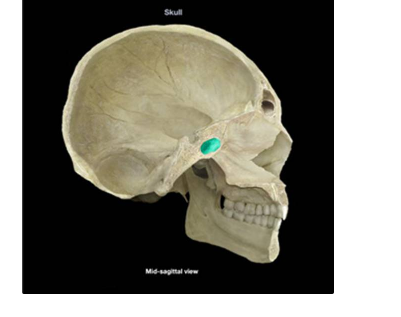
frontal sinus, middle meatus
structure and where it drains
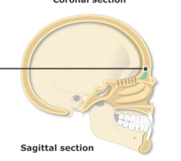
maxillary sinus, middle meatus
structure and where it drains
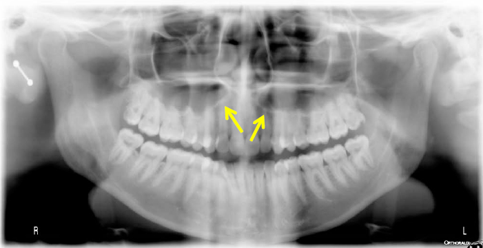
ethmoid air cells, middle meatus (ethmoid bulla)
structure and where it drains
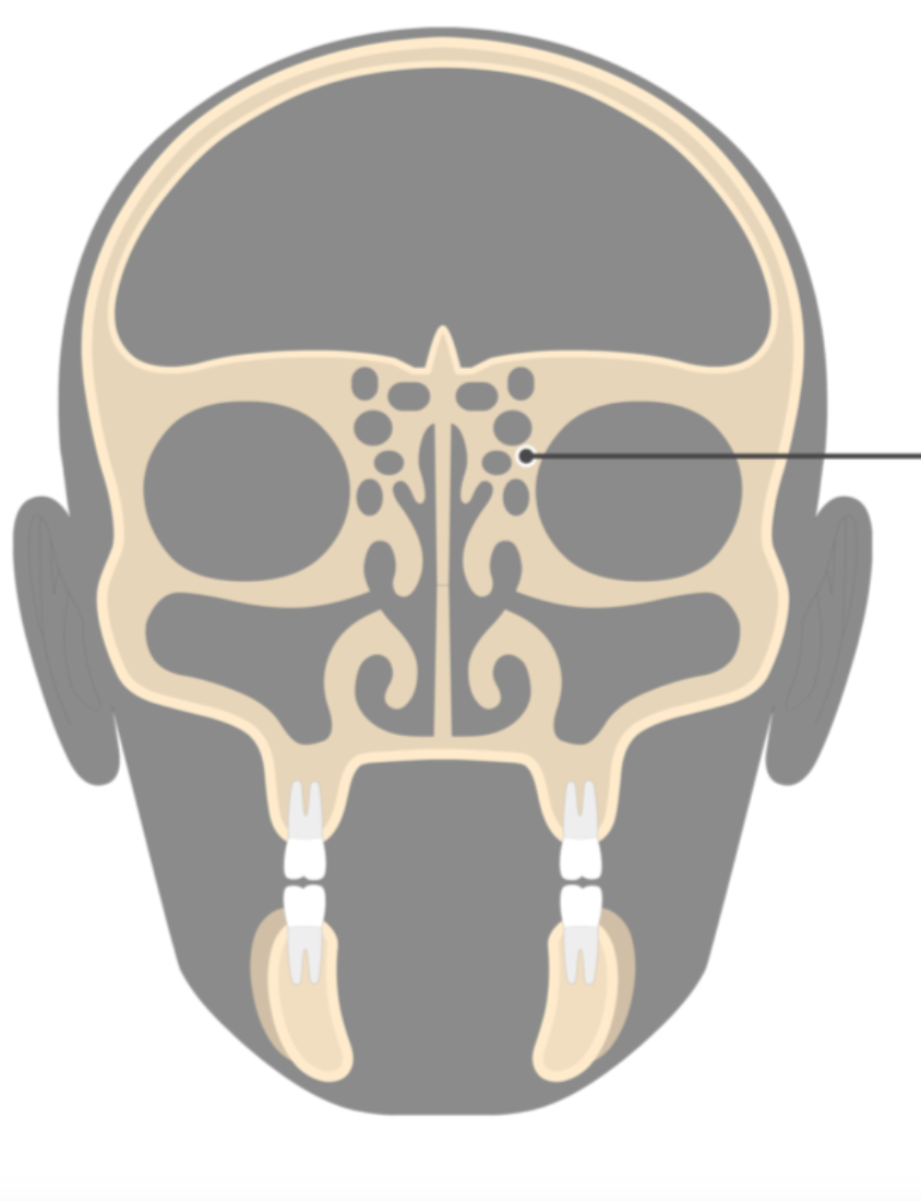
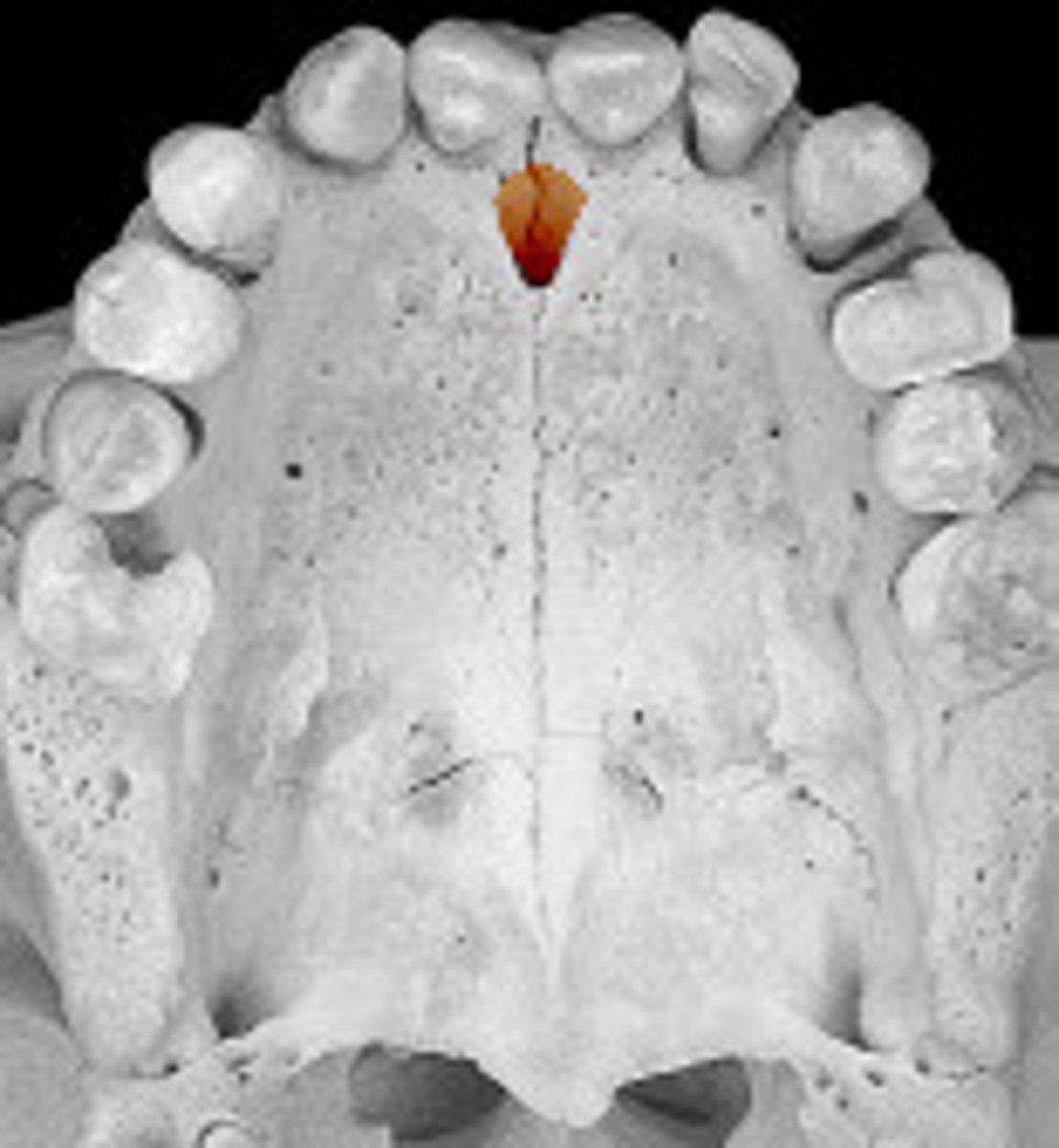
incisive fossa/foramen
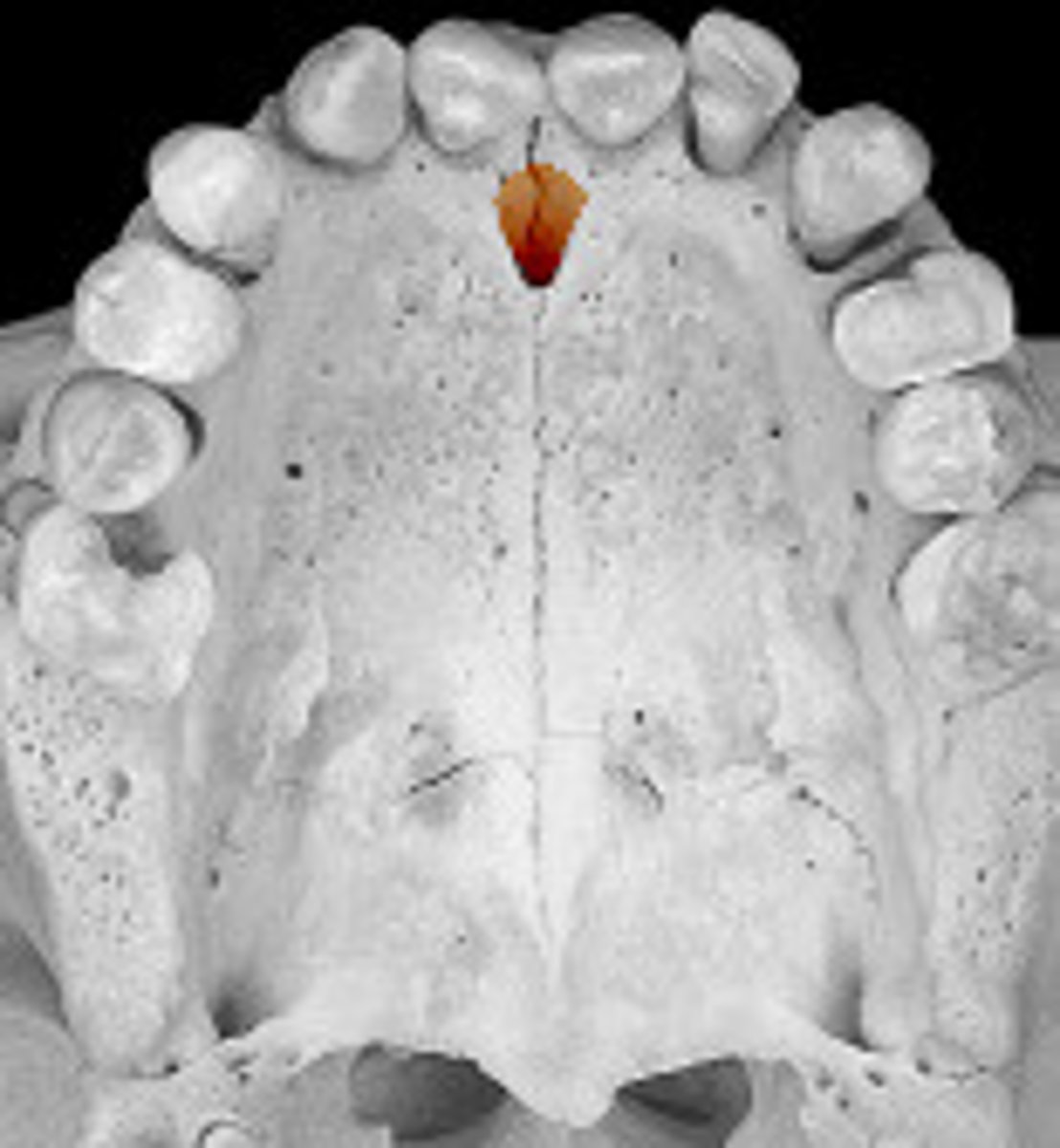
nasopalatine (CN V2)
what nerve travels through this opening
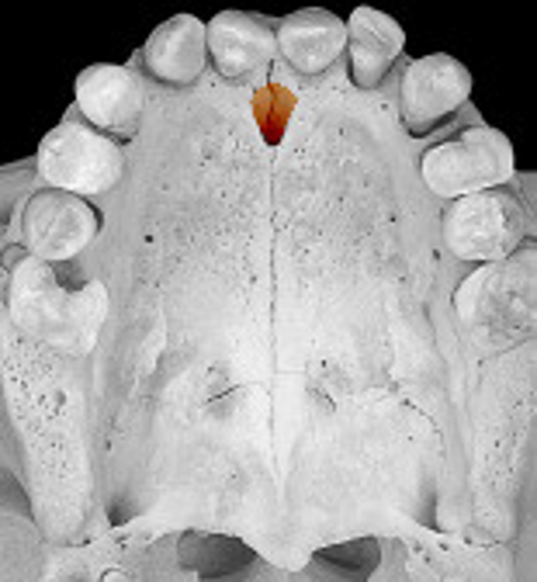
Sphenopalantine artery
what artery travels through this opening
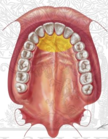
nasopalatine
what nerve innervates this portion of the palate
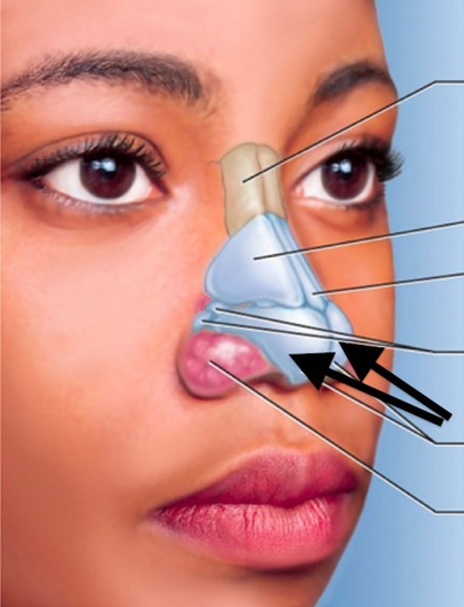
naris
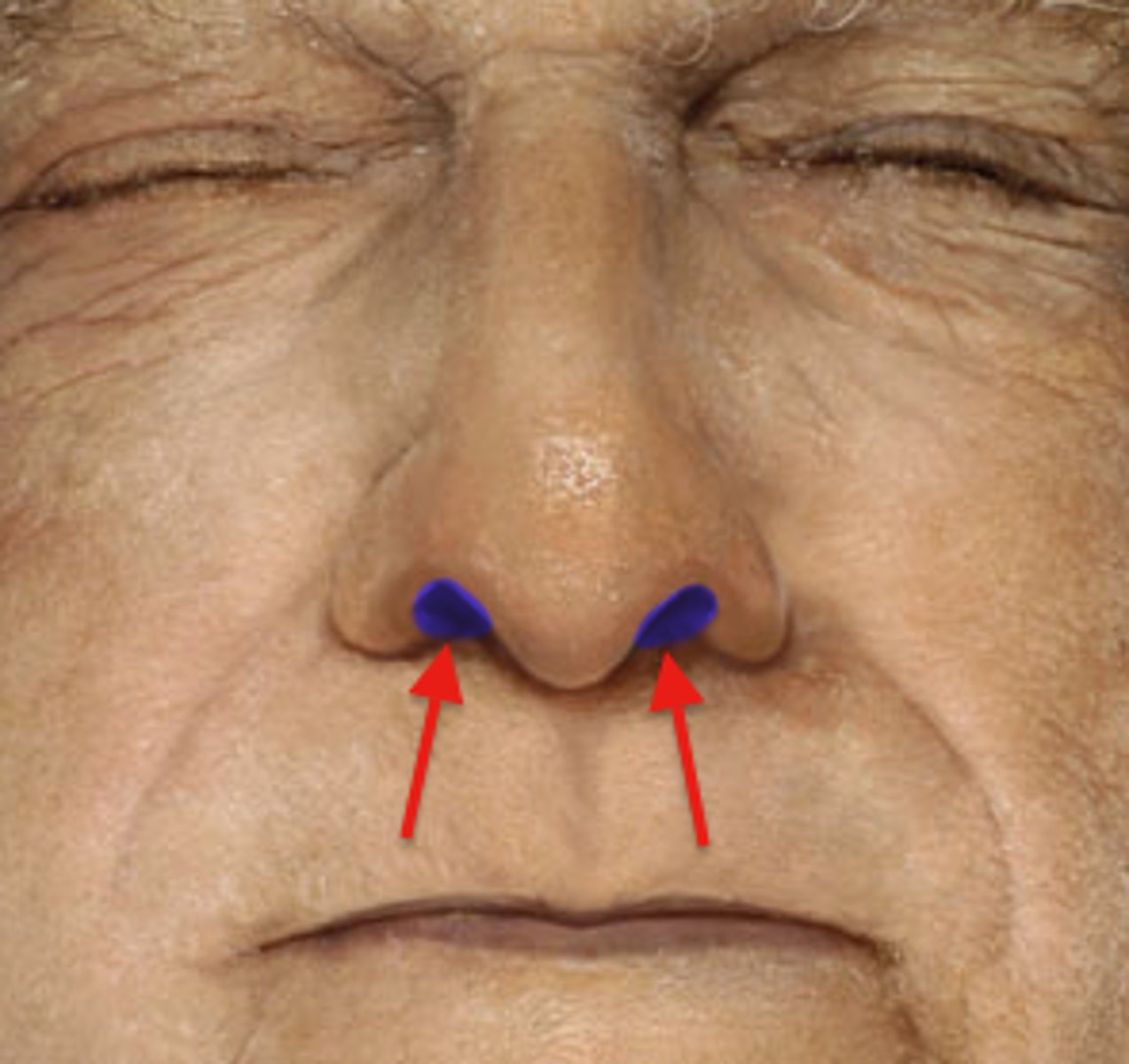
nasal vestibule
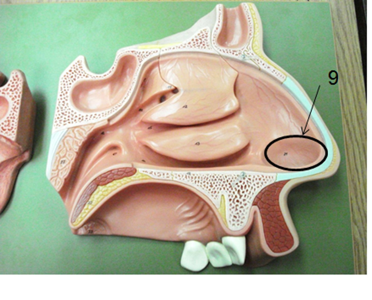
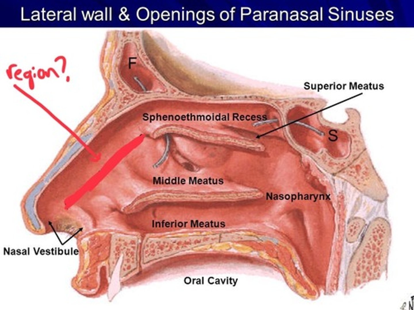
nasal atrium
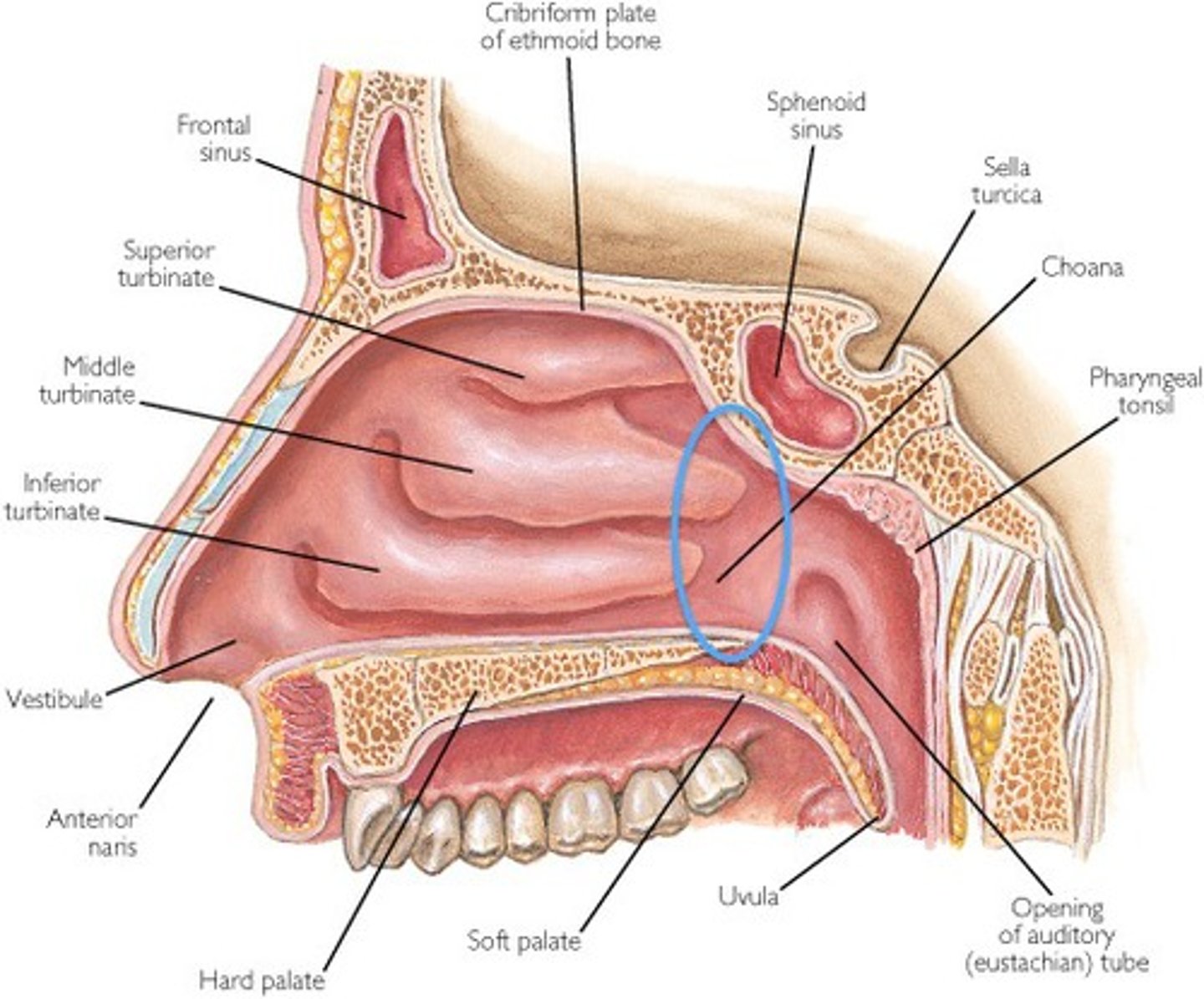
choanae
alar cartilage
cartilage that supports the apex of the nose and helps shape the nares; it is connected to the septal cartilage and connective tissue of the alae
