Rad Technique 2
1/29
There's no tags or description
Looks like no tags are added yet.
Name | Mastery | Learn | Test | Matching | Spaced | Call with Kai |
|---|
No analytics yet
Send a link to your students to track their progress
30 Terms
Dual Focus Tubes
0.5 mm/1.0 mm
0.6 mm/1.2 mm
1.0 mm / 2.0 mm
0.3 mm/ 1.0 mm (angiography and magnification procedures)
0.1 mm/ 0.3 mm (mammography)
Microfocus Tubes
FSS of 0.3mm or less
Large focus
Normal imaging
Higher mA can be impressed for larger/dense body parts
Higher heat capacity at the anode
Shorter exposure times to minimize motion
Small focus
–A given amount of electrons cover smaller area on anode
–Cannot use high mA due to limited heat capacity
–Used for magnification radiography where detail is required
–For smaller and thinner body parts where large quantity of x-ray photons are not necessary
X-ray beam is Poly… and hetero…
polyenergetic and heterogenous
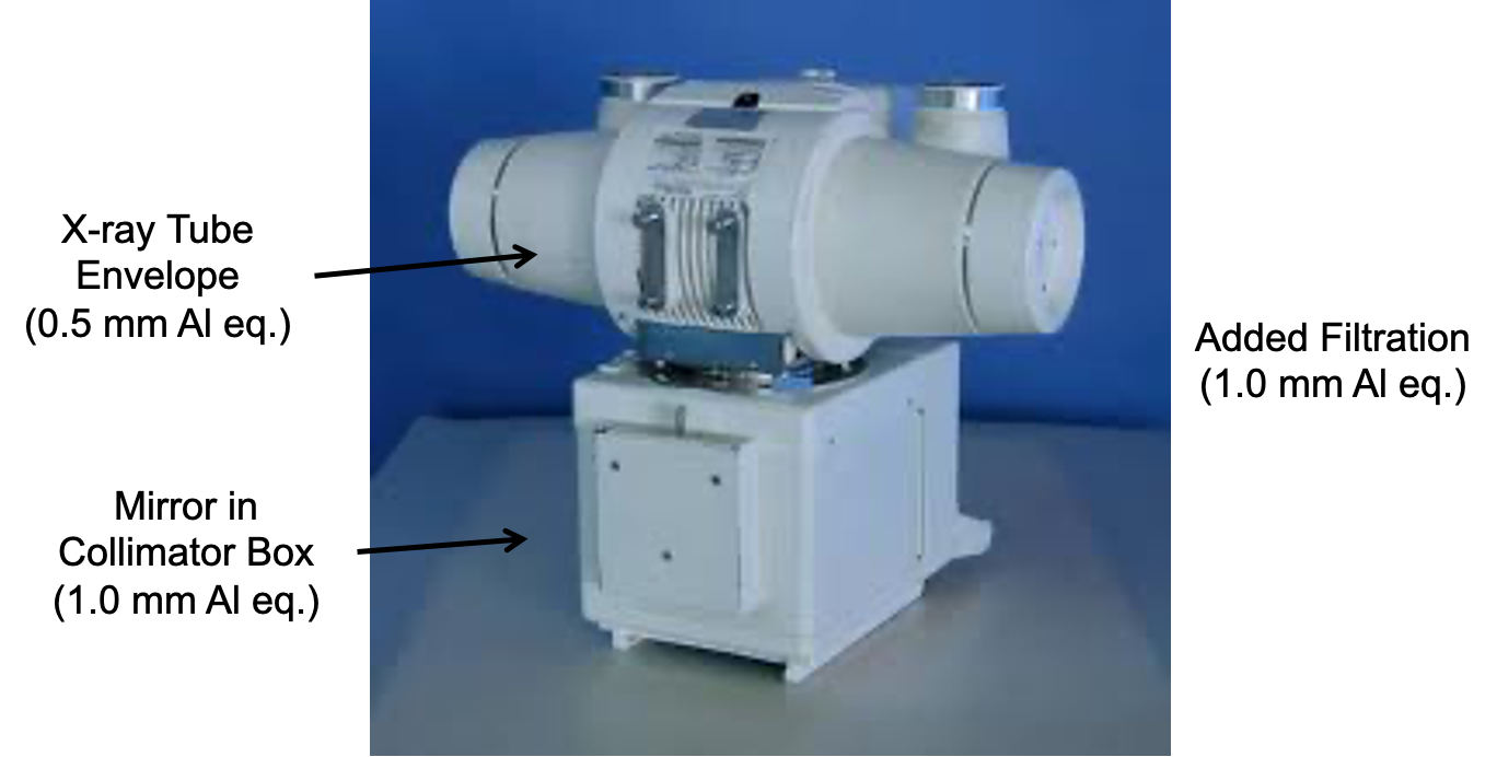
Filtration
“hardens” the beam
Inherent
Added
***Total filtration equals 2.5 mm of Al equivalency
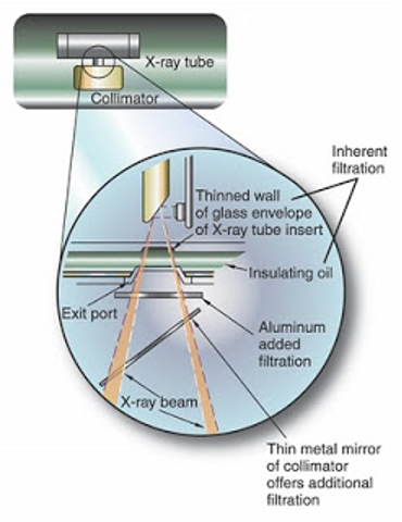
Filtration - Inherent
glass or metal envelope (0.5 mm Al equivalent)
Filtration - Added
mirror in collimator box (1.0 mm Al equivalent)
Another 1.0 mm Al equivalent is added between the x-ray tube housing and the collimator
Half-Value Layer (HVL)
Improves the quality of the beam
The amount of material that reduces the intensity of the x-ray beam to half its original value
Used for both shielding and filtration
Increase in HVL increases the quality of the beam
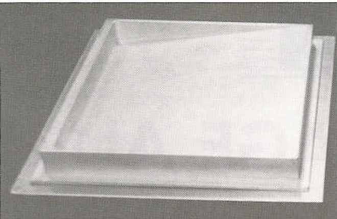
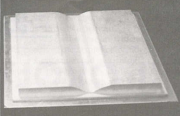
Compensation filter
–Different shapes of Al mounted under the variable light aperture (collimator)
–Balances intensity of x-ray beam for a more uniformed exposure to the image receptor
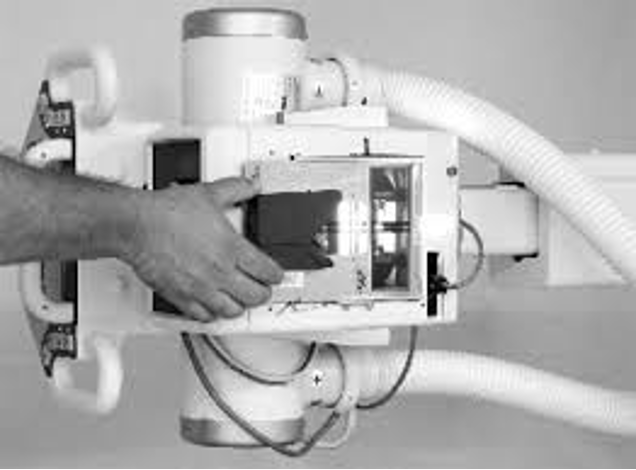
2 types of Compensation filter
Wedge filter
Trough filter

what type of?
what type of?
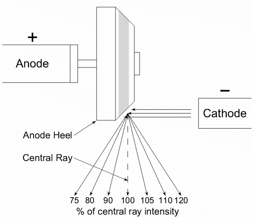
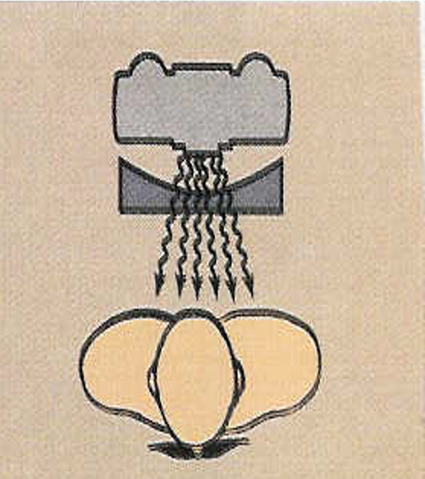
Anode Heel Effect
phenomenon in which the intensity of the x-ray beam is greater towards the cathode side of the tube. The loss of intensity is a result of the electrons traveling farther to the anode heel portion of the target
High-Voltage Generators influences…
radiation quantity and quality produced in the x-ray tube
3 types of High-Voltage Generators
1.Single phase (half-wave and full-wave rectified)
2.Three phase (half-wave and full-wave rectified)
3.High frequency
Single Phase Generators Half-Wave Rectified
•Half of electrical cycle wasted
•X-ray produced only half the time
•100% ripple
•Used in mobile radiography and dental
Single Phase Generators Half-Wave Rectified 2
•Voltage wave form same as half-wave but there is no dead time
•X-rays are emitted continuously as a pulse
•Required exposure time is half that for half-wave
•100% ripple
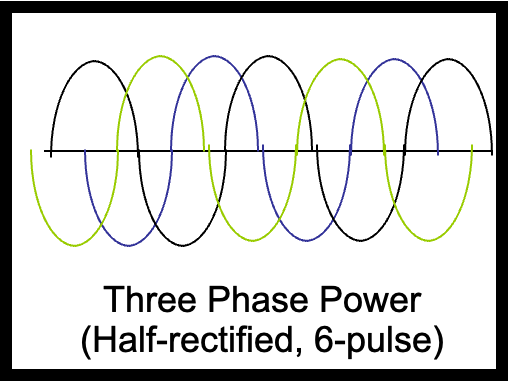
Three-Phase Generators
Commercial power generally delivered as 3 phase
Phases 120o apart
More efficient than single phase
More x-rays produced for a given mAs, average energy is greater
X-ray production is near constant rather than pulsed
14% ripple
High-Frequency Generators
•Developed in 1980’s, increasingly used
•Near constant potential
•LESS than 1% ripple
•Used in highly sophisticated systems: mammography and CT
Voltage Ripple
•Variation in peak voltage waveform
•Single phase- 100% ripple
•3 phase/6 pulse- 14% ripple
•3 phase/12 pulse- 4% ripple
–Less ripple- greater efficiency
•shorter exposure times may be used with 3 phase
3 phase comparison to 1 phase
•3 phase more efficient than 1 phase
•3 phase requires more complex circuitry
•3 phase more expensive to install
Patient Factors
•Patient’s size, shape, and physical condition greatly influence the required radiographic technique
•
–Size and shape (body habitus)
–Anatomy density (thickness)
–Pathology
–Composition
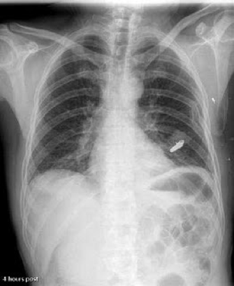
•Chest v. Abdomen
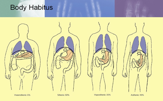
Sthenic- “strong, active”. Average size
2. Hyposthenic- thin but healthy in appearance
3. Hypersthenic- large framed and usually overweight
4. Asthenic- small, frail, emaciated, and often elderly
Thickness
–The thicker the patient, the more x-rays are needed to penetrate the body to expose the image receptor
–kV (quality of x-rays) is based on body part thickness
–Do not guess thickness. Use calipers
–Generally kV is fixed, while mAs is varied to achieve optimum density on film
Composition
•Measuring the thickness of a part doesn’t automatically release the technologist from exercising some additional judgment when selecting the proper technical factors
•
–Ex: the chest and the abdomen may have the same thickness, but vary considerably in composition

•Chest:
–Lung (air), Ribs (bone), heart (muscle)
–High subject contrast: mostly black and white, with some grays in between
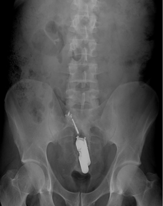
•Abdomen:
–Stomach, bowels, liver, kidney, spleen, pancreas, diaphragm, urinary bladder, etc.
–Muscle, fat, and water very similar in composition
–Many shades of gray

Radiopaque vs. Radiolucent
•Radiopaque
–Difficult to penetrate
–Bone, metal
–
•Radiolucent
–Easy to penetrate
–Air, soft tissue

Pathology
–Type, size, and composition will influence radiographic technique
–Obtaining good patient history and previous exams will help assessment
–Destructive process causing tissue to be more radiolucent. Ex: osteoporosis
–Constructive in increase mass density and composition. Ex: osteopetrosis