apk2100c chapter 5 - the integumentary system
 Call Kai
 Learn
Learn Practice Test
 Spaced Repetition
 Match
Match Flashcards
 Knowt Play
1/83

There's no tags or description
Looks like no tags are added yet.
Name | Mastery | Learn | Test | Matching | Spaced | Call with Kai | Chat |
|---|
No analytics yet
Send a link to your students to track their progress
84 Terms
The integumentary system
skin
accessory organs
Skin (integument)
largest organ of body
Accessory organs
hair
nails
oil and sweat glands
Regions of skin
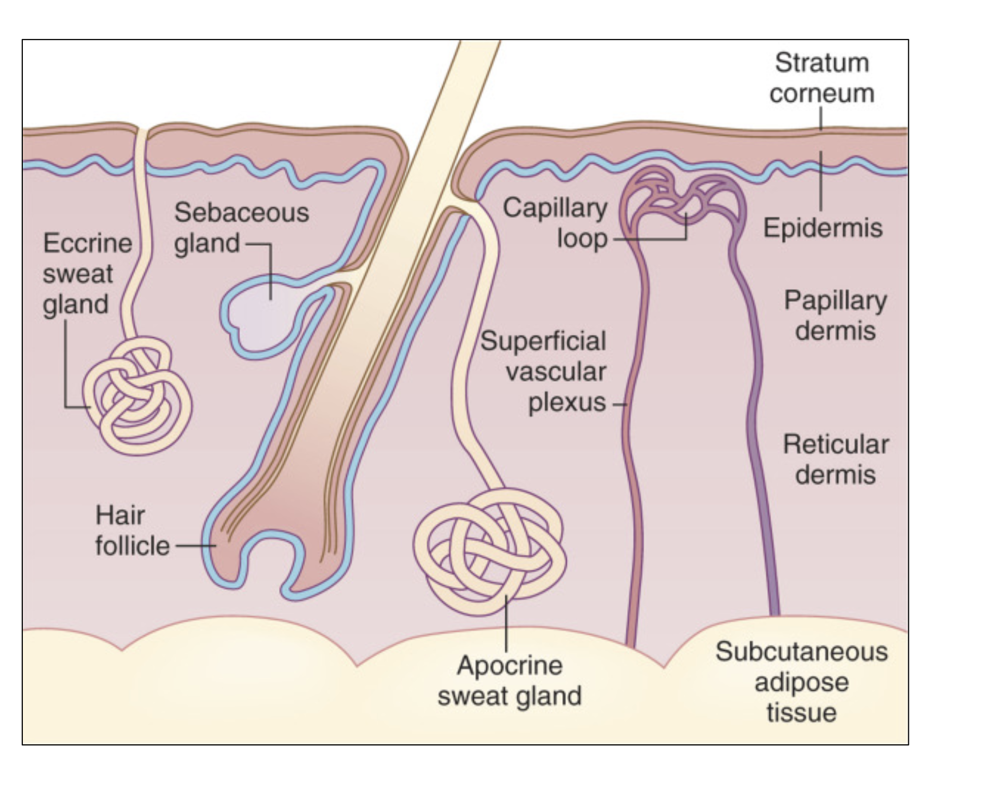
epidermis (top layer)
dermis (deep to epidermis)
hypodermis (underneath, not technically part of the skin - association with layers on top)

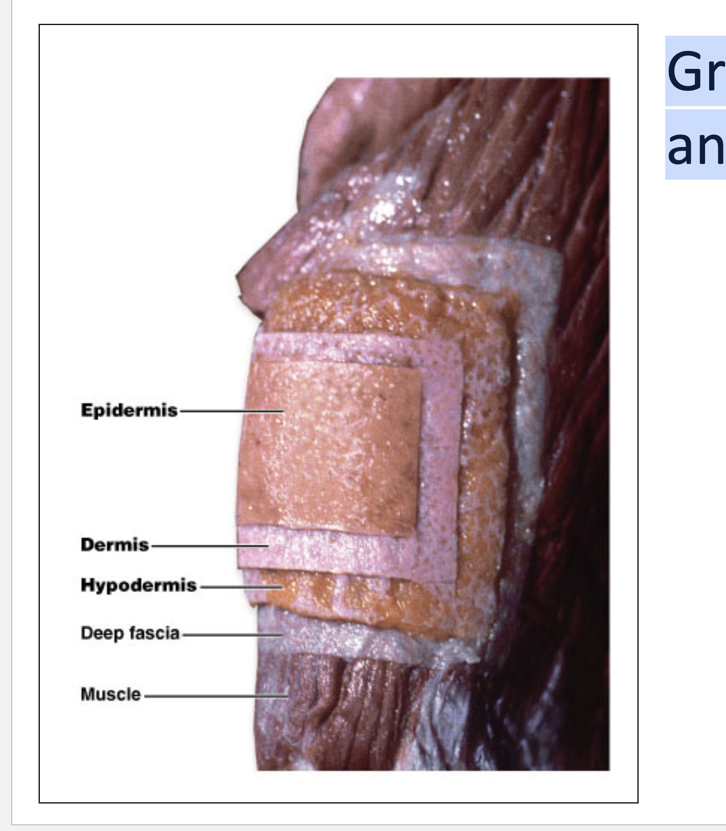
Gross Structure of the Skin and Underlying Anatomy
epidermis then dermis
hypodermis (has lot of adipose tissue)
Functions of skin
thermoregulation
protection
sensory reception
excretion
vitamin D production
thermoregulation
vessels, capillaries
ability to sweat
disipate heat
protection
natural physical barrier from external vs internal
sensory reception
lots of nerve endings that will sense signals
(tactile, pressure, cold temp, etc.)
excretion
certain solutes, salts, water content can leave the body
vitamin d production
happens in the skin
epidermis
Most superficial layer
avascular
keratinized stratified squamous epithelium (hard)
contains 4 principal cells
keratinocytes - over 90 percent
melanocytes - pigment
langerhans cells (dendritic cells) - immune function
merkel cells (tactile epith cells) - pressure, touch, tactile senses
avascular
no blood vessels ( in epidermis )
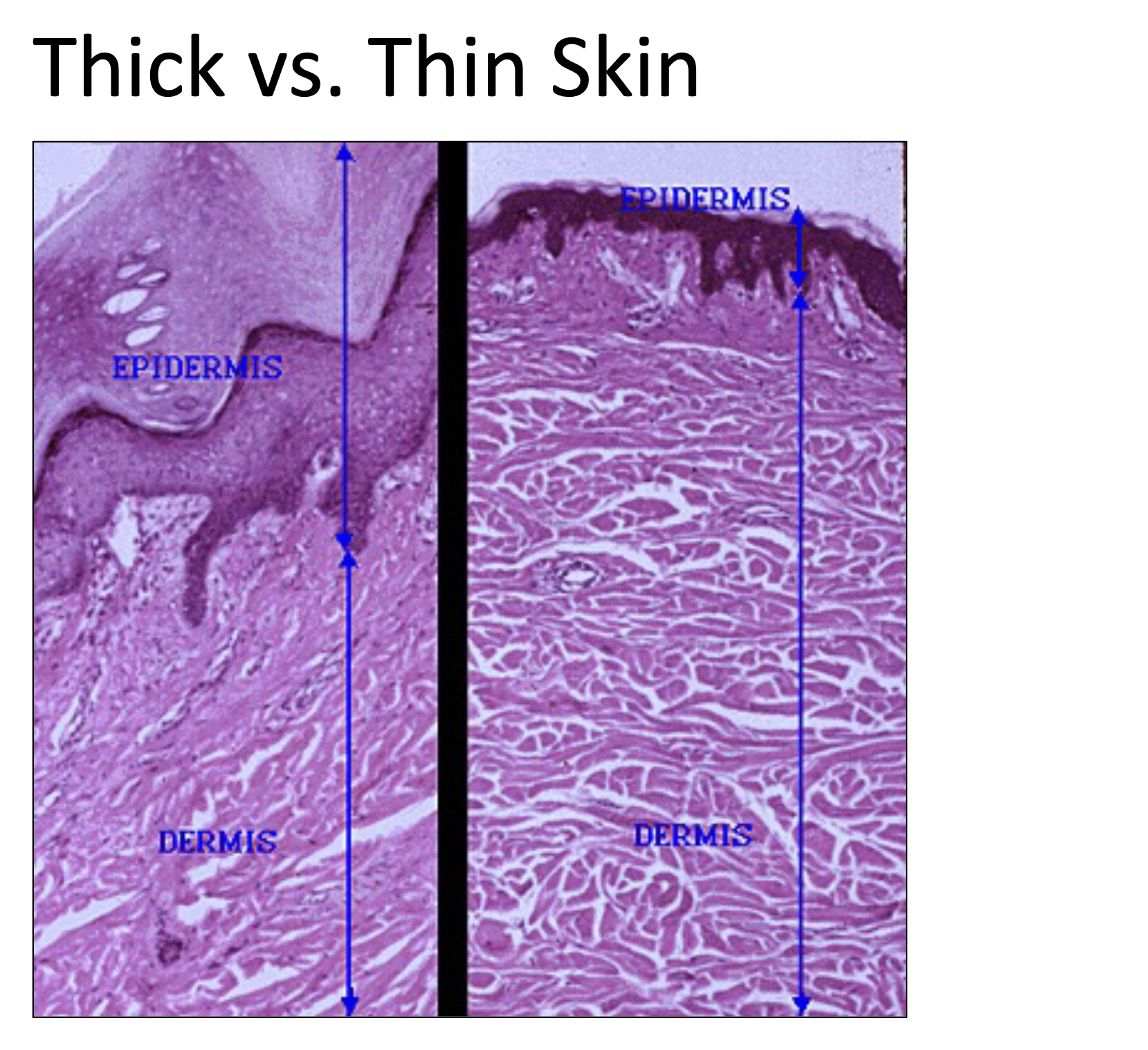
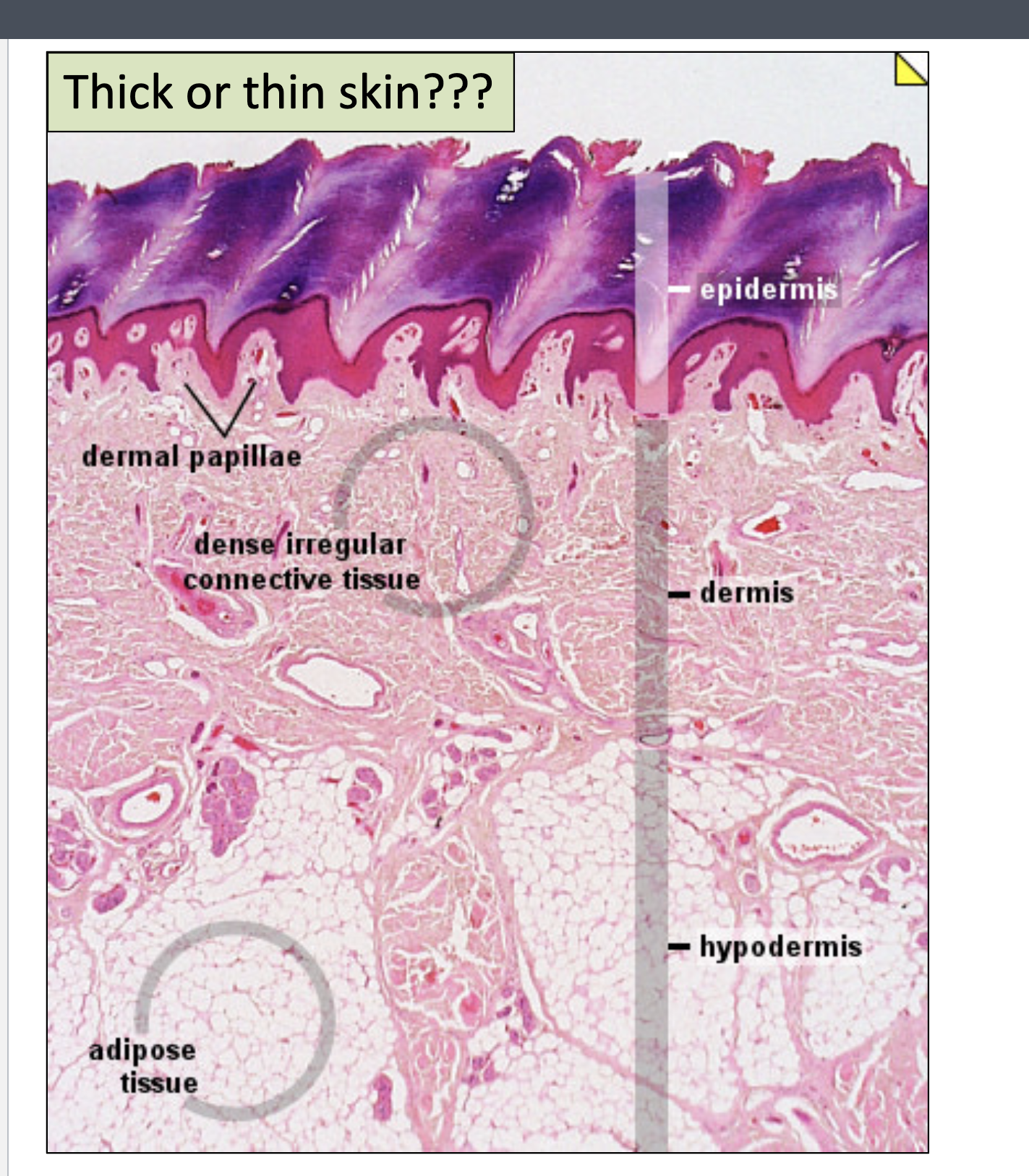
Thin skin
keratinized stratified squamous ET
areas of skin where little friction develops
most of body
FOUR epidermal layers
thin epidermis

Thick skin
areas where friction commonly develops
palms, feet, fingertips
FIVE epidermal layers
thick/larger epidermis (keratinized layer)
Stratum
layer
Stratum basale (TE)
Thin epidermis
Most basal layer → Deepest epidermal layer
One row of actively mitotic cells (keratinocytes)
A lot of cell division happening
pushing up → bumping cells upward
Melanocytes (produced melanin) → deposits melanin into keratinocytes
see dendritic cells - touch sensation
Melanocytes
dictates skin pigment - produces melanin
Stratum spinosum (TE)
thin epidermis
several layers of keratinocytes unified by desmosomes
cells contain thick bundles of intermediate filaments made of pre-keratin
tonofilaments
pushed up from basale
Statum granulosum (TE)
Thin epidermis
one to five layers of flattened cells
organelles deteriorating
cytoplasm full of lamellar granules (release lipids - makes skin waterproof) and keratohyaline granules
Stratum corneum (TE)
thin epidermis
most superficial layer
20-30 layers of dead cells → flat membranous sacs filled with keratin
glycolipids in extracellular space
Layers of thin epidermis

Does the thick epidermis have the same layers as the thin epidermis?
Has all four, but one extra layer called the stratum lucidum
stratum corneum = more thicker in thick epidermis
Stratum lucidum
only in thick epidermis
slightly translucent
thin, clear layer of dead skin cells
Epidermis is replaced how often?
Every month
ex. tanning (epidermis)
ex. tattoo = deposited into deeper skin (places that hav blood vessels)
What are the 3 pigments that impart varying colors and tones to skin and hair?
Melanin (produced in hair and skin)
Carotene (orange color - baby eating carrots)
Hemoglobin (in blood, red color)
Two types of melanin
Pheomelanin
Eumelanin
Pheomelanin
imparts yellow to red colors
particularly noticeable in hair
Eumelanin
impacts a brown to black color (overrides of pheomelanin)
Number of melanocytes for everyone is…
the same
Why are there color differences (although there is the same number of melanocytes)?
Due to the amount of pigment produced (melanin) and transferred to keratinocytes
Light skin
melanocytes producing less melanin
melanin clumped near nucleus
breaks down quickly
Dark skin
Melanocytes produce more melanin
melanin spread through keratinocytes
breaks down slowly (harder)

Abundant pheomelanin in hair
Minimal eumelanin in skin

Abundant eumelanin in skin and hair
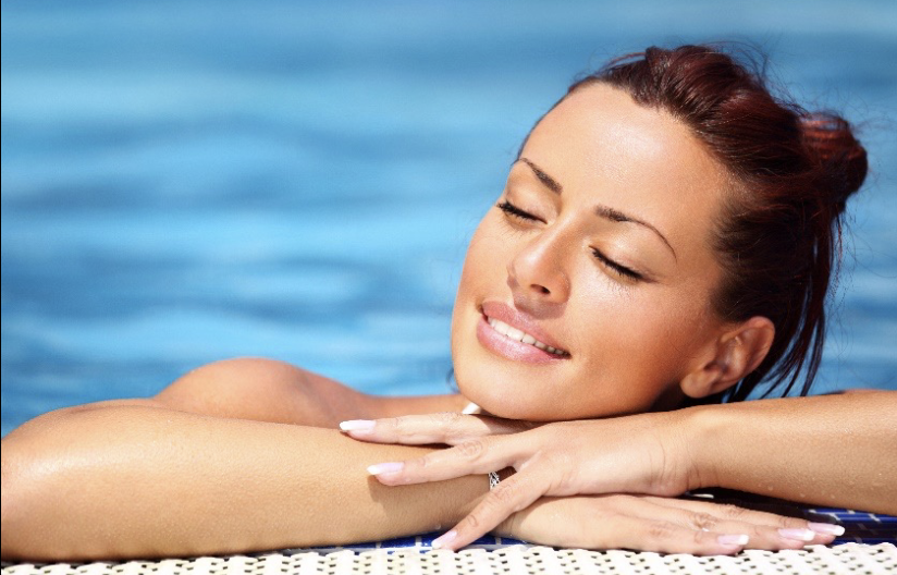
Moderate pheomelanin and eumelanin in skin
Abundant eumelanin in hair
How do melanocytes produce protein (melanin)?
Takes the amino acid tyrosine and converts it to melanin
Melanocytes transfer melanin granules into keratinocytes
melanin keratocytes like to surround itself around nucleus (shield against UV rays)
What do melanin place itself between?
The sun and the cell’s nucleus
melanin granules act as shield against UV rays to prevent cancer
Albinism
Cannot produce any melanin due to a genetic disorder
Inability to produce the tyrosinase enzyme
translucent hair, skin, and eyes → can have visual issues
display pink or red eyes → due to hemoglobin in blood vessels
What tissue is in the dermis
Mostly connective tissue → very tough
What structures are in the dermis?
blood vessels → vascular region of skin
lymphatic vessels
nerves
Regions of dermis
Papillary layer of the dermis = superficial → areolar loose connective tissue proper
Reticular (network) layer of the dermis = deeper → dense irregular connective tissue proper
Papillary layer of dermis
superficial → areolar loose connective tissue proper
Reticular layer of dermis
deeper → dense irregular connective tissue proper
swirling pattern of collagen fibers
what is dermal papillae
Feature of dermis
small projections of papillary dermis into overlying epithelium above
dermal papillae function
Enhances surface area for nutrients/gas exchange (to go up to the epidermis that is avascular)
Strengthens the dermal-epidermal junction → tightly bound
does NOT make fingerprints
Epidermal ridges
Ridges are part of fingerprints
dermal papillae don’t match the epidermal ridges
Dermal ridges
Form fingerprints
Underlying mounds of tissue and protrudes upward
Cleavage lines
Separations or less dense regions of collagen bundles (space between bundles) in the reticular dermis
create an invisible cleavage line in the skin
Incisions made along lines heal more quickly and gape less than incisions made across (important for surgery)
run longitudinally
Hypodermis
Not a part of the skin (below the skin)
Subcutaneous layer of CT
What is hypodermis made of?
Mostly adipose tissue loose CT proper
varies in thickness
cushion and padding
protects underlying structures, insulates, and stores energy
What does the hypodermis contain?
Large blood vessels, lymphatic vessels, and nerves whose branches extend into dermis
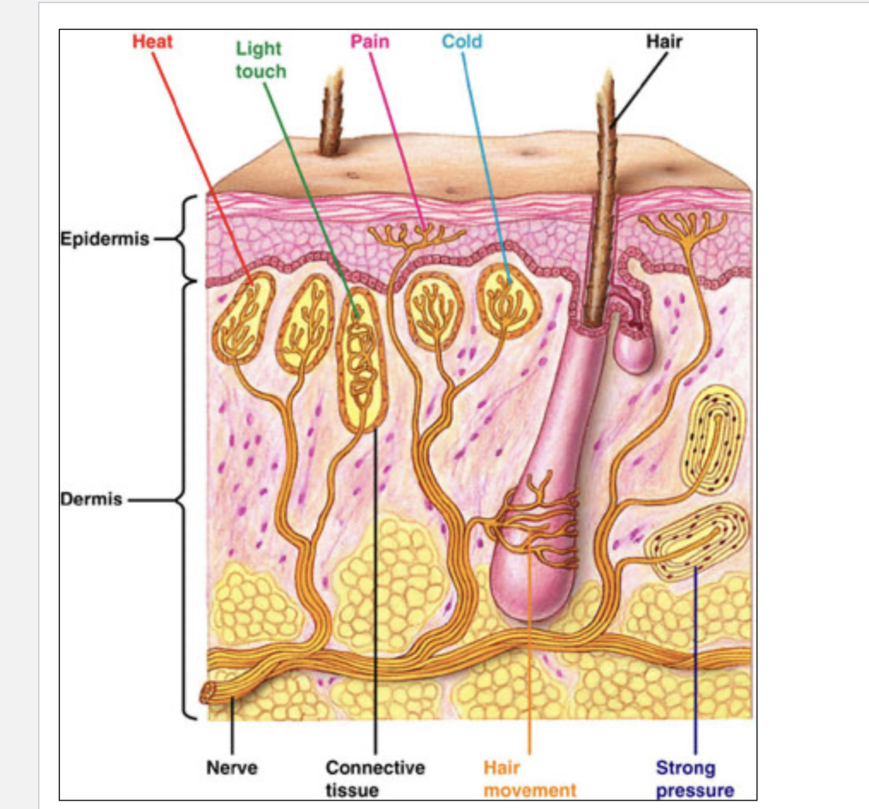
Cutaneous sensation (how do we feel things with our skin)
many sensory structures embedded in the dermis (nerve endings)
some free endings - sensory structures derived from the epidermis
hair follicles
sweat glands
oil glands
some structures are from underlying nerves, which transmit information to the CNS
in epidermis so you can detect pain right away

Appendages of skin
derived from epidermis but mostly located in dermis
nails
hair and hair follicles
sebaceous (oil) glands
sweat glands
Nails
scale like modification of epidermis containing dead keratinocytes full of HARD keratin
Superficial nail structures
free edge of the nail
lunule
lateral nail fold
nail plate
eponychium (cuticle)
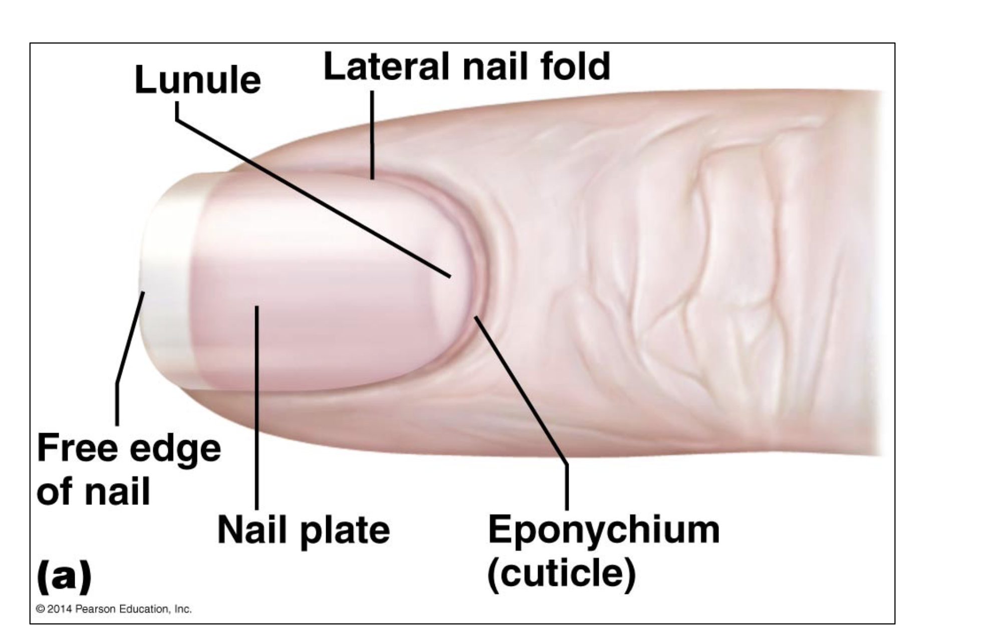
free edge of nail
part you cut off
nail plate
directly over digit of nail
pink part of nail - transparent due to dead keratinocyte so see blood
lunule
half moon shape white area
only visible part of the nail matrix - cells proliferating for nail growth (thick) → highly mitotic cells
lateral nail fold
around nail and will turn into the cuticle (eponychium)
eponychium
cuticle
Deep nail structures
nail bed
phalanx (bone of fingertip)
nail root

Nail bed
made up of epidermal cells under nail plate
Nail root
covers the nail matrix
Hair
flexible columns of dead keratinocytes full of HARD keratin
shaft of hair
above the surface
root of hair
below surface
Hair medulla
inner core
Hair cortex
around medulla
Hair cuticle
superficial layer
hair follicle
Sheath around the hair
made up of external and internal root sheath
external root sheet
most superficial to hair
derived from the stratum basale
internal root sheath
closest to hair
just outside of hair cuticle
derived from stratum corneum of the epidermis
invaginates downward to turn into this
Hair bulb
End of hair root - enlarged
Has a connective tissue papilla = blood vessels come into matrix
matrix cells = highly mitotic allowing for hair growth

Arrector pili muscle - hair
Directly attached to the hair follicle
This muscle contracts, pulling hair up = goosebumps
Smooth muscle - involuntary
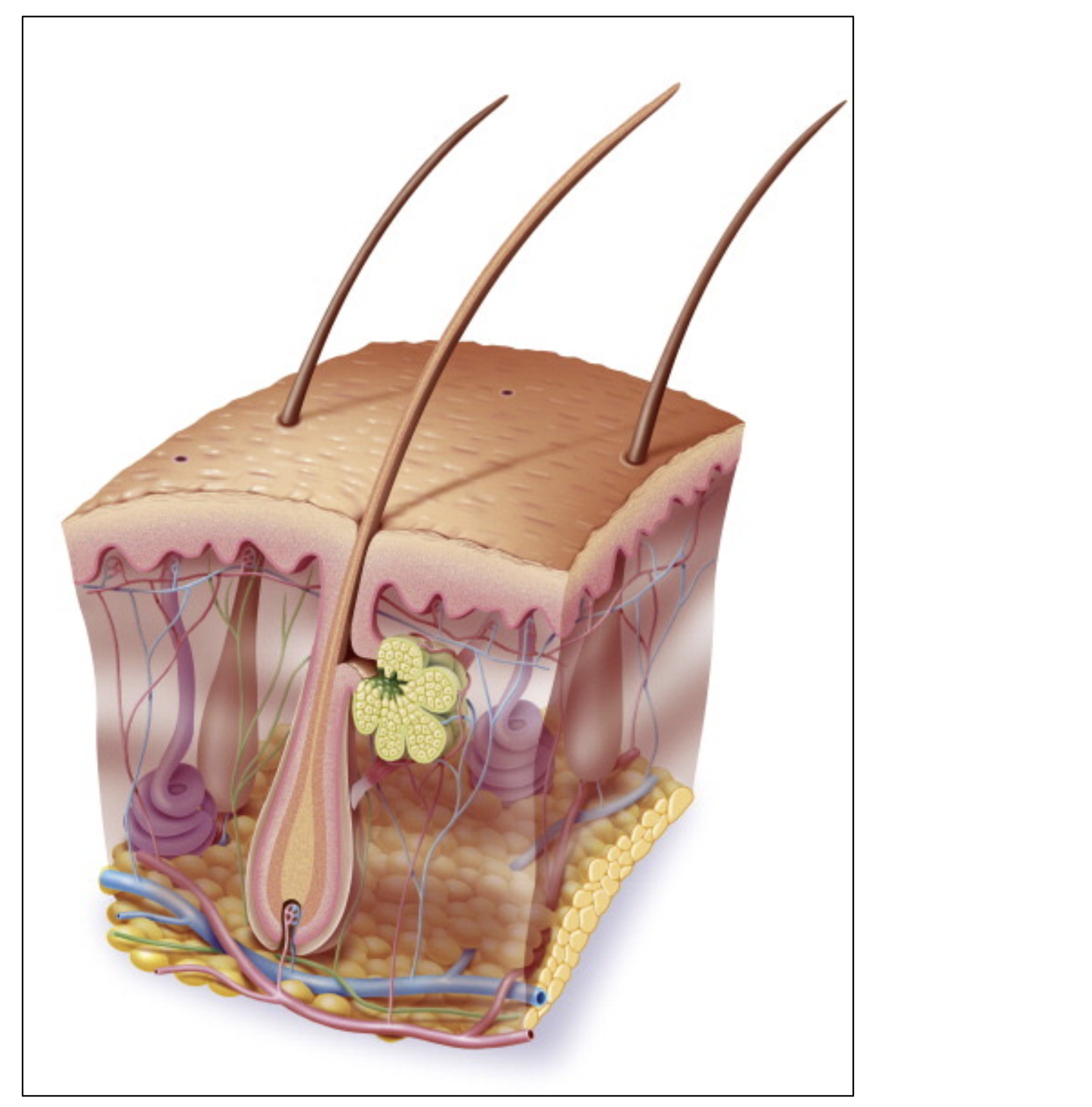
Sebaceous gland
Body’s natural moisturizer
produces oil = sebum
Product dumped into the hair follicle
Sebum
Keeps hair/skin soft
protects against brittle hair/skin
Protects the epidermis from cracking
Collects dirt/dust
impedes water loss across the skin
Where do we have sebaceous glands?
Located everywhere except plantar and palmar regions (palms of hand and soles of feet)
Glassy membrane
Outside of the root sheath of hair
Thin transparent tissue
Anchors self to basement membrane
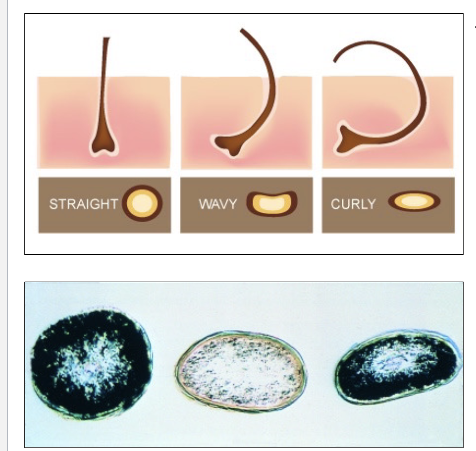
Types of hair
straight (circle cross section)
wavy
curly (oval cross section)
What are the two types of sweat glands (sudoriferous)
Eccrine sweat glands
Apocrine sweat glands
Eccrine sweat glands
palms, soles, forehead (predominantly)
true “sweat”; watery
thermoregulatory function
duct opens directly onto skin surface
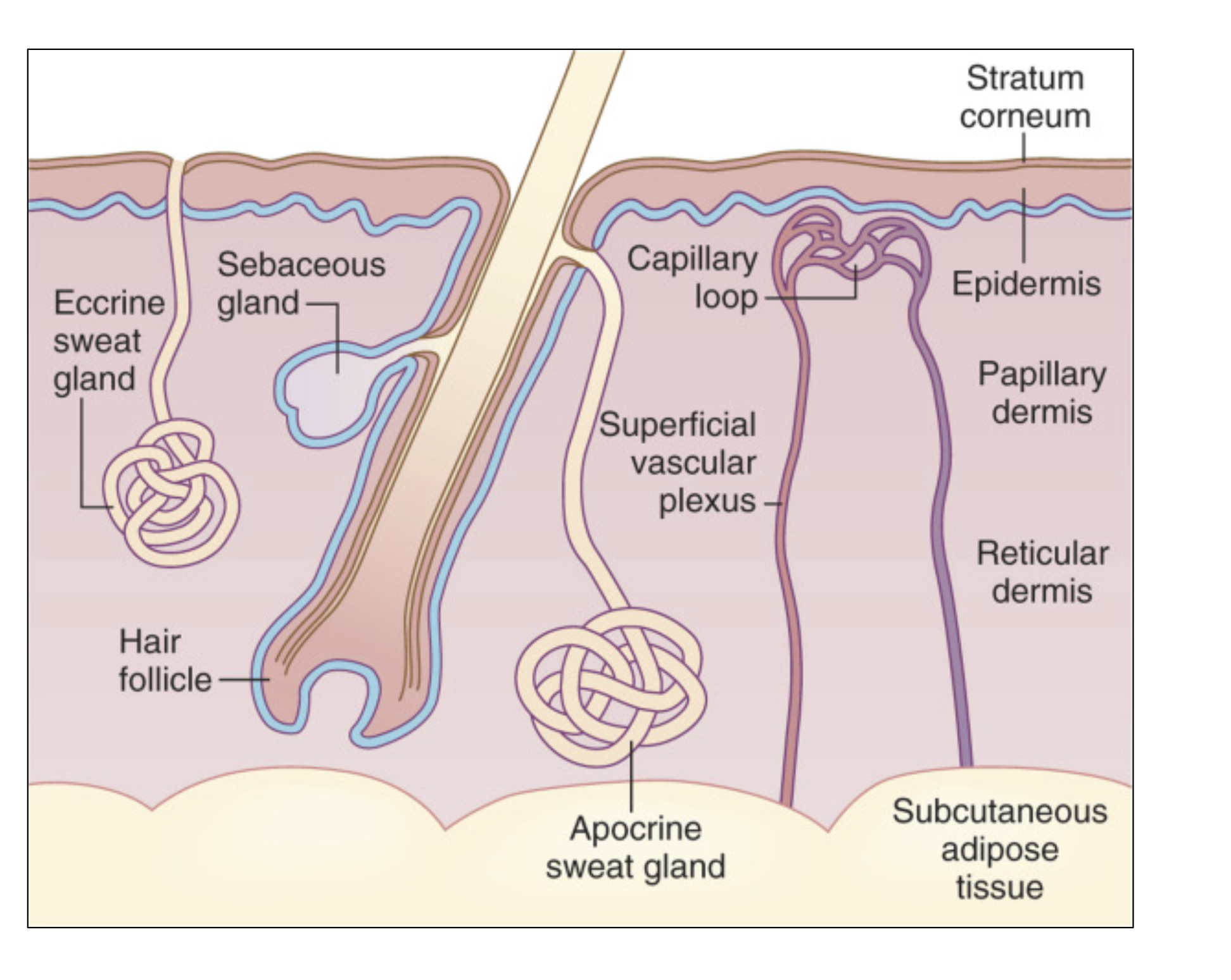
Apocrine sweat glands
axillary, anal, genital areas only
duct opens to the hair follicle (similar to sebaceous) + mixes with sebum/stuff
dumps into hair follicle
milky “sweat”
body odor
no thermoregulatory function