EKG Readings
 Call Kai
 Learn
Learn Practice Test
 Spaced Repetition
 Match
Match Flashcards
 Knowt Play
1/29

There's no tags or description
Looks like no tags are added yet.
Name | Mastery | Learn | Test | Matching | Spaced | Call with Kai | Chat |
|---|
No analytics yet
Send a link to your students to track their progress
30 Terms
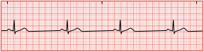
Sinus bradycardia is a normal EKG tracing, but there is a heart rate of less than 60/min.
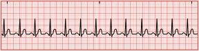
Sinus tachycardia is a normal EKG except for the heart rate greater than 100/min.
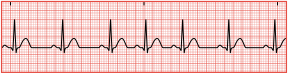
Sinus dysrhythmia is a slight irregularity in the rhythm.
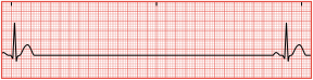
Sinus arrest is a break in the normal EKG pattern.
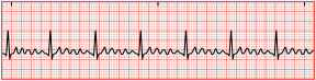
Atrial flutter is a condition in which the atria are contracting at a rate much faster than the ventricles are contracting.
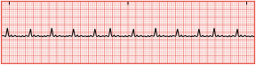
Atrial fibrillation in there is no organized contraction of the atria. They are in a quivering state.
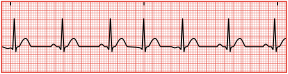
Premature junctional complex (PJC) is an early impulse that occurs before the next expected beat. The P wave could occur before, after, or even buried within the QRS complex, which causes an irregularity in the rhythm.
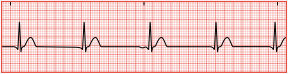
Junctional escape rhythm can result in an absent P wave, but if recognizable, it will be inverted. The heart rate will not exceed 60/min.
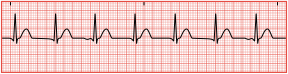
Accelerated junctional rhythm is the same as the escape rhythm, but the rate is 60 to 100/min. It is unlikely that the patient will exhibit signs of decreased cardiac output.
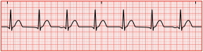
Junctional tachycardia rhythm is the same as the escape and accelerated rhythm, but the heart rate will be between 100 and 150/min.
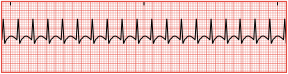
Supraventricular tachycardia (SVT) or narrow complex tachycardia will have a heart rate greater than 150/min. Due to the rapid nature of the heart rate, P waves are usually not visible.
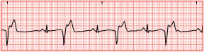
Premature ventricular complexes (PVCs) occur when a P wave is not visible, and the QRS complex is often wider than normal with an unusual shape. PVCs are classified as occasional if there are one to five in 1 minute, and frequent if there are six or more per minute.
Unifocal: Single early PVC indicates one irritable area.
Multifocal: PVCs with multiple shapes indicate more than one irritable area.
Interpolated PVC: PVC occurs with no interruption in the normal rhythm.
Bigeminy: PVCs occur every second beat.
Trigeminy: PVCs occur every third beat.
Quadgeminy: PVCs occur every fourth beat.
Coupling: Two PVCs occur back to back.
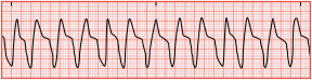
Ventricular tachycardia (VT) is three or more PVCs in a row with a ventricular rate greater than 100/min. There are no noticeable P waves in the tracing. The QRS complexes are wide and unusual, with the T wave deflected in the opposite direction.
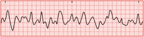
Ventricular fibrillation (VF) is an emergency state in which the ventricles are not contracting, but quivering—and there is no cardiac output. There are no discernible waves noted throughout the tracing.
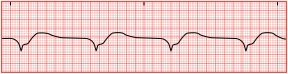
Idioventricular rhythm occurs when only the ventricular pacemaker is functioning. The ventricular rate will range from 20 to 40/min, and there are no discernible P waves. The QRS complex is wide with an unusual appearance. If the heart rate ranges between 40 and 100/min but all other aspects of the idioventricular rhythm are present, this is an accelerated idioventricular rhythm.
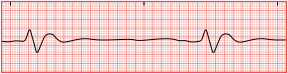
Agonal rhythm results when all the pacemakers of the heat (SA node, AV node, Purkinje fibers) have failed. The tracing shows a wide, unusual QRS complex with no P or T wave. The ventricular rate is less than 20/min.
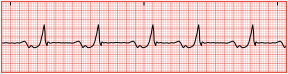
Bundle branch block occurs when there is interference somewhere in one of the bundle branches.
In a left bundle branch block (LBBB), the current will move through the right bundle branch for right ventricular contraction. But the current that would normally go down the left bundle moves to the left ventricle via the septum, which results in an abnormal right to left stimulation.
When there is a right bundle branch block (RBBB), the septum is depolarized normally and the left ventricle is still activated by the left bundle branch. Because the right side is blocked, the left ventricle is able to send impulses through the myocardium to the right ventricle in order to depolarize the right ventricle.
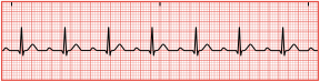
First-degree atrioventricular block represents a delay in conduction from the SA node to the AV node. The impulse still travels through the normal pathway but is delayed. As a result, the PR interval will be greater than the normal 0.20 seconds.
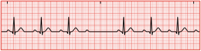
Second-degree atrioventricular block, type I, is also known as Mobitz I or Wenckebach. There will be missing QRS complexes. The PR interval will get progressively longer until a QRS is dropped, and then the pattern repeats itself.
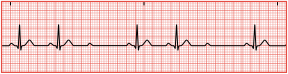
Second-degree atrioventricular block, type II, is also known as Mobitz II, this is the classic form of heart block. The PR interval remains constant, but during the tracing, it is noted that a P wave is present with no QRS complex or T wave.

Third-degree atrioventricular block—also known as complete heart block (CHP)—occurs when all electrical impulses that originate above the ventricles are blocked. There is no pattern to the cardiac cycle as the atria and ventricles are contracting independently. The atria contract at a normal rate but the ventricles contract at 20 to 40/min, depending on where the pacemaker site is originating.
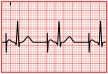
The pacing spike is an artifact in the EKG tracing that is a result of the electrical impulse produced by an artificial pacemaker. It will appear as a thin spike at the P wave, QRS complex, or both, depending on the specific pacing needed and programmed. If atrial pacing is used, a normal P wave will follow the single thin line. The QRS configuration is altered and gives the appearance of a bundle branch block if ventricular pacing is incorporated, but the spike will appear prior to the complex.

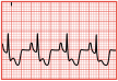
Myocardial ischemia is exhibited through ST segment depression of 1 mm or greater, occurring in two contiguous leads or T wave inversion.
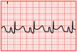
When ST elevation is noted, the injury is current, and tissue has not yet died. Without intervention, the tissue will die. This, along with symptoms of chest pain, is referred to as ST elevation myocardial infarction (STEMI), and a complete blockage of a coronary artery has occurred.
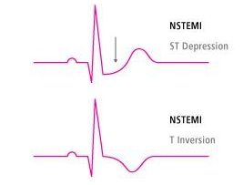
A myocardial infarction that does not have ST segment elevation is referred to as non-STEMI (or NSTEMI). Pathologic Q wave changes are indicative of infarction and necrosis if noted in two or more continuous leads. The Q wave will measure 0.04 seconds and will be equal or greater than one-third the height of the R wave.
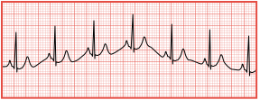
A wandering baseline is exactly what the name implies—the recording demonstrates wavelike up-and-down movements throughout the tracing.
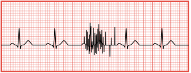
Muscle movement on an EKG tracing is referred to as a somatic tremor. Somatic tremor exhibits as uneven spikes on the EKG tracing. The tremors are caused by multiple factors.
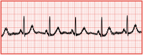
AC interference (60 cycle interference) is a type of artifact that is recognized as uniform sharp spikes on the EKG tracing.
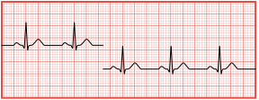
An interrupted baseline is demonstrated by a tracing that is not continuous. A break in the baseline or a fully nonrecorded lead can be noted.
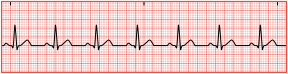
Normal sinus rhythm is indicated by the following.
P wave is present.
P wave is upright and rounded.
P wave has an amplitude less than 2.5 mm.
P wave duration is less than 110 milliseconds.
QRS complex is usually narrow.