IB Bio HL Form and Function Quiz
1/99
Earn XP
Description and Tags
Name | Mastery | Learn | Test | Matching | Spaced | Call with Kai |
|---|
No analytics yet
Send a link to your students to track their progress
100 Terms
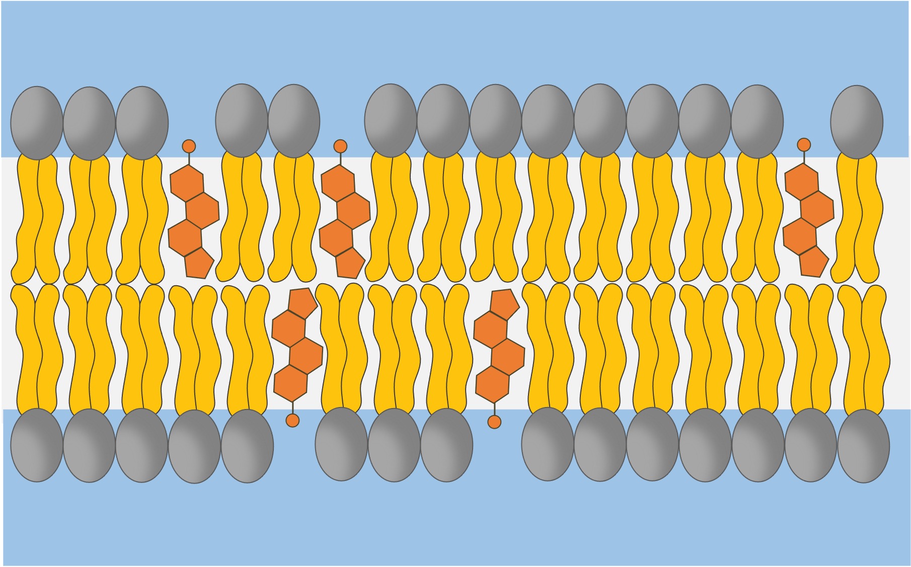
What does cholesterol do in a membrane?
Cholesterol helps to stabilize the fluidity of cell membranes, maintaining their integrity and flexibility across varying temperatures.
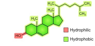
What is the structure of cholesterol in a membrane?
Lipid molecule with a hydrophobic steroid ring structure and a small hydrophilic hydroxyl group
Where is cholesterol found in the membrane?
In between phospholipids, with the hydrophilic hydroxyl groups exposed to water and the hydrocarbon rings and tail within the hydrophobic core of the membrane
Membrane fluidity refers to the ______ of bilayer
viscosity
What does cholesterol do at high temps?
It stabilizes the membrane by reducing movement of phospholipid fatty acids and and reduces fluidity by making it less permeable to small molecules.
What does cholesterol do at low temps?
It prevents the membrane from becoming too rigid by disrupting the packing of phospholipid fatty acids, thus maintaining fluidity.
What is the structure of a cholesterol molecule?
Membrane fluidity is affected by:
the composition of phospholipids:
fatty acid length: a longer tail means more phospholipid interaction, meaning less fluidity
temperature changes: more fluid at higher temps, less fluid at lower temps
Draw the structure of a membrane
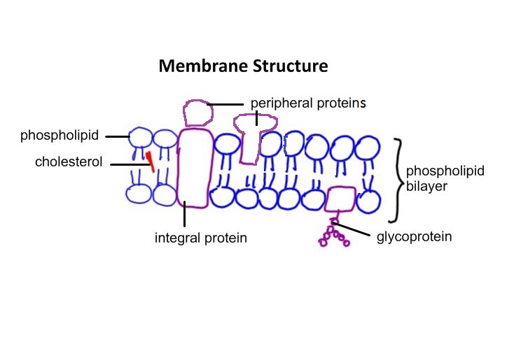
Define the purpose of a membrane
Semi-permeable (allows certain molecules through but not others) and thus encloses the contents of the cell, separating the intracellular components from the external environment, allowing for the control of internal conditions within the cell and the maintenance of homeostasis
Why are lipids ideal for membrane’s purposes?
Amphipathic nature, with hydrophilic (water-attracting) and hydrophobic (water-repelling) properties. This allows them to form bilayers in aqueous environments, creating a semi-permeable membrane that can separate the interior of the cell from the external environment.
Provides fluidity and flexibility to the membrane while also being a barrier to protect cellular components.
Evaluate whether lipids alone are suitable for this purpose
Purpose: Provide a semi-permeable bilayer, which lipids, particularly unsaturated ones, allow to be fluid
They are not suitable alone
Selective permeability: won’t allow large/polar molecules through, must use facilitated diffusion or active transport
Cell communication: membrane proteins are required for signalling, ie receptor proteins that bind to hormones or other signaling molecules
Enzymatic activity: must be done by enzymes imbedded in membrane
Cell recognition: done by glycoproteins and glycolipids
Stability and shape: achieved by cytoskeletal elements
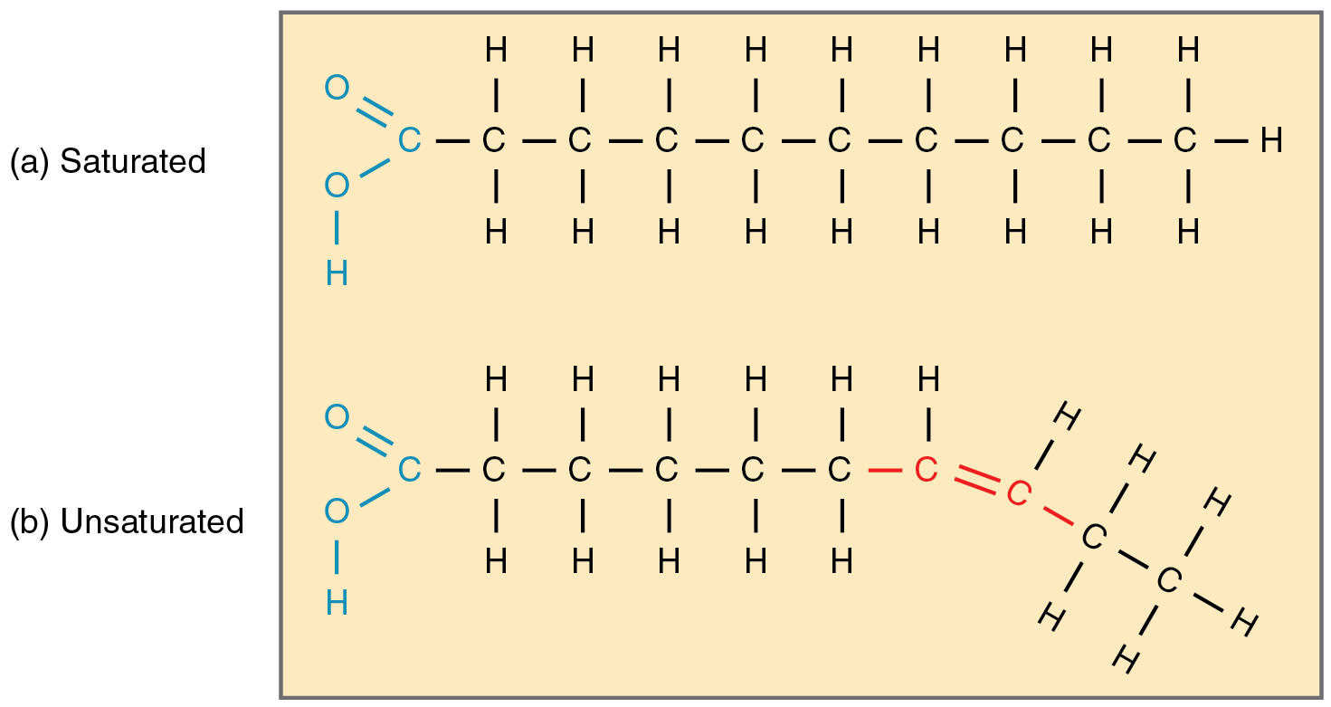
What does an unsaturated fat vs saturated fat look like?
Importance of membrane fluidity:
enable molecules to diffuse through the membrane towards areas of the cell where they are needed
facilitate the interaction between proteins, which is crucial for cell signaling
enable membranes to fuse with one another during vesicle formation, endocytosis and exocytosis
ensure the even distribution of membrane molecules between daughter cells during cytokinesis
Phospholipid tails are made of molecules called _____.
fatty acids
Fatty acids all have a ________ (carbon and hydrogen atoms arranged in a chain).
hydrocarbon chain
What happens if the phospholipid tails are longer?
There will be more interactions between the tails and the membrane will be less fluid
What happens if the tails are made of saturated fatty acids (ie, no double bonds between adjacent carbon atoms)
Tails will be relatively straight, decreasing membrane fluidity
What happens if the tails are made of unsaturated fatty acids (ie, DOES have double bonds between adjacent carbon atoms)
There will be a "bend" in the molecule, and these kinks will create space in the membrane and increase membrane fluidity
At low temperatures, organisms increase ______ of fatty acids, ie, in chickpea (Cicer arietinum), low-temperature results in a 31% increase in the degree of ______ of fatty acids.
unsaturation
At high temperatures, organisms increase _______ of fatty acids, ie Arabidopsis plants grown at 36°C have more double bonds compared to plants grown at 17°C
saturation
Fluidity refers to:
the viscous flow of phospholipids in the cell membrane and organelles of the endomembrane system
What are 4 things that membrane fluidity is affected by?
Temperature
Fatty acid length
Fatty acid saturation
Presence of cholesterol
At higher temperatures, the membrane has more or less fluidity than at lower temperatures?
more
_____ fatty acid tails allow for more interactions between phospholipids, leading to less fluidity.
Longer
What are the steps of endocytosis (engulfing materials into the cell)
The plasma membrane folds inward forming a cavity that fills with extracellular fluid, dissolved molecules, food particles, foreign matter, pathogens, or other substances.
The plasma membrane folds back on itself until the ends of the in-folded membrane meet. This traps the fluid inside the vesicle.
The vesicle is pinched off from the membrane as the ends of the in-folded membrane fuse together.
The vesicle breaks away from the cell membrane and moves into the cytoplasm.
The cell membrane has gotten smaller.
What are some examples of endocytosis?
Macrophages (a type of white blood cell) can engulf pathogens when fighting infection
Single-celled organism like amoeba can engulf other microorganisms as a food source
Proteins (such as antibodies) are taken from the mother’s blood at the placenta to be given to the fetus
What are the steps of exocytosis?
Vesicles containing molecules are transported from within the cell to the cell membrane.
The vesicle membrane attaches to the cell membrane.
Fusion of the vesicle membrane with the cell membrane releases the vesicle contents outside the cell.
Cell membrane has grown larger
Examples of exocytosis
Release of neurotransmitters from a presynaptic membrane
Secretion of hormones from endocrine glands, such as insulin and glucagon from the pancreas
Removal of excess water from the contractile vacuole of some unicellular organisms
Release of cortical granules from egg cell during fertilization, preventing polyspermy
What are ligand-gated ion channels?
Type of transmembrane protein that opens/closes in response to the binding of a specific chemical messenger, or ligand, alters the channel's conformation, allowing ions to flow across the membrane, leading to depolarization/repolarization of the cell.
What are mechanically-gated ion channels?
Mechanically-gated ion channels are transmembrane proteins that open or close in response to mechanical stimuli, such as pressure or stretch, causing conformational changes in the channel, allowing specific ions to flow across the membrane, leading to depolarization of the cell and the transmission of signals.
What are nicotinic receptors?
Type of cholinergic receptor that respond to the neurotransmitter acetylcholine and nicotine.
What are voltage-gated ion channels?
Type of transmembrane protein that open or close in response to changes in membrane potential (voltage).
Where are ligand-gated channels used?
Cellular signaling, particularly in neurons where they are involved in synaptic transmission.
Where are mechanically-gated ion channels used?
Various physiological processes, including sensation. For example, they are involved in the action potential generation in sensory neurons responsible for touch and hearing.
Where are voltage-gated ion channels used?
The generation and propagation of action potentials in neurons and muscle cell, cell signaling, excitability, and muscle contraction, making them crucial for physiological processes
How do voltage-gated ion channels work?
When the membrane potential reaches a particular threshold, voltage-gated sodium channels open, allowing Na+ ions to flow into the cell, causing depolarization. This is followed by the opening of voltage-gated potassium channels, which allow K+ ions to exit the cell, leading to repolarization.
What is the nicotinic receptor’s role in the nervous system?
They are ligand-gated ion channels that open when acetylcholine binds, allowing Na⁺ ions to flow into the cell. This causes depolarization and leads to the generation of an action potential: in autonomic nervous system, they mediate synaptic transmission between neuron and at neuromuscular junctions, they trigger muscle contraction.
Where are nicotinic receptors found?
Autonomic nervous system (e.g., ganglia), neuromuscular junctions, and the central nervous system.
What are muscarinic receptors?
Type of cholinergic receptor that respond to acetylcholine and the compound muscarine, used for opening ion channels or triggering second messenger systems
Where are muscarinic receptors found?
Parasympathetic target tissues: Smooth muscle, cardiac muscle, and glands.
Central Nervous System: Important in modulating brain functions.
What is the function of muscarinic receptors?
Cardiac muscle: Decreases heart rate and contraction strength.
Smooth muscle: Stimulates contraction (e.g., in the digestive tract).
Glands: Increases secretion (e.g., saliva, sweat).
What are cotransporter proteins?
type of membrane transport protein that moves two molecules or ions across a cell membrane at the same time: one with the concentration gradient and one against
Cotransporters use _______ active transport
indirect
How do cotransporters move particles across the membrane using indirect active transport?
Establishmen of concentration gradient: ATP energy is used to pump ion across the membrane through a pump protein (low to high)
Ion moves back through the membrane: via the cotransporter protein, it moves with its concentration gradient (from higher to lower concentration)
New molecule gets a ride: moves against the concentration gradient (from lower to higher concentration) by utilizing the energy released with the movement of the first ion with its gradient
What is the function of a sodium-dependent glucose cotransporter?
Couples the movement of sodium ions down their concentration gradient with the movement of glucose against its concentration gradient into cells.
How does a sodium-dependent glucose cotransporter use indirect active transport?
The sodium-potassium pump actively pumps sodium ions out of the cell (using ATP), creating a sodium gradient.
Sodium ions move back into the cell via the cotransporter protein, traveling down their gradient.
As sodium moves through, glucose is transported into the cell against its gradient, utilizing the energy released by sodium's movement.
Where are sodium-dependent glucose cotransporters used in the body?
Kidneys: Reabsorbs glucose in the proximal tubule, preventing glucose loss in urine.
Small Intestine: Absorbs glucose into the body after digestion in the gut.
Two main functions of the nucleus
Stores the cell's genetic material
The nucleus coordinates the cell's activities via gene expression: ie growth, metabolism, protein synthesis, and division.
nucleoplasm
The semifluid matrix found inside the nucleus
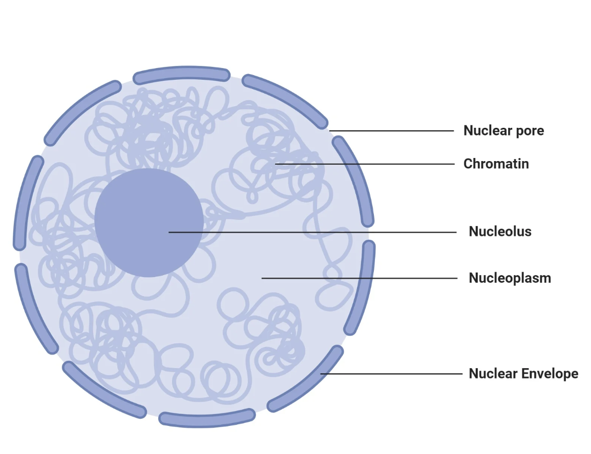
chromatin
DNA within the nucleus; the less condensed form that organizes to form chromosomes during prophase of mitosis or meiosis.
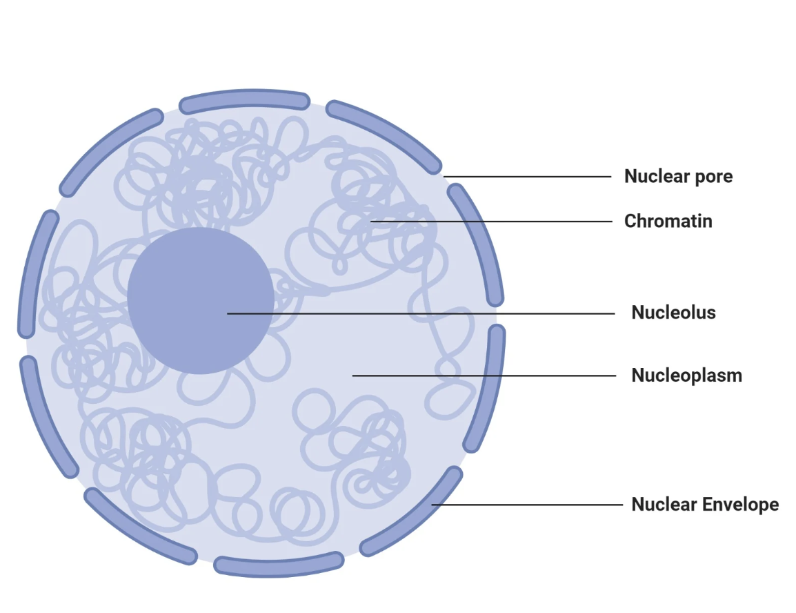
nucleoli
organelles that synthesize ribosomes.
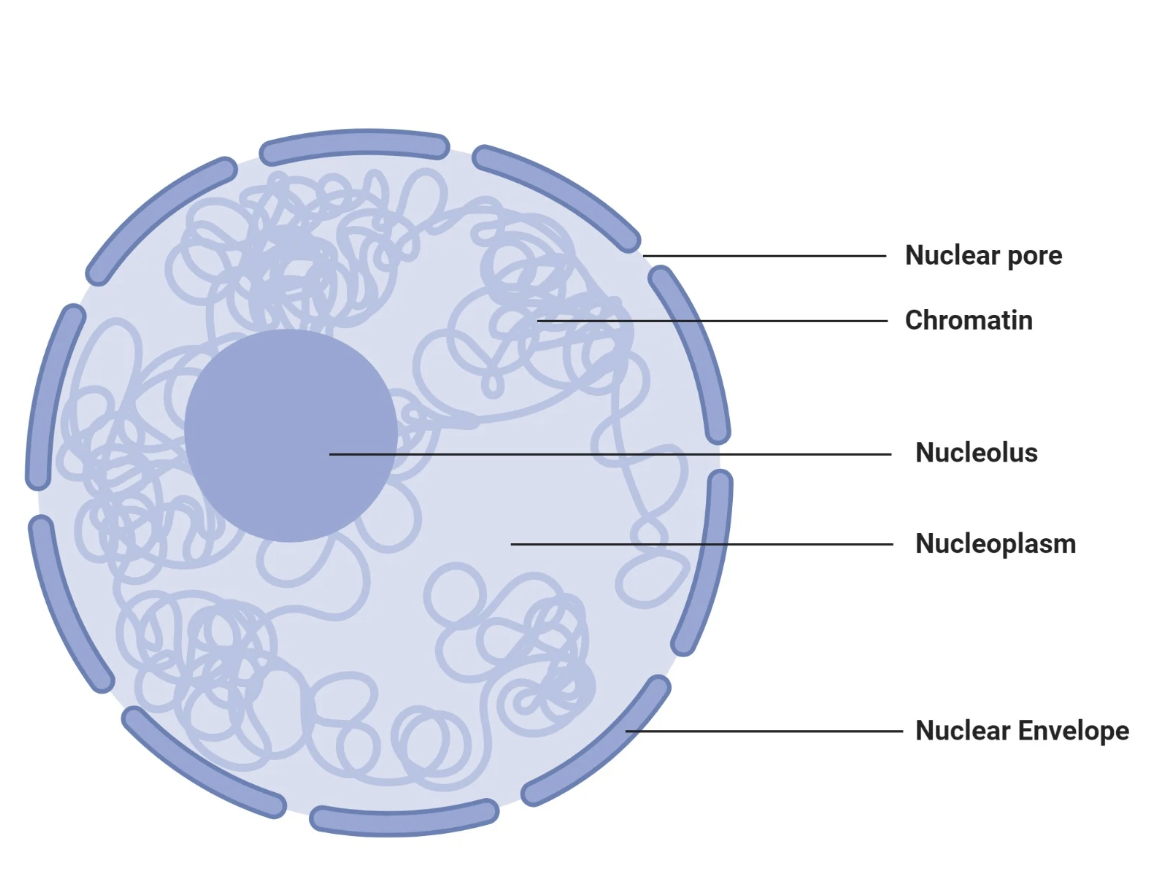
nuclear pores
Act as gateways allowing selective transport of molecules between the nucleus and the cytoplasm.
Ensure proteins and RNA move in and out efficiently to maintain cell function, ex:
histones
helicase
RNA polymerases
mRNA
Why are nuclear pores allowing molcules into the cell important for protein synthesis?
Proteins necessary for genome structure and function are synthesized in the cytoplasm and imported into the nucleus through nuclear pores. Examples include:
Histones: Organize DNA into chromatin.
Helicases and DNA polymerases: Unwind and replicate DNA.
RNA polymerases: Transcribe DNA into RNA.
Transcription factors: Regulate gene expression.
Splicing factors: Process RNA transcripts.
Why are nuclear pores allowing molecules out of the cell important for protein synthesis?
Molecules formed in the nucleus need to leave for the cytoplasm to function in translation:
mRNA and tRNA: Products of transcription, exported for translation in the cytoplasm to produce proteins.
Ribosomal subunits: Assembled in the nucleolus, exported to the cytoplasm or endoplasmic reticulum (ER) to facilitate protein synthesis.
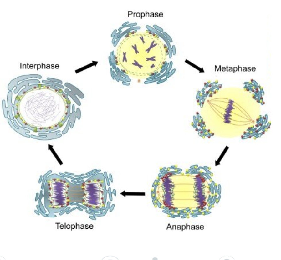
During ________, the nuclear membrane and endoplasmic reticulum are fragmented into vesicles. The vesicles are moves to the edge of the cell.
prophase
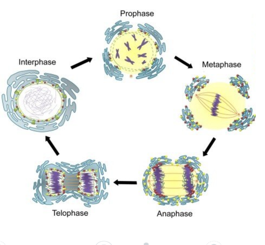
During ______, the vesicles are moved around the new sets of daughter chromosomes and the nuclear membrane and endoplasmic reticulum are reformed.
telophase
Cells can increase the SA:V ratio by:
Forming long, thin extensions
Having a thin, flat shape
Forming invaginations or microvilli
Why do long, thin extensions increase the surface area to volume ratio (SA:V)?
Because they significantly expand the cell's surface area while adding only a small amount to its overall volume.
What are examples of a long and thin extension to increase SA:V
Phagocytic cell of mammal immune system
Thin branches of a cell of a chordate nervous system
Epidermis cell forming a root hair in a plant
What are examples of a flat and thin adaptation to increase SA:V by maximizing the amount of surface area exposed relative to the volume it occupies?
Red blood cells (erythrocytes) exchange and transport O2
Thinness of Type 1 pneumocyte cells in the lung alveoli reduces distances for diffusion of gasses
Thin, flat epithelial cells form the tube of a capillary vessel for maximizing gas and nutrient exchange with tissue cell
What is an invagination?
Cavity formed when a surface folds inward to create a cavity.
What is a microvilli?
small finger-like projections that are tiny invaginations of the cell membrane of a single cell
Examples of microvili using increased surface area with increasing volume
Microvilli on the surface of cells in the small intestine increase the surface area for absorbing nutrients from digested food
Microvilli on the surface of cells in the kidney proximal convoluted tubule increase the surface area for reabsorbed useful substances before the are excreted in urine
What is the alveolar epithelium?
thin layer of specialized cells lining the alveoli in the lungs:
type 1 pneumocytes
type 2 pneumocytes
facilitates gas exchange between the air and the blood.
What are type 1 pneumocytes, and how are they adapted for gas exchange?
Large, flattened cells that makeup 95% of the alveolar surface.
Adaptations:
Thin (0.15 µm), reducing diffusion distance for gases.
Large surface area for efficient gas exchange.
Joined by tight junctions to prevent fluid leakage into alveoli.
What are type 2 pneumocytes, and how do they support lung function?
cuboidal cells scattered among type 1 cells.
Functions:
Secrete surfactant to reduce surface tension and prevent alveolar collapse.
Aid in repair and replacement of type 1 cells when damaged.
Adaptations:
Contain secretory vesicles for surfactant production.
Metabolically active with many mitochondria.
What are capillary cells, and how are they adapted for gas exchange?
Line the blood vessels surrounding the alveoli.
Adaptations:
Extremely thin, minimizing diffusion distance for O₂ and CO₂.
Closely associated with the alveolar epithelium to form a shared basement membrane.
What are erythrocytes, and how are they adapted for oxygen transport?
Erythrocytes (red blood cells) transport oxygen from the lungs to tissues and CO₂ back to the lungs.
Adaptations:
Biconcave shape increases surface area for gas exchange.
Lack of a nucleus and organelles maximizes space for hemoglobin.
Flexible membrane allows them to squeeze through narrow capillaries.
What are the overall adaptations of the alveoli for efficient gas exchange?
Large surface area provided by numerous alveoli.
Thin barrier: Alveolar epithelium, shared basement membrane, and capillary endothelium minimize diffusion distance.
Rich capillary network for maintaining steep concentration gradients.
Surfactant from type 2 pneumocytes reduces surface tension and prevents alveolar collapse.
What are the structures that make up the lungs in descending order?
Trachea —> bronchi —> bronchioles —>alveoli
What is passive transport?
Movement of substances across the membrane without energy input, relying on concentration gradients.
What are the 3 types of passive transport?
Simple diffusion
Facilitated diffusion
Osmosis
What is simple diffusion (passive transport)?
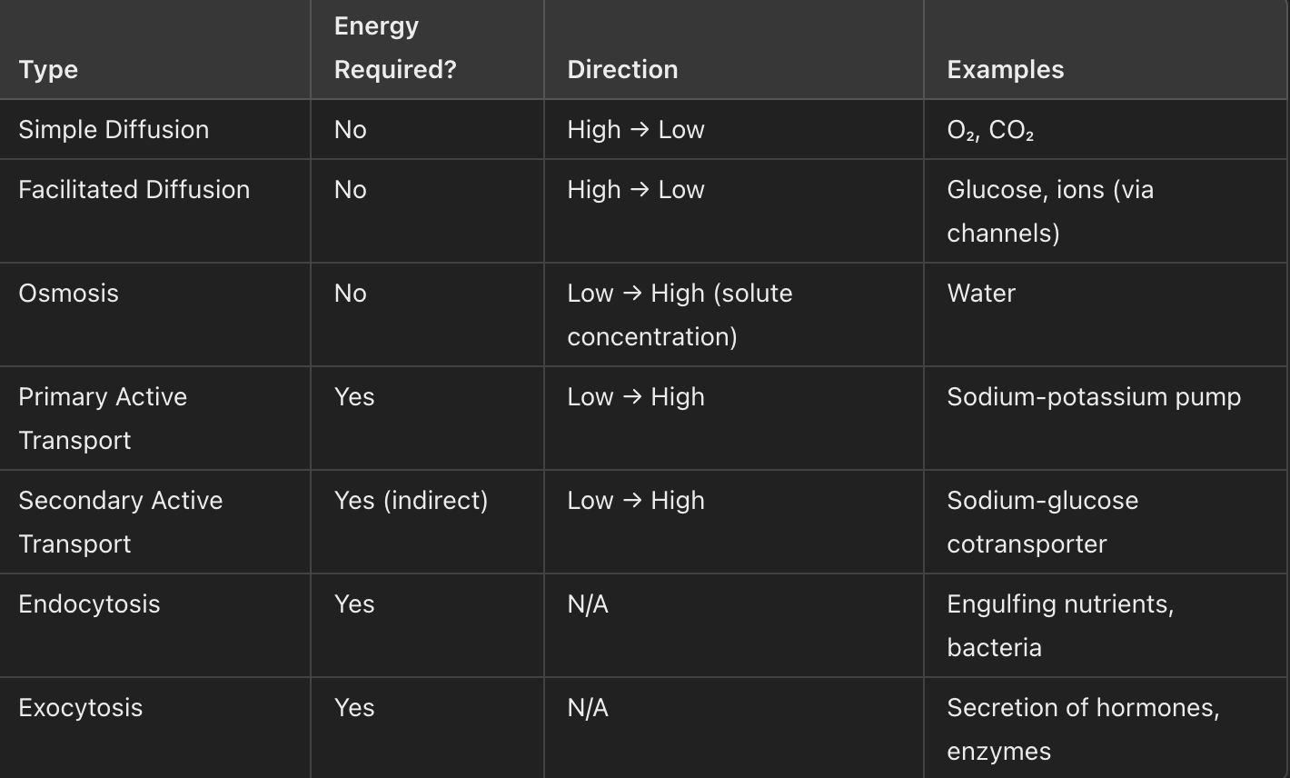
Molecules move directly across the lipid bilayer from a region of high concentration to low concentration.
Example: Oxygen (O₂) and carbon dioxide (CO₂).
No membrane proteins required.
What is facilitated diffusion (passive transportation)?
Movement of molecules through channel proteins or carrier proteins in the membrane, from high to low concentration.
Example: Glucose transport via carrier proteins, ion movement via channel proteins.
What is osmosis?
Diffusion of water molecules across a semi-permeable membrane from an area of low solute concentration (high water concentration) to high solute concentration.
Example: Water moving into a cell in a hypotonic solution.
Compare and contrast the 3 types of diffusion (passive, active, and bulk)
What is active transport?
Movement of substances against their concentration gradient (low to high concentration).
Requires energy (ATP).
Includes:
Primary active transport
Secondary active transport
What is primary active transport?
Uses energy directly from ATP to pump molecules.
Example: Sodium-potassium pump (3 Na⁺ out, 2 K⁺ in).
What is secondary active transport (cotransport)?
Uses existing concentration gradient set up through primary active transport to move molecules
Types:
Symport: Substances move in the same direction (e.g., sodium-glucose cotransporter).
Antiport: Substances move in opposite directions (e.g., sodium-calcium exchanger).
What is bulk transport?
Movement of large molecules or particles via vesicles, requiring ATP.
Includes:
Endocytosis (into the cell).
Exocytosis (out of the cell).
What is endocytosis?
A form of bulk transport where substances are brought into the cell via vesicles.
Example:
Phagocytosis: Engulfing large particles.
Pinocytosis: Engulfing fluids.
What is exocytosis?
A form of bulk transport where substances are released out of the cell by vesicle fusion with the membrane.
Example: Secretion of neurotransmitters or hormones.
The layer of leaf cells from top to bottom is:
cuticle —> upper epidermis —> palisade mesophyll —> spongy mesophyll —> lower epidermis —> cuticle
What is the function of the cuticle?
Waxy layer covering the surface of the leaf.
Reduces water loss by evaporation.
Provides a barrier against pathogens
Like the cuticle of your nail protects the fleshy part from being affected by water or germs
What is the role of the upper epidermis?
Transparent layer that allows light to pass through to the photosynthetic tissues.
Secretes the cuticle to reduce water loss.
U for Upper and Unobstructed—lets light in!
What is the function of the palisade mesophyll?
Primary site of photosynthesis.
Contains tightly packed cells rich in chloroplasts to maximize light absorption
P for Photosynthesis Powerhouse: Visualize the palisade as tall soldiers standing upright to capture sunlight, like soldiers outside a palace.
What is the role of the spongy mesophyll?
Loosely packed cells with air spaces for gas exchange (CO₂ in, O₂ out).
S for space for gas exchange; lungs are spongy and so are the “lungs” of the plant
What is the function of the lower epidermis?
Contains stomata and guard cells for gas exchange.
Helps regulate water loss through transpiration.
L for Letting gases in and out
What are stomata, and what is their function?
Small pores found mostly on the lower epidermis.
Allow gas exchange (CO₂ enters, O₂ and water vapor exit).
staying open for gas exchange
What are guard cells, and what is their function?
Specialized cells that surround stomata.
Control the opening and closing of stomata to regulate gas exchange and water loss.
"Guards control the opening of the fortress (the stomata)!”
What are vascular bundles, and what do they contain?
Xylem: Transports water and minerals from roots to leaves.
Phloem: Transports sugars (products of photosynthesis) to other parts of the plant.
XY to the sky" (xylem transports water upward) and "PHlow down" (phloem transports food downward).
How does the structure of the leaf maximize photosynthesis?
Large surface area: Maximizes light capture.
Thin structure: Short diffusion distance for gases.
Chloroplasts in palisade mesophyll: Capture light efficiently.
Air spaces in spongy mesophyll: Facilitate gas exchange.
Transparent upper epidermis: Allows light to pass through.
What is the function of ribosomes, and where are they synthesized?
Ribosomes are synthesized in the nucleolus.
Their primary function is to synthesize proteins based on mRNA instructions.
Explain the role of the rough and smooth endoplasmic reticulum in protein synthesis and lipid metabolism.
Rough ER: Involved in protein synthesis (via ribosomes attached to it) and the initial stages of protein folding.
Smooth ER: Involved in lipid and carbohydrate metabolism, and detoxification, and it synthesizes steroid hormones.
It does not send proteins directly out of the nucleus—this is the function of the Golgi apparatus.
What are some adaptations of the leaf structure that help maximize photosynthesis?
The spongy mesophyll has air spaces for gas exchange
Stomata allow gas exchange (CO₂ in and O₂ out)
Chloroplasts are more abundant on the upper side to capture light efficiently.
How does the palisade mesophyll contribute to photosynthesis?
The palisade mesophyll packed with chloroplasts and densely arranged to maximize light absorption for photosynthesis.
What is the function of the stomata, and how do guard cells regulate their opening and closing?
Stomata allow gas exchange (CO₂ in, O₂ out) and transpiration (water vapor out).
Guard cells regulate the stomata by changing shape, which controls their opening and closing based on environmental factors (light, CO₂ concentration, water availability).
What are the steps involved in the process of transcription?
RNA polymerase binds to the promoter region on DNA.
It separates the DNA strands.
It synthesizes a complementary mRNA strand from the 3' to 5' DNA strand.
The mRNA is processed and leaves the nucleus to be translated.