Shoulde&Elbow muscles + muscle types

0.0(0)
Studied by 0 people
call kaiCall Kai
Locked
learnLearn
examPractice Test
spaced repetitionSpaced Repetition
heart puzzleMatch
flashcardsFlashcards
GameKnowt Play
Card Sorting

1/20

encourage image

There's no tags or description

Looks like no tags are added yet.

Last updated 3:03 PM on 9/20/26
Name
Mastery
Learn
Test
Matching
Spaced
Call with Kai
Chat

No analytics yet

Send a link to your students to track their progress

21 Terms

1
New cards

humeral fracture locations and nerve damage

  1. Humeral (surgical neck): Axillary n., posterior humerus circumflex a.(*both travel through Quadrangular space)

  2. Spiral groove/radial groove: Radial n., deep brachial a (both in triangular interval)

  3. Supracondylar region: Median n. (*anterior elbow joint; lat hand/digits 1-3)

*if fracture distal ends displaces anteriorly

  1. Medial epicondyle: Ulnar n. (*”funny bone”; compress against med epi; med hand/digits 4-5)


<ol><li><p><strong>Humeral (surgical neck)</strong>: <mark data-color="blue" style="background-color: blue; color: inherit;">Axillary n., posterior humerus circumflex a</mark>.(*both travel through Quadrangular space)</p></li><li><p><strong>Spiral groove/radial groove:</strong> <mark data-color="blue" style="background-color: blue; color: inherit;">Radial n., deep brachial a</mark> (both in triangular interval)</p></li><li><p><strong>Supracondylar region:</strong> <mark data-color="blue" style="background-color: blue; color: inherit;">Median n.</mark> (*anterior elbow joint; lat hand/digits 1-3)</p></li></ol><p><mark data-color="red" style="background-color: red; color: inherit;">*if fracture distal ends displaces anteriorly</mark></p><ol><li><p><strong>Medial epicondyle: </strong><mark data-color="red" style="background-color: red; color: inherit;">Ulnar n</mark>. (*”funny bone”; compress against med epi; med hand/digits 4-5)</p></li></ol><p></p>
2
New cards

list the muscle types and skeletal muscle types

3 Types of Muscles

  1. Cardiac muscle

  2. Smooth muscle

  3. Skeletal muscle

7 Types of Skeletal Muscles

  1. Flat

  2. Fusiform

  3. Pennate (Uni, bi, and multipennate)

  4. Convergent

  5. Quadrate

  6. Circular (sphincteral)

  7. Multiheaded/multibellied


3
New cards

3 types of muscle

Cardiac

-the myocardium (*heart muscle)

-autonomic control (*beats on own)

Smooth

-in the GI tract

-autonomic control (*digest on own)

Skeletal

-part of the locomoter system

-somatic/voluntary control
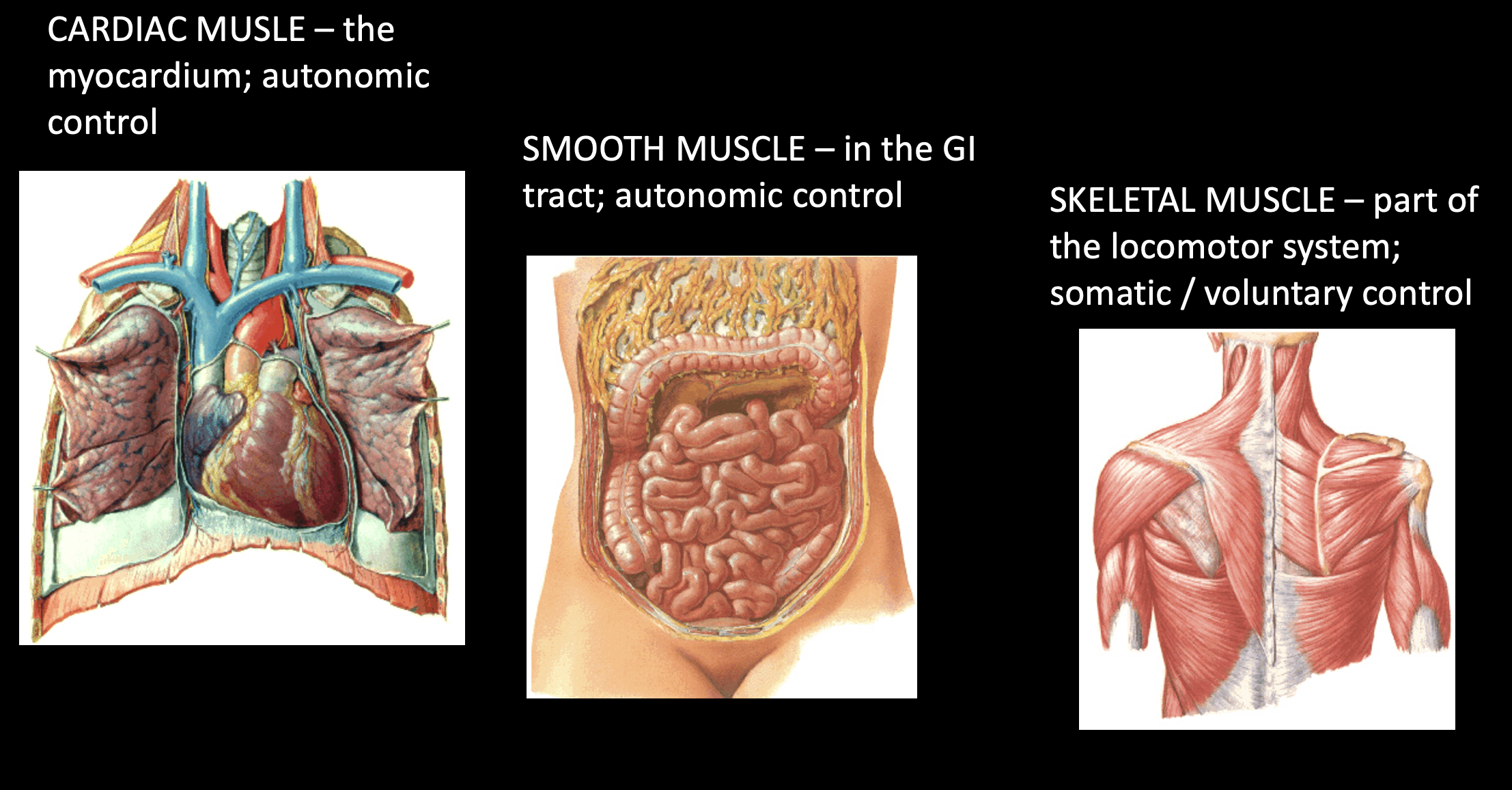
<p><u>Cardiac</u></p><p>-the myocardium (*heart muscle)</p><p>-autonomic control (*beats on own)</p><p><u>Smooth</u></p><p>-in the GI tract</p><p>-autonomic control (*digest on own)</p><p><u>Skeletal</u></p><p>-part of the locomoter system</p><p>-somatic/voluntary control</p>
4
New cards

flat muscle

-parallel fibers, often with a falt tendinous sheath (aponeurosis)

-ex=abdominal obliques; rhomboids
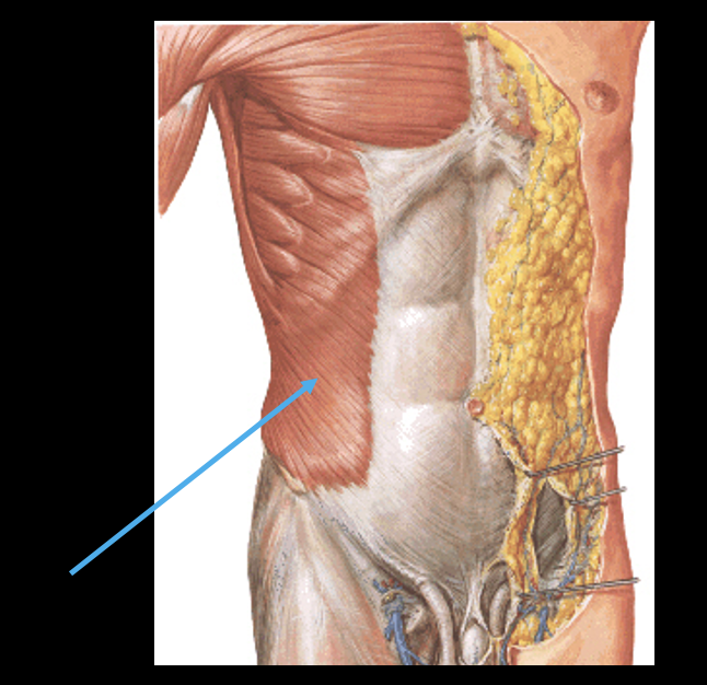
<p>-parallel fibers, often with a falt tendinous sheath (aponeurosis)</p><p>-ex=abdominal obliques; rhomboids</p>
5
New cards

Fuisiform muscle

-thick belly, tapers both ends

-these are very common muscle types

-ex= Biceps brachii

<p>-thick belly, tapers both ends</p><p>-these are very common muscle types</p><p>-ex= Biceps brachii</p>
6
New cards

Pennate muscles

-more muscle fibers to surface area increases force of contraction (*mechanical advantage)

-unipennate: 1 central tendon and fibers off 1 side; ex=extensor digitorum

-bipennate: fibers off both sides 45 degrees; ex=dorsal interossei

-multipennate: multiple plumes in body; ex=deltoid
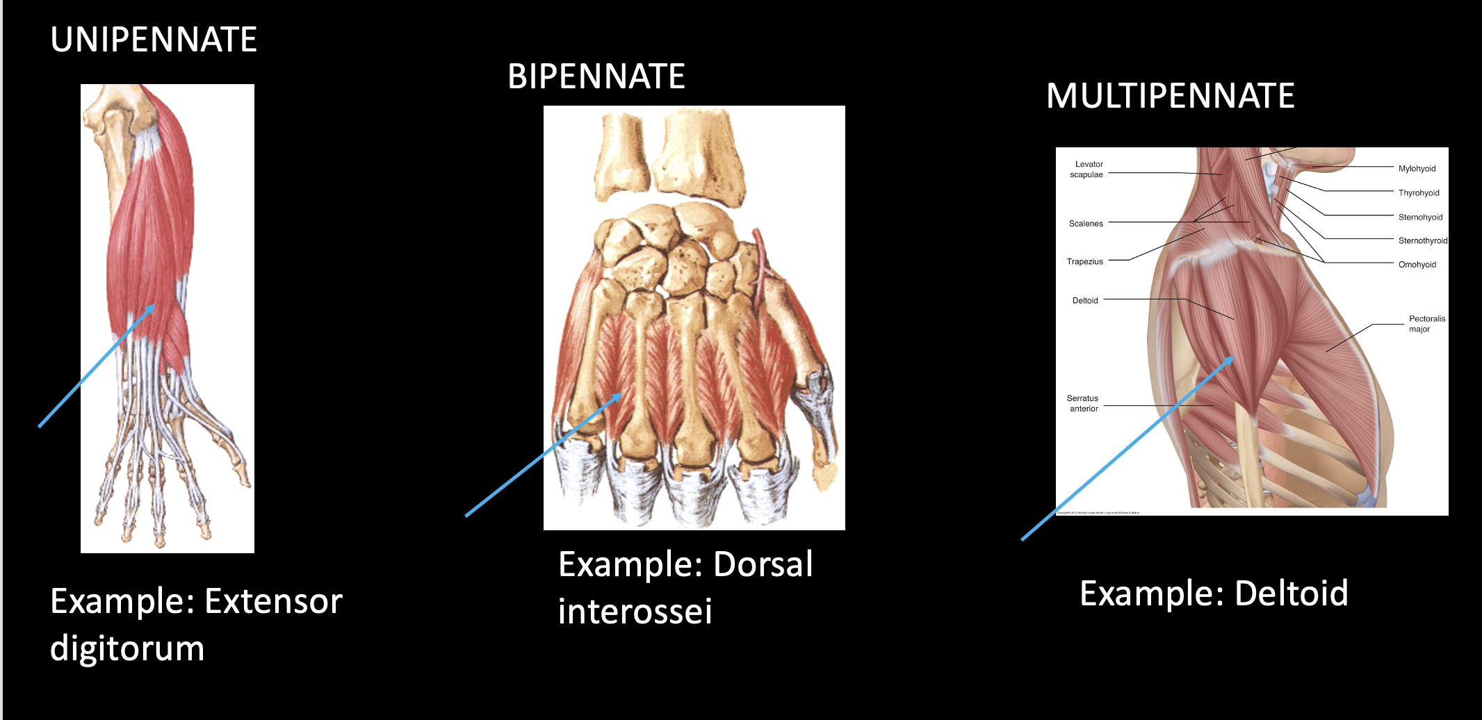
<p>-more muscle fibers to surface area increases force of contraction (*mechanical advantage)</p><p><strong>-unipennate:</strong> 1 central tendon and fibers off 1 side; <mark data-color="red" style="background-color: red; color: inherit;">ex=extensor digitorum</mark></p><p><strong>-bipennate:</strong> fibers off both sides 45 degrees; <mark data-color="red" style="background-color: red; color: inherit;">ex=dorsal interossei</mark></p><p><strong>-multipennate:</strong> multiple plumes in body; ex=deltoid</p>
7
New cards

Convergent muscle

-originate from a broad area, converge to a single tendon

-ex=latts (force exerted from multiple directions bc extend w/ posterior origin, IR w/ anterior origin, etc.)

<p>-originate from a broad area, converge to a single tendon</p><p>-ex=latts (force exerted from multiple directions bc extend w/ posterior origin, IR w/ anterior origin, etc.)</p>
8
New cards

Quadrate muscle

-four equal sides

-ex=pronator quadratus
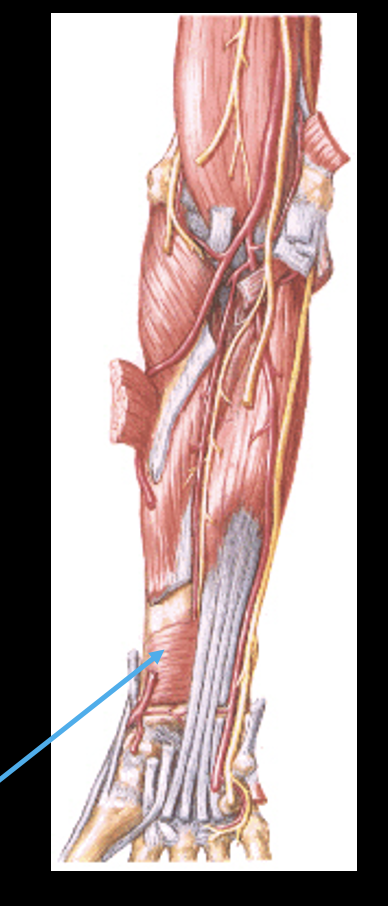
<p>-four equal sides</p><p>-ex=pronator quadratus</p>
9
New cards

circular muscle

-constrict a body opening

-ex=orbicularis oris; orbiculars oculi

<p>-constrict a body opening</p><p>-ex=orbicularis oris; orbiculars oculi</p>
10
New cards

multiheaded muscle

-multiple points of origin, usually one insertion

-ex=triceps brachii

*note: muscles can fall into multiple categories, but if pennate only 1 type of pennate

<p>-multiple points of <em>origin</em>, usually one insertion</p><p>-ex=triceps brachii</p><p>*note: muscles can fall into multiple categories, but if pennate only 1 type of pennate</p>
11
New cards

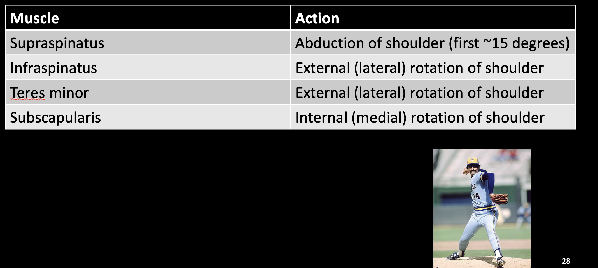
shoulder muscle table

knowt flashcard image
12
New cards

list the rotator cuff muscles

-supraspinatus

-infraspinatus

-teres minor

-subscapularis

<p>-supraspinatus</p><p>-infraspinatus</p><p>-teres minor</p><p>-subscapularis</p>
13
New cards

rotator cuff functions

knowt flashcard image
14
New cards

rotator cuff injury

-supraspinatus tendon most commonly torn rotator cuff tendon

<p>-supraspinatus tendon most commonly torn rotator cuff tendon</p>
15
New cards

suprascapular artery and nerve

-Artery is superior to transverse scapular ligament
-Nerve is inferior

 *“Army over Navy!”

<p><span style="font-family: &quot;Calibri Light&quot;;"><strong>-Artery is superior to transverse scapular ligament<br>-Nerve is inferior</strong><br>&nbsp;*“Army over Navy!”</span></p>
16
New cards

muscle table: elbow flexion and extension
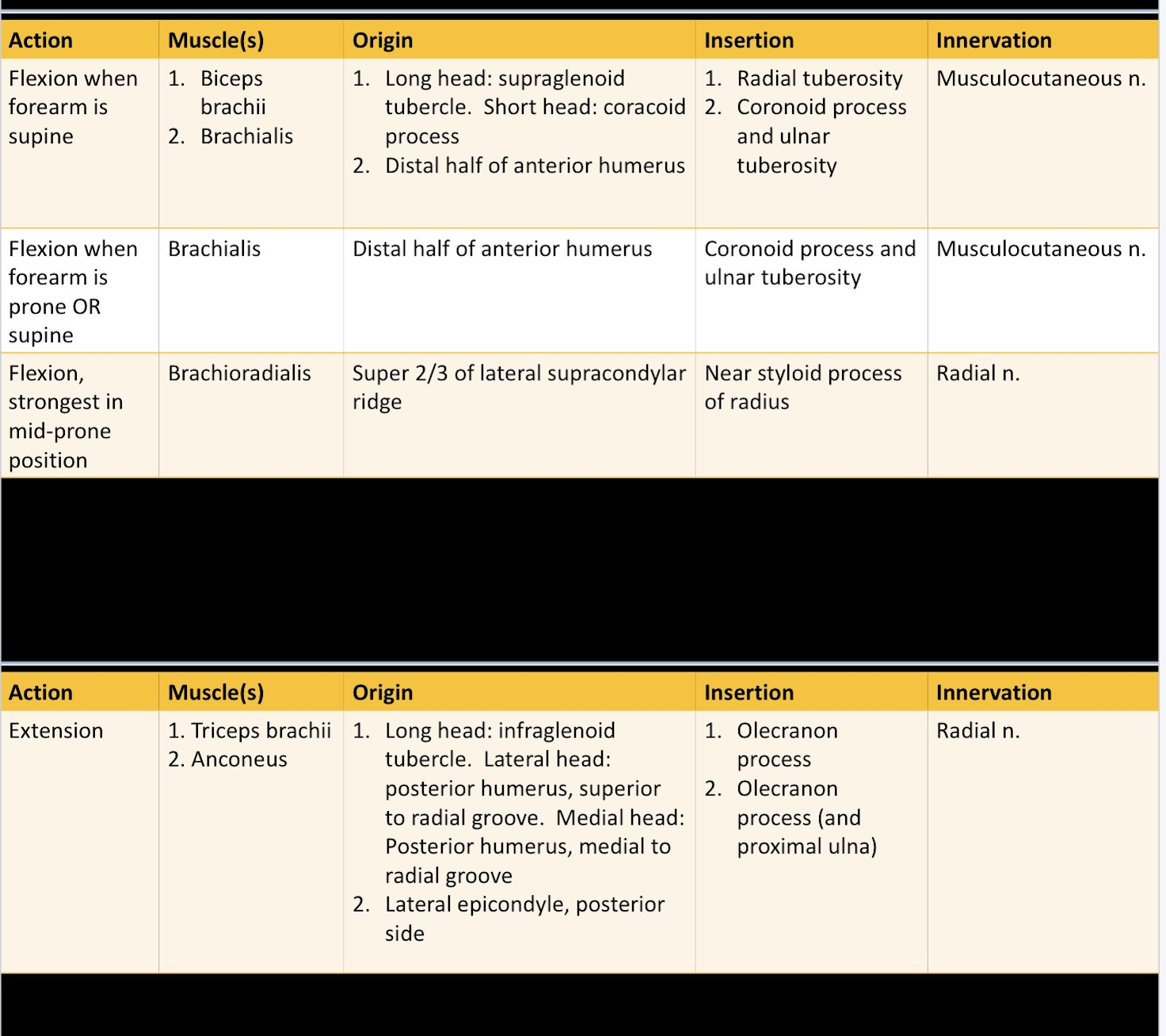
knowt flashcard image
17
New cards

biceps + triceps origin and the shoulder joint

knowt flashcard image
18
New cards

proximal radio-ulnar joint

-Head of radius sits in the radial notch of the ulna

-The annular ligament holds the radial head in place

<p><mark data-color="blue" style="background-color: blue; color: inherit;">-Head of radius sits in the radial notch of the ulna</mark></p><p>-The <strong>annular ligament</strong> holds the radial head in place</p>
19
New cards

distal radio-ulnar joint

-head of the ulna sits in the ulnar notch of the radius

<p>-head of the ulna sits in the ulnar notch of the radius</p>
20
New cards

-what type of synovial joint are the radio-ulnar joints?

-what actions are possible at the radio-ulnar joints

-what forearm bone stays fixed during pronation and supination?

-pivot joints

-pronation and supination

-ulna

21
New cards

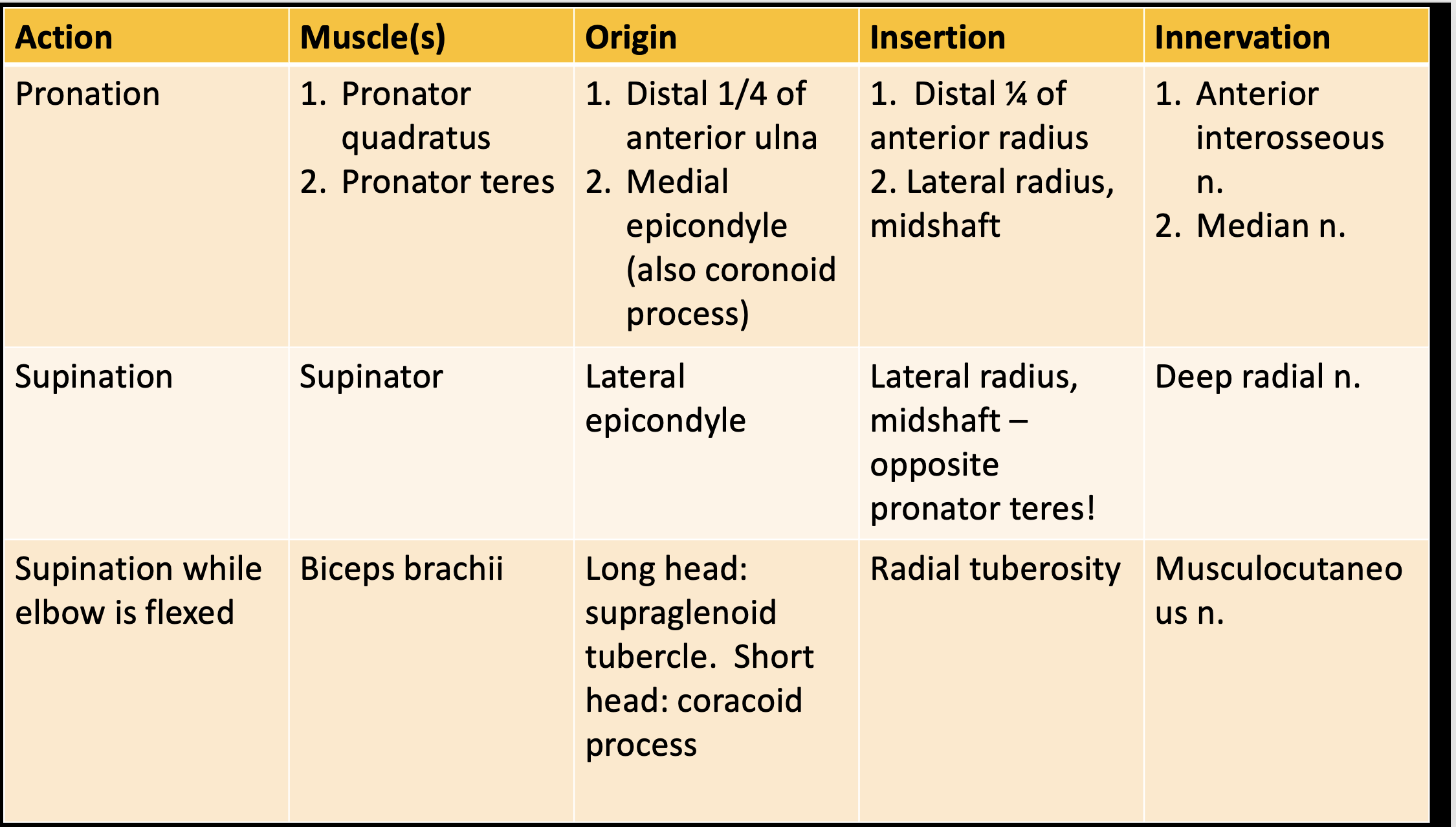
pronation and supination muscle table

knowt flashcard image