Psych 230 Sheep Brain Dissection Quiz
 Call Kai
 Learn
Learn Practice Test
 Spaced Repetition
 Match
Match Flashcards
 Knowt Play
1/116

There's no tags or description
Looks like no tags are added yet.
Name | Mastery | Learn | Test | Matching | Spaced | Call with Kai | Chat |
|---|
No analytics yet
Send a link to your students to track their progress
117 Terms
Central Nervous System (CNS)
brain and spinal cord
Peripheral Nervous System (PNS)
all the nerves that extend past brain and spinal cord (CNS)
Superior/Dorsal View

Anterior/Rostral View
side of brain your face is on
Posterior/Caudal View

Inferior/Ventral View
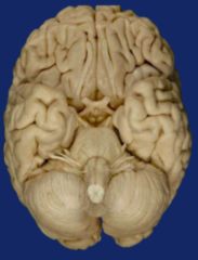
Lateral
Toward side of brain
Medial
Toward center of brain
Coronal Cut of Brain
moving from front to back
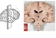
What direction does the coronal cut divide the brain into?
rostral to caudal
Sagittal Cut of Brain
moving from left to right
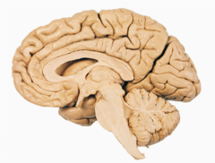
Horizontal Cut of Brain
moving from top to bottom
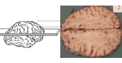
What direction does the horizontal cut divide the brain into?
dorsal to ventral
What protects the brain?
meningies, including:
dura mater (outermost, toughest), arachnoid (space within this layer is filled with CSF, and pia mater (adheres tightly to surface of brain)
Frontal Lobe Location

Parietal Lobe Location

Temporal Lobe Location

Occipital Lobe Location

Cerebellum Location
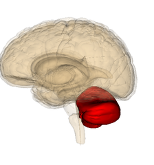
Frontal Lobe Responsibilities
higher cognitive processing, executive function
Parietal Lobe Responsibilities
touch, proprioception, speech
Temporal Lobe Responsibilities
speech, memory, hearing
Occipital Lobe Responsibilities
vision
Cerebellum Responsibilities
motor learning and balance
Gyrus/Gyri
ridges or raised portions of the convoluted cortex (hills)
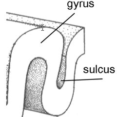
Sulcus/Solci
furrows of the convoluted cortex (valleys)
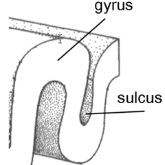
Fissure
large groove that divides the brain into lobes
Longitudinal Fissure
separates the left and right hemispheres
Central Sulcus
separates the frontal and parietal lobes
Lateral Fissure
separates the frontal lobe from the temporal lobe, as well as the temporal lobe from the parietal lobe
Bundles of Axons in the PNS
nerves
Bundles of Axons in the CNS
tracts
Olfactory Bulbs Location

Optic Nerve Location

Optic Chiasm Location
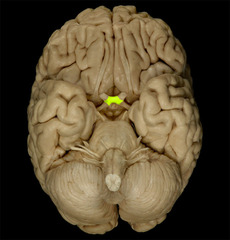
Hypothalamus Location
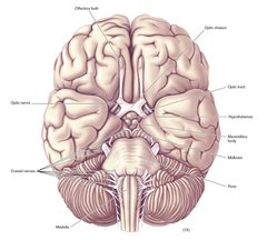
Pons Location
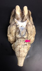
Medulla Location

Spinal Cord Location
No picture for this one but it is located caudal to the pons and medulla
Infundibulum Location
rostral to hypothalamus, caudal to optic tract

Optic Tract Location
caudal to optic chiasm, rostral to infundibulum
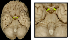
Mammillary Bodies Location
caudal to hypothalamus, rostral to cerebral peduncle

Cerebral Peduncle Location
caudal to mammillary bodies
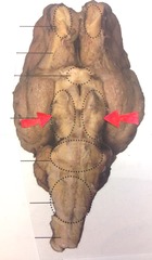
Superior Colliculus Location (Dorsal View)
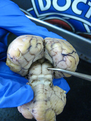
Inferior Colliculus Location (Dorsal View)
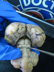
Pineal Gland Location (Dorsal View)

4th Ventricle Location (Dorsal View)

Ventricles
hollow spaces in the brain that produce and circulate cerebral spinal fluid (CSF)
Lateral Ventricle Location

Third Ventricle Location (Sagittal View)

Cerebral Aqueduct of Sylvius Location
no good pic for this one but rostral to the 4th ventricle and caudal to the 3rd ventircle
4th Ventricle Location (Sagittal View)
no good pic but ventral to the cerebellum and rostral to the central canal
Central Canal Location
in spinal cord

Third Ventricle Location (Ventral View)
no good picture but very close to hypothalamus and infundibulum
What was the first cut we made on the brain?
A midsagittal cut along the longitudinal fissure
Corpus Callosum Location

Pineal Body/Gland Location

Superior Colliculus Location (Sagittal View)
ventral to pineal body, dorsal to interior colliculus
Inferior Colliculus Location (Sagittal View)

Arbor Vitae Location

Hypothalamus Location (Sagittal View)
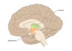
Thalamus Location (Sagittal View)
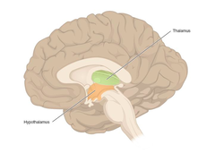
Optic Chiasm Location (Sagittal View)

Fornix Location (Sagittal View)

Olfactory Bulb Location (Sagittal View)
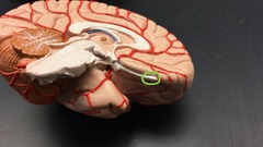
Cingulate Gyrus Location (Sagittal View)

Septum Pellucidum Location (Sagittal View)

Mammillary Body Location (Sagittal View)

What would happen if the frontal lobe was damaged?
Issues with executive function such as personality and behavioral changes
What would happen if the parietal lobe was damaged?
Issues with perception, speech, and recognition.
What would happen if the temporal lobe was damaged?
Issues with speech and hearing, as well as memory.
What would happen if the occipital lobe was damaged?
Issues with vision (this is why vision can go black when a concussion is sustained)
Cingulate Gyrus Function
emotion and attention
Corpus Callosum Function
connects the left and right hemispheres
Septum Pellucidium Function
seperates left and right lateral ventricle
Lateral Ventricle Function
contains choroid plexus that produces CSF
Fornix Function
major axon projections from the hippocampus
Thalamus Function
relay station for sensory input except olfaction
Optic Chiasm Function
point at which visual info crosses over to the contralateral side before entering the occipital lobe
Hypothalamus Function
maintenance of homeostasis, 4 Fs: flight, fight, food, family
Mammillary Body Function
Memory
Pineal Gland Function
produces hormones, specifically melatonin
Cerebral Aqueduct function
connects third and fourth ventricles
Pons Function
has nuclei important for sleeping, swallowing, bladder control and sleep
Arbor Vitae Function
Lots of white matter to bring sensory and motor information to and from the cerebellum
4th ventricle Function
cushion the brain from trauma
Medulla Function
responsible for vital functions (usually autonomic) in the body (ex: sneezing, coughing, vomiting), monitor fluids
Brain Stem Function
connects your brain and spinal cord, regulates vital functions like breathing and balance
What makes CSF?
the choroid plexus
What was the second type of cut we performed on the sheep brains?
6 coronal cuts
Corpus Callosum Location (coronal view)
Major white matter tract

Lateral Ventricle Location (Coronal Location)

Caudate Nucleus (Coronal View)

Putamen Location (Coronal View)

Thalamus Location (Coronal View)
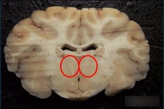
Third Ventricle Location (Coronal View)

Fornix Location (Coronal View)

What provides input to the thalamus?
sensory stimuli
Amygdala Location (Coronal View)

Body of Fornix Location (Coronal View)
