Chamber Quantification
1/20
There's no tags or description
Looks like no tags are added yet.
Name | Mastery | Learn | Test | Matching | Spaced |
|---|
No study sessions yet.
21 Terms
The RA is at its largest volume in which phase of the cardiac cycle?
end-systole
The LA filling through the pulmonary veins takes place during which phase of the cardiac cycle?
Ventricular systole
Left atrial linear dimension in 2-D PLAX is measured according to the following standards:
At end-systole, leading-edge to leading-edge
LA volume index is the result of the ratio between:
LA volume/BSA (body surface area)
One of the major physiologic functions of the LA is pumping blood from the LA to the LV during diastole. What percentage of the entire LV filling is delivered during atrial contraction?
20-30%
The MV flow Doppler profile represents the pattern of the blood flow from the LA to the LV during diastole. Which one of the following measurements assesses LA contractility?
A-wave max velocity or VTI
The A-wave time (duration) represents which one of the following parameters on the MV inflow Doppler profile?
Length of atrial systole
Relevant structures to the LA function are all of the following, EXCEPT:
IVS
Which one of the following views is recommended to accurately measure the RV free wall thickness?
SC 4-Ch
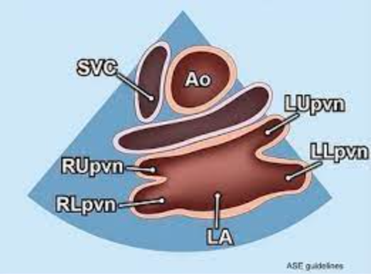
The image below visualizing the LA and all four pulmonary veins represents which one of the following echocardiographic views?
Suprasternal SAX, “crab view”
Right atrial linear dimensions and area are measured in AP 4-Ch view in which of the following phases of the cardiac cycle?
End systole
Right atrial area is used to calculate which one of the following chamber dimensions and indexes?
RA volume and RAVI
The normal pressure in the RA is the lowest in all four cardiac chambers.
True or False
True
The RA pressure is estimated by evaluating the status of which vessel in which view?
IVC in SC LAX@IVC
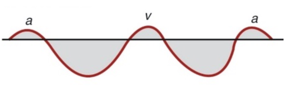
The following image represents the Doppler flow pattern for
Pulmonary vein flow
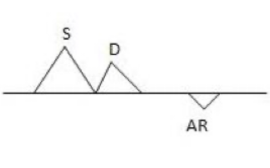
The following image represents the Doppler flow pattern for
Hepatic vein flow and IVC
Eustachian valve
Venous valve at the junction of IVC and RA, attached to the wall or floating, may be mistaken for a thrombus
Chiari network
Mobile, membrane-like structure serving as a valve for coronary sinus and located near the orifice of the IVC
Crista terminalis
C-shaped ridge located in the endocardial aspect of the RA with the SA node at its superior segment
Fossa ovalis
Embryologic remnant of the foramen ovale that closes after birth
The right ventricle can be visualized in all of the following echocardiographic views:
PLAX
RVIT
RVOT
PSAX@Ao
PSAX@MV
PSAX@PM
Ap 4-Ch
Ap 5-Ch
Ap 3-Ch
SC 4-Ch