(A) Canine and Feline Skeletal Disorders
 Call Kai
 Learn
Learn Practice Test
 Spaced Repetition
 Match
Match Flashcards
 Knowt Play
1/153

There's no tags or description
Looks like no tags are added yet.
Name | Mastery | Learn | Test | Matching | Spaced | Call with Kai | Chat |
|---|
No analytics yet
Send a link to your students to track their progress
154 Terms

what is it (Osteoarthritis)
synovial fluid
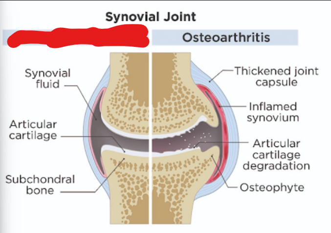
what is it (Osteoarthritis)
normal

what is it (Osteoarthritis)
articular cartilage

what is it (Osteoarthritis)
subchondral bone
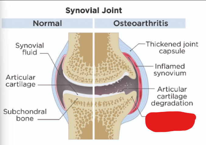
what is it (Osteoarthritis)
osteophyte

what is it (Osteoarthritis)
articular cartilage degradation
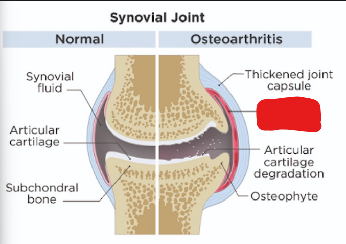
what is it (Osteoarthritis)
inflamed synovium

what is it (Osteoarthritis)
thickened joint capsule

what is it (Osteoarthritis)
ostearthritis
Osteoarthritis aka (Osteoarthritis)
degenerative joint disease
DJD stands for (Osteoarthritis)
degenerative joint disease
Osteoarthritis is a progressive — — of — (Osteoarthritis)
inflammatory disease of joints
how does Osteoarthritis occur (5) (Osteoarthritis)
the process often starts in the joint (articular) cartilage that covers the smooth joint (articular) surfaces of the bones
Something upsets the normal remodeling process of the cartilage causing removal to outpace repair
This leads to progressive thinning of the protective cartilage layer and can lead to exposure of the underlying bone
result in bone-to-bone contact replacing the normal cartilage-to-cartilage contact.
The ensuing trauma to the bone surfaces causes the smooth articular surfaces to become roughened.
Osteoarthritis leads to (5) (Osteoarthritis)
buildup of fluid within the joint
osteophytes around the joint
bone spurs around the joint
narrowing of the joint space
pain.
what causes Osteoarthritis (7) (Osteoarthritis)
Aging
Trauma
Bone and joint malformation
Infection
Immune mediated
Idiopathic
immune system dysfunction
clinical signs of Osteoarthritis in dogs (9) (Osteoarthritis)
Pain
Lameness
Decreased activity
Stiffness when raising from rest
Behavioral changes
Joint distention
Muscle atrophy
thickening and scarring of joint membranes
Difficulty navigating stairs or jumping on couches
clinical signs of Osteoarthritis in cats (11) (Osteoarthritis)
hesitation to jump
difficulty with stairs
reduced activity
more time spent resting
more time sleeping
temperament changes
irritability
reluctance to be handled or petted
matted
scruffy coat
reduction in grooming
define atrophy (Osteoarthritis)
shrinkage
how to diagnose Osteoarthritis (4) (Osteoarthritis)
Radiographs
Arthroscopy
CT
MRI
what would be seen in a physical exam for Osteoarthritis (4ish) (Osteoarthritis)
Changes in gait
changes in posture
Abnormal palpable changes to joints such as swelling or crepitus
Pain
define crepitus (Osteoarthritis)
any grating, crackling, popping, or crunching sound and sensation that happens in a joint during movement
how to treat Osteoarthritis (Osteoarthritis) (7)
Weight reduction
Physical rehabilitation
Low impact activities
Pain management
Joint support
Use of non-slip rugs and ramps to get on beds, couches or cars
surgery
Canine Hip Dysplasia Is an abnormal — of the — joint
laxity, coxofemoral
what are the 2 forms of canine hip displasia
Acetabular Hip Dysplasia
Femoral Hip Dysplasia
define Acetabular canine Hip Dysplasia
Inability of the femoral head to join into the developing acetabulum causes damage to the dorsal rim
define Osteophytes
extra growths of bone tissue that form along the edges of bones and joint
what causes canine hip displasia (12)
Inherited
high calorie diet
excessive growth
difference between the muscle mass and bone development
Failure of surrounding soft tissue to maintain the joint congruity
Puppies of dysplastic parents
larger breeds are affected more
Genetic predisposition
Environment
Failure of the soft tissues of the hip to maintain joint congruity between the surfaces of the hip joint, resulting in bony changes within the joint
Young dogs between 5 and 8 months of age
mature animals with chronic disease
define polygenic
a trait or health condition that is controlled by the combined action of multiple genes rather than a single gene
clinical signs of canine hip dysplasia (general) (4)
Difficulty in rising
stiffness that diminishes as the animal warms up on exercise
Pain is elicited on palpation of the dorsal pelvic area or over the hip joint
may affect mobility significantly
the severe form of canine hip dysplasia affects what age range
5 - 8 months old
clinical signs of the Severe form of canine hip dysplasia (12)
sudden onset of lameness
pain
difficulty rising - will use front legs to push itself up
Decreased activity
Some are unable to stand or walk
Bunny hop gait in hind limbs
thigh and gluteal muscles atrophy
On palpation dogs are painful in the hip joint
Reduced range of motion
Crepitation
Pain during flexion and extension
Joint laxity
define joint laxity
your ligaments are loose, allowing your joints to have a wider or looser range of motion than normal
define Crepitation
An audible creaking, cracking sound
what age (super general) does the chronic form of canine hip dysplasia affect
older dogs
clinical signs of the chronic form of canine hip dysplasia (8)
Gradual onset because of development of osteoarthritis
Lameness unilateral or bilateral
Difficulty rising
Atrophy of gluteal
thigh muscles apparent
Gait changes to waddling
there is decreased range of motion (ROM) of hip
a waddling gait
pain
how to diagnose canine hip dysplasia (2)
Ortolani test
Radiographs
how to do Ortolani test (4)
Done in either dorsal or lateral recumbency
rotate femoral joint
Will feel the joint clicking in and out
how to conservatively treat canine hip dysplasia (4)
Moderate exercise
Weight control
Anti Inflammatory medications:
Nutriceuticals
what are the 3 most commonly performed surgeries for canine hip dysplasia
Femoral head osteotomy
Triple pelvic osteotomy
Total hip replacement
what does FHO stand for
Femoral head osteotomy
in femoral head osteotomy what does removing the femoral head do
Removal allows formation of a “false joint” from surrounding soft tissue
what aftercare is required after a Femoral head osteotomy
Vigorous exercise is required after surgery to increase muscle strength and limb function.
what does TPO stand for
Triple pelvic osteotomy
define osteotomy
a surgical procedure where a doctor cuts a bone to shorten, lengthen, or change its alignment
what is done in a Triple pelvic osteotomy (2ish)
Bone cuts into the pelvis so the acetabulum can be rotated over the femoral head and provide better coverage.
The bones are then stabilized with a bone plate.
A Triple pelvic osteotomy can not be performed if …
arthritis is apparent on x-rays
what happens in a total hip replacement
A cobalt chrome shaft and head are implanted into the femoral shaft and placed into an artificial acetabular cup.
how to prevent canine hip dysplasia (3)
Healthy weight
Supplements for joint health
Selective breeding
what is the Average age of onset for panosteitis (range)
6 - 8 months
Panosteitis causes degeneration of — bone
medullary
Panosteitis causes thickening of — bone
endosteal
what long bones are mostly affected by Panosteitis (5)
ulna
humerus
radius
femur
tibia
what causes Panosteitis (8)
Idiopathic
High protein diets
over nutrition
viruses
genetic predisposition
metabolic disease
allergies
hormonal excess
clinical signs of Panosteitis (6)
Intermittent lameness
Anorexia
Fever
Weight loss
Lethargy
Reluctance to move
define Intermittent lameness
may appear to the client to shift from leg to leg.
how to diagnose Panosteitis (2)
Pain response on palpation of long bone(s) affected
Radiology
how to treat Panosteitis (5)
Self-limiting
Rest
Most dogs return to normal state within 1 year
Analgesics for pain
anti-inflammatory drugs for pain
how to prevent Panosteitis (1ish)
Diets with lower calcium, fats, and proteins to avoid rapid growth
what does OCD stand for
Osteochondritis Dissecans
define Osteochondrosis
degeneration/aseptic necrosis of bone and cartilage, then reossification
define Osteochondrosis/Osteochondritis Dissecans
a dissecting flap and inflammation
define Endochondral ossification
is the process by which cartilage on the ends of long bones ossifies, or changes to bone.
what happens if there is no synovial fluid (4)
cells are not nourished,
cells degenerate
cells die
causing cracks in the cartilage
what happens If cartilage does not properly calcify
it thickens and prevents synovial fluid from reaching cartilage cells beneath it
what causes OCD (5)
Hereditary component
fast growing dogs
high calorie
nutrient dense diets
Trauma
Joints most commonly affected by OCD are (4)
shoulder joint
stifle
hock
elbow
clinical signs of OCD
lameness
atrophy of muscles
Dog exhibits pain on extension and flexion of affected joint
what age does OCD affect (range)
3 - 18 months
what percentage of OCD is bilateral
75%
how to diagnose OCD (4)
history
radiographs with cartilage flap
radiographs with joint mice
contrast study
define joint mice
loose cartilage pieces
define arthrography
Arthrography is an imaging test using contrast dye and X-rays, CT, or MRI
how to treat OCD (5)
weight reduction
restricted exercise
NSAID’s
nutraceuticals
Surgery
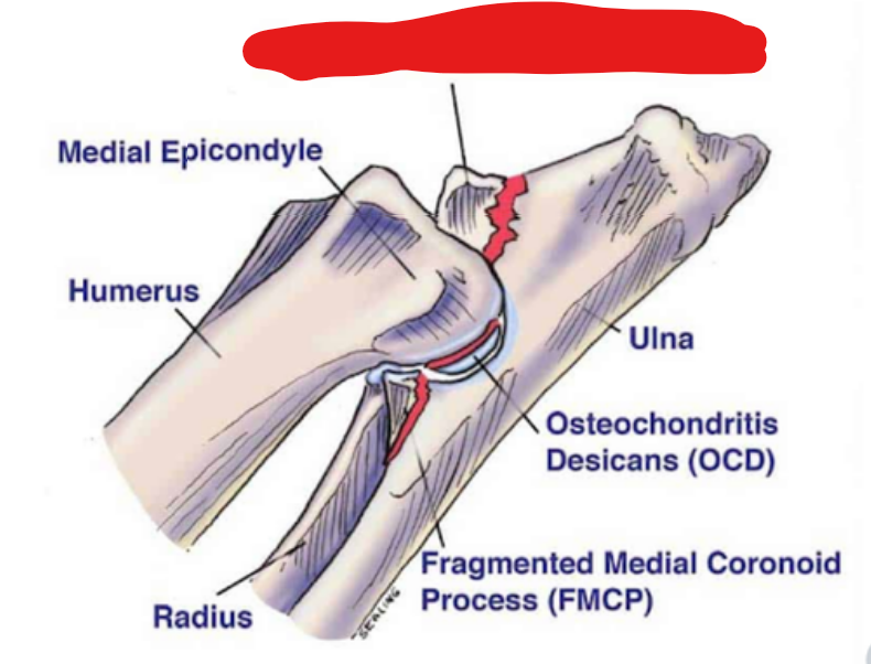
what is it
Ununited anconeal process

what is it
medial epicondyle
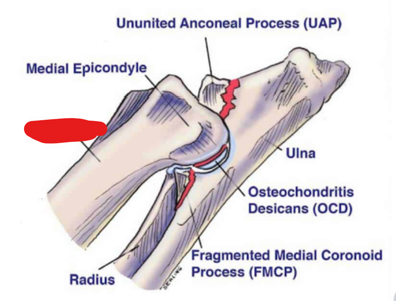
what is it
humerus

what is it
radius

what is it
fragmented medial coronoid process
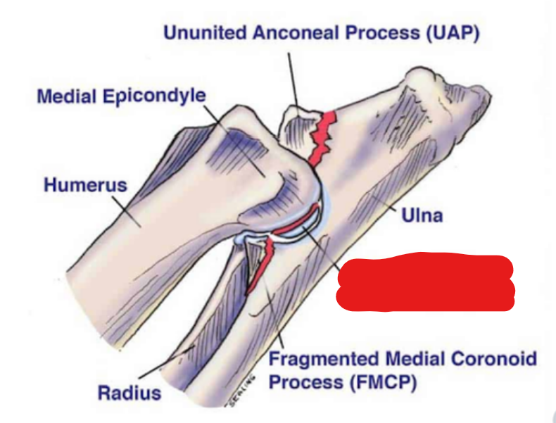
what is it
osteochondritis desicans
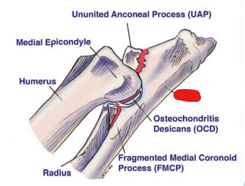
what is it
ulna
what does UAP stand for
Ununited anconeal process
what does OCD stand for
osteochondritis desicans
what does FMCP stand for
fragmented medial coronoid process
clinical signs of UAP (6)
Mild lameness
shortened stride
Lameness
Pain on full extension
pain on palpation of elbow
decreased ROM
what percentage of UAP is bilateral
60%
the head-bob goes — when the dog is weightbearing on the lame side
up
the head-bob goes — when the dog is weightbearing on the sound side
down
how to diagnose UAP (2)
History
flexed lateral Radiographs
how to treat UAP (7)
DJD the process can be stabilized by screwing it back in place
surgical removal of the ununited anconeal process.
Weight reduction
Restricted exercise
NSAID’s
Chondroprotectives
Nutraceuticals
what does CCL stand for
Cranial Cruciate Ligament
what does the CCL do
Ligament stabilizes the stifle joint
what causes a ruptured CCL (4ish)
Middle aged, obese, inactive dogs that hyperextend the stifle during activity
Dogs in sports
Trauma
Abnormal conformation
clinical signs of ruptured CCL (10)
Acute hind limb lameness
Medial joint swelling
Effusion
Crepitation
Excessive cranial laxity of the proximal tibia
Increased internal tibial rotation
Often NWB
bilateral sitting symmetry
partial tear may present as stiffness
The tibia usually rotates internally when the animal tries to bear weight
NWB stands for
Non-Weight-Bearing
how to diagnose ruptured CCL (4)
Gait observation
Radiographs
cranial drawer test
Tibial compression test
how to treat ruptured CCL (8)
Exercise restriction
analgesia
weight control
physio
Custom knee braces
TPLO
Simitri
Tibial Tuberosity Advancement
after surgery for a ruptured CCL what is the instructions for owner (2)
The pet requires restricted exercise for the first 3 - 4 weeks after surgery.
Restricted exercise means cage rest with leash walking for elimination.
what causes patellar luxations (3)
Breed dispositions
Anatomical abnormality
Hereditary disorder
what are some anatomical abnormalities that cause Patellar luxations (5)
a reduced coxofemoral angle
lateral bowing of the femur
internal rotation of the tibia
shallow trochlear groove
hypoplasia of the medial femoral condyle
how do the anatomical abnormalities cause patellar luxations (2)
cause the pull of the tendon to be off-line
one rim of the trochlear groove may not be high enough to hold the patella securely in place