HLS anatomy 1 : thoracic duct and spleen
1/20
There's no tags or description
Looks like no tags are added yet.
Name | Mastery | Learn | Test | Matching | Spaced |
|---|
No study sessions yet.
21 Terms
Thoracic duct begin from …(1)… anteriorly to bodies of …(2)..,…(3).. vertebrae between the right crus of the diaphragm and the aorta
(1) cisterna chyli
(2) L1
(3) L2
(T/F)
the thoracic duct is is a thin-walled vessel which has a beaded appearance due to presence of many valves
True
course and relations of thoracic duct before the crossing into left side?
1- It enters the thorax through the aortic opening of the diaphragm between the aorta (on the left) and azygos vein (on the right).
2-In the posterior mediastinum, it ascends Between the aorta (on the left) and azygos vein (on the right) , Behind right border of esophagus , In front of the vertebral column, posterior intercostal arteries, and hemiazygos veins.
At the level of …(1)… , It crosses the median plane from right to left behind the esophagus.
T5
N.B : in the superior mediastinum thoracic duct ascends behind left border of esophagus.
thoracic duct curved behind the carotid sheath at the level of ….(1)…
C6
(T/F)
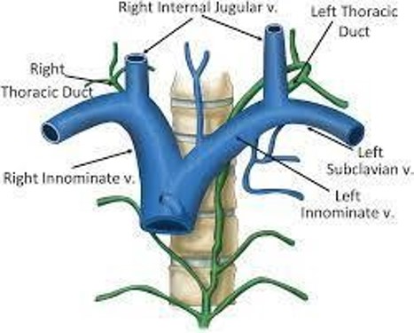
Thoracic Duct end into the junction of the left subclavian and left internal jugular veins
True
- According to Tributaries of Thoracic Duct
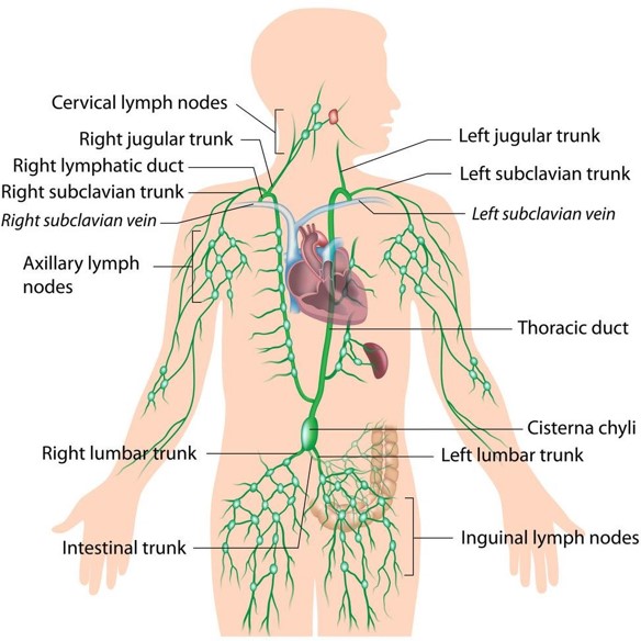
…(1)…drains the left upper limb
Left jugular lymph trunk drains …(2)…
…(3),,,, drains the left 1/2 of the thoracic cavity
…(4)… receives lymphatic from the lower part of the body through Intestinal lymph trunk and Right and left lumbar lymph trunks
(1) Left subclavian trunk
(2) the left 1/2 of the head and neck
(3) Left broncho-mediastinal lymph trunk
(4) Cisterna chili
there is 2 more Tributaries :
- Efferent from the posterior mediastinal lymph nodes
- Efferent from the posterior intercostal lymph nodes.
(T/F)
spleen lies in the left hypochondriurm
true
The surface anatomy of the spleen?
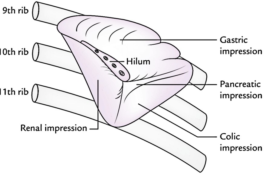
❖The long axis of the spleen lies along the long axis of the 10th rib
❖Its upper border is parallel to the superior border of the 9th rib
❖Its lower border is parallel to the inferior border of the 11th rib
❖The anterior end normally lies just behind the left midaxillary line
❖The posterior end lies one and half inches lateral to the 10th thoracic spine.
to place the spleen in the correct anatomical position , Hold the spleen in your left hand with its convex surface applied to (1) , the round posterior end towards (2) , the broad anterior end towards (3) and the notched upper border applied to (4)
(1) The palm
(2) the wrist
(3) tips of fingers
(4) to the thumb
(T/F)
The spleen has Ends, Posterior end (tapering) directed upwards, backwards and medially
and Anterior end (broad) directed downwards, forwards and laterally
True
Notching of the upper border of the spleen is an indication of ….
fetal lobulation
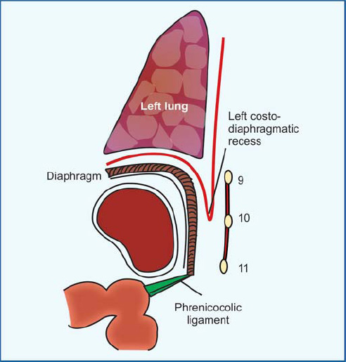
the diaphragm separates the spleen (Diaphragmatic surface) from …(1)… and …(2)…
(1) 9th, 10th and 11th ribs and the intercostal structures.
(2) Left pleura and the left lung
the 5 impressions in the Visceral surface of the spleen?
A. Gastric impression
B. Renal Impression
C. Colic impression
D. Pancreatic impression
E. Hilum of the spleen
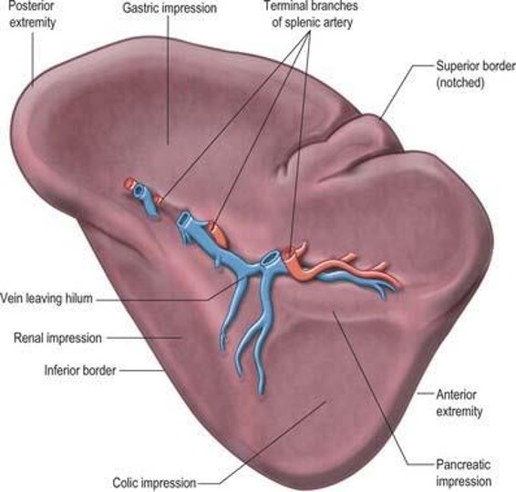
Hilum of the spleen transmits (1) , (2) and (3)
1- Terminal branches of the splenic artery (5-6 branches)
2- Tributaries of the splenic vein
3- Autonomic nerves and lymphatic
(T/F)
The spleen is completely surrounded by peritoneum
false
The spleen is almost completely surrounded by peritoneum except at the hilum
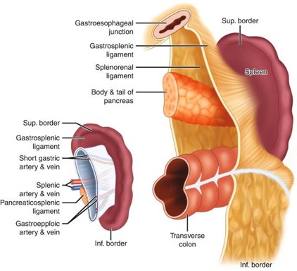
Gastrosplenic ligament Contents?
1- Short gastric vessels
2- Left gastro-epiploic vessels
3- Sympathetic plexus around the arteries
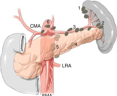
4- Pancreatico-splenic lymph nodes
5- Extraperitoneal fatty tissue
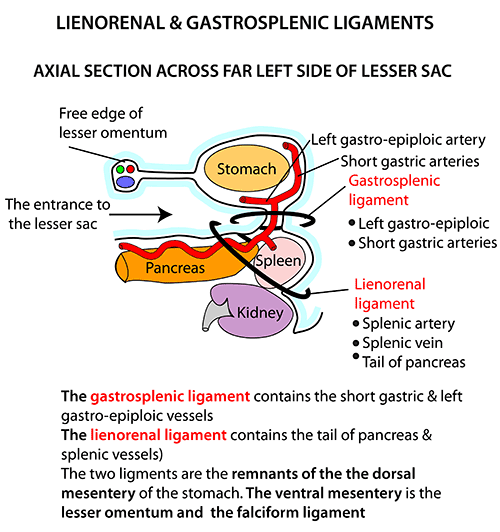
Lienorenal ligament Contents?
1- Tail of pancreas
2- Splenic vessels
3- Sympathetic plexus around the splenic artery
4- Pancreatico-splenic lymph nodes
5- Extraperitoneal fatty tissue.

Arterial supply of the spleen?
Splenic artery which is the largest branches of the coaliac trunk

(T/F)
splenic vein ends by joining the superior mesenteric vein to form the portal vein
True
Lymphatic drainage of the spleen?
To the pancreatico-splenic lymph nodes
N.B : Red bulb of the spleen has no lymphatic