Malignant Renal Masses
 Call Kai
 Learn
Learn Practice Test
 Spaced Repetition
 Match
Match Flashcards
 Knowt Play
1/29

There's no tags or description
Looks like no tags are added yet.
Name | Mastery | Learn | Test | Matching | Spaced | Call with Kai | Chat |
|---|
No analytics yet
Send a link to your students to track their progress
30 Terms
RCC may also be referred to as
hypernephroma or adenocarcinoma of the kidney
primary cancer means that cancer
begins in the organ
RCC is a primary form of cancer, meaning this form of cancer begins in the kidney, specifically originating from the —-
renal tubular epithelium
things that increase the risk for renal cell carcinoma
smoking, hypertension, obesity, and tuberous sclerosis
there is a strong association between RCC and
von Hippel-Lindau disease
patients who have — are especially suseptible to develop RCC as well
acquired renal cystic disease from long term dialysis
frequenct symptoms of RCC manifest late in the disease when the tumor is
moderately large
RCC tumor can spread into the
renal vein and IVC
RCC that spreads to the renal vein and IVC is termed
tumor thrombus
RCC may be referred to as a hypernephroma. It has a strong link with —.
smoking
clinical findings of renal cell carcinoma
anorexia
flank pain
gross hematuria
hypertension
palpable mass
smoker
weight loss
sonographic findingd of renal cell carcinoma
hypoechoic, isoechoic, or hyperechoic solid mass on the kidney
can have a complex cystic appearance as well
check the renal vein and IVC for tumor invasion
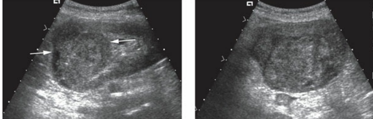
Renal cell carcinoma. Longitudinal (left) and transverse images (right) of the left kidney showing a heterogeneous solid lesion (arrows) in the superior pole.
transitional cell carcinoma of the kidney is a
malignant tumor that is most often found in teh area of the renal pelvis
TCC may also be found in the
ureter and urinary bladder
like RCC, patients who — are also at an increase risk for TCC
smoke
TCC can cause
focal dilation of the calices, and small lesions can be dificult to identify with sonography
clinical findings of renal transitional cell carcinoma
gross hematuria
pain secondary to renal obstruction
history of smoking
sonographic findings of renal transitional cell carcinoma
hypoechoic or isoechoic mass within the renal sinus
varying degrees of hydronephrosis may be present
TCC found in the kidney is most often located within the
renal pelvis
metastases to the kidneys are most often from the
lungs or breast
loss often metastases to the kidneys are from
prostate, pancreas, and melanoma
RCC can also metastasize from the
contralateral kidney
lymphoma and — of the kidney will appear sonographically similar
leukemia
lymphoma and leukemia can either result in a — or be seen as —
multiple, hyperechoic masses; bilateral renal enlargement with a decrease in overall renal echogenicity
lymphoma, which is most likely in teh form of non-hodgkin lymphoma, typically affects
both kidneys
patients who have a history of — should be thoroughly evaluated for renal metastasis
primary malignancy
clinical findings of other renal malignancies
history of primary cancer (often lung or breast)
hematuria
fever
weight loss
sonographic findings of other renal malignancies
bilateral, hypoechoic masses with lymphoma
lymphoma or leukemia can manifest as an enlarged kidney
metastases have avrying sonographic findings but are most often solid tumors that are hypoechoic or hyperechoic