Exam 1 Flashcards - ACBS 400A
 Call Kai
 Learn
Learn Practice Test
 Spaced Repetition
 Match
Match Flashcards
 Knowt Play
1/158

There's no tags or description
Looks like no tags are added yet.
Name | Mastery | Learn | Test | Matching | Spaced | Call with Kai | Chat |
|---|
No analytics yet
Send a link to your students to track their progress
159 Terms
What is bone made up of?
both collagenous proteins (majority) & non-collagenous proteins
non-collagenous proteins: proteoglycans (necessary for cell proliferation, mineral depositions, remodeling) & glycoproteins (necessary for mineral regulation)
minerals
water
L1
Define epiphysis.
end part of a long bone
initially growing separately from shaft
L1 - bones
Define diaphysis.
shaft or central part of long bone
contains mostly compact bone
L1 - bones
Define metaphysis.
region where epiphysis joins diaphysis
L1 - bones
Define epiphyseal/ growth plate.
hyaline cartilage plate in the metaphysis at each end of a long bone
L1 - bones
Define medullary space.
hollow part of bone that contains bone marrow
L1 - bones
Define trabecular/ cancellous/ spongy bone.
hierarchical, spongy, and porous material composed of hard & soft tissue components
found at epiphyses and metaphyses of long bone and in vertebral bodies
L1 - bones
Define compact/ cortical bone.
hard, dense outer later of all bones throughout body
L1 - bones
Define periosteum.
dense outer later of bone that contains vascular connective tissue enveloping bones expect at surfaces of joints
L1 - bones
Defie endosteum.
membrane lining inner surface of bony wall and bone marrow
L1 - bones
Define osteoblasts.
mononuclear cells that form new bone
come from mesenchymal cells in bone marrow
primary ossification: formation of 1-cell thick layer covering surfaces undergoing remodeling
L1 - bones
Define osteoclasts.
large multi-nucleated cells at none surface and actively resorb bone
cytoplasm dense with non-membrane bound ribosomes and lysosomes
have ruffled edge on area in bone contact → very mobile
L1 - bones
Define osteocytes.
inactive and entrapped osteoblasts within bone matrix
maintains bone tissue
L1 - bones
Define osteoprogenitor.
germ cell aka stem cell that turns into other cells in bone
L1 - bones
Describe the structure of compact bones.
compact bone: where bony matrix is solid & filled with organic ground substance & inorganic salts
primary component of long bones to provide strong structural support
ground substance: rich in mineralized calcium phosphate compound called hydroxyapatite
osteon: cylindrical structures made up of layers of bone tissue called lamellae that surround a central canal
Haversian/ central canals: any minute tube which forms a network in bone and contain blood vessels that run parallel to length of bone
Volkmann/ perforating canals: channels that contain smaller blood vessels that run perpendicular to Haversian canals
connect Haversian canals to periosteum & medullary cavity
Lacunae: small areas of holes in bone matrix where osteocytes reside
Canaliculi: tunnels that form connections between neighboring osteocytes → where cytoplasmic projections go
L1 - bones

Describe the structure of trabecular bone.
trabecular bone: light, porous bone enclosing many large spaces in a honeycomb or spongy appearance (bone matrix)
most often found in long bones, ribs, skull, and vertebrae
often in epiphysis and metaphysis but can be in medullary space
forms matrix for bone marrow
trabeculae: thin columns and plates of bone that create spongy structure along stress lines
provides flexibility and improves resistance to fracturing
stores bone marrow, location of blood cell genesis, storage for minerals
L1 - bones
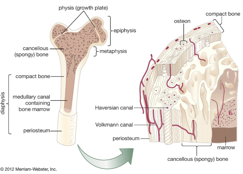
Describe the similarities and differences between periosteum and endosteum.
Similarities:
both vascular connective tissue layers
both contain osteoblasts and fibroblasts
Differences:
periosteum: covers all bones except at articular surfaces of long bones (at cartilage)
has two layers (fibrous outer layer & inner connective tissue layer)
mature osteoblasts primary cells
nourishes compact bone & site of attachment for tendons and ligaments
endosteum: present in all bones
singular loose connective tissue later
fibroblasts & hematopoietic cells primarily found here
used for growth, repair, and remodeling of bones
L1 - bones
Describe the differences between red bone marrow and yellow bone marrow.
Red bone marrow
contains hematopoietic tissue that creates blood cells
small % of marrow
in young animals, located throughout most flat & long bones
will recede from long bones with maturity in trabecular spaces
in adults, found in flat bones of vertebrae, sternum, ribs pelvis, along proximal (top) ends of humerus and femur
Yellow bone marrow
contains mostly adipose tissue
majority of marrow has potential to convert back to rend marrow under severe necessity
located primarily in long bones
L1 - bones
What minerals and inorganic salts make up bone?
Minerals:
calcium (Ca2+), phosphorus (phosphate ion), magnesium (Mg2+), sodium (Na+), potassium (K+), chloride (Cl-), fluoride (F-)
most abundant: Ca2+ and phosphorus
Salts:
citrate, hydroxyl, carbonate anions
General structure of bone mineral: hydroxyapatite crystals - (Ca10(PO4)6(OH)2)
L1 - bones
What type of collagens make up bones?
primarily composed of type I collagen
synthesized mostly in osteoblasts
osteoblasts make procollagen → hydroxylation with vit C (required) on proline and lysine groups → 3 collagen fibrils braid together to form tropocollagen → leaves osteoblasts
tropocollagen: braided collagen fibrils that have improved strength and rigidity
5 form microfibrils
collagen matrix: microfibrils laid end to end in staggered formation in same direction in single bone metabolic unit
L1 - bones
Describe collagen breakdown.
hydroxyproline: neutral heterocyclic protein amino acid that’s commonly found in bone & gelatin
digestion of bone collagen causes concentrations to rise in blood → used as diagnostic indication of bone destruction
hydroxylysine: amino acid unique to collagen-like peptides
has covalent cross-link norleucine across adjacent collagen fibrils → stabilizes fibrils
creates banding pattern on collagen fibrils
L1 - bones

What is the function of osteocalcin?
protein found in bone
osteocalcin: protein found in extracellular matrix of bone and in serum of circulating blood
produced solely by osteoblasts - vitamin K often present
inhibits haphazard formation of hydroxyapatite precipitates and mineralization
hemoattractant of leukocytes → also attracts osteoclasts to areas in need of resorption and remodeling
L1 - bones
What are the types of bone formation?
intramembranous ossification: bone formation within primitive connective tissue
endochondral ossification: pre-existing cartilage is turned into bone
ectopic bone formation: pathologic process in which connective tissues that aren’t usually turned into bone are ossified
L2 - bone formation & remodeling
Define mesenchyme.
intramembranous ossification
tissues characterized by primordial cell types surrounded by large amount of extracellular matrix (loose connective tissue)
condenses & becomes vascularized ding embryonic development
L2 - bone formation & remodeling
Define woven bone.
intramembranous ossification
early bone where collagen fibrils remain randomly interwoven
disorganized arrangement of collagen fibers
weaker and more flexible
rapidly formed
L2 - bone formation & remodeling
Define osteoid.
unmineralized, organic portion of bone matrix that forms prior to the maturation of bone tissue
can be found in woven and lamellar bone
L2 - bone formation & remodeling
Describe the process of intramembranous ossification.
begins in embryo development
formation of ossification spicules: mesenchymal cells form collagen fibrils → form matrix → conversion to osteoblasts
formation of extracellular matrix and trabeculae: osteoblasts combine fibrils with Ca2+ → calcification leads to formation of trabeculae
osteoid vascularization development: fibrils go from random to organized assortment → formation of lamellar layers around blood vessels
osteons and compact bone formation: outer layer connective tissue condenses into peroiranium → osteoblasts turn into fibroblasts
L2 - bone formation & remodeling
Define hyaline cartilage.
translucent bluish white type of cartilage present in joints, respiratory tract, and immature skeleton
L2 - bone formation & remodeling
Describe the process of endochondral ossification.
includes: bones in extremities, base of skull, vertebral column, pelvis
starts as hyaline cartilage
mesenchyme differentiates into chondrocytes → lay out in shape of being being formed
chondrocytes secrete hyaline matrix of type II collagen
osteoprogenitor cells in perichondrium lining in cartilage model is activated
deposit thin collar of woven bone on outside of model
L2 - bone formation & remodeling

What are centers of ossification?
within hyaline cartilage & are distinguished by enlargement of chondrocytes in particular areas of cartilaginous bone
lacunae expand with thin strands of cartilage matrix separating adjacent ones → remaining matrix accumulates Ca2+ phosphate
calcification causes hypertrophied chondrocytes do apoptosis
blood vessels form from new periosteum and invade these areas → new osteoprogenitor & other cells follow vessels → form osteoblasts that congregate on calcified cartilage spicules → deposit bone matrix
in horses & cows
starts in diaphysis of long bones by 3rd month of fetal development
secondary centers in epiphyses of long bones later
expansion of epiphyseal ossification won’t replace area’s cartilage (articular surfaces)
transverse disk of epiphyseal cartilage remains between sections (epiphyseal plate)
chondrocytes in area arranged into columns that allows growth in length of bone
L2 - bone formation & remodeling
Define zone of proliferation.
formed by chondrocytes that are further from diaphysis
L2 - bone formation & remodeling
Define zone of maturation.
formed by chondrocytes that have stopped dividing and have become enlarged
L2 - bone formation & remodeling
Define zones of hypertrophy.
formed by hypertrophy and vacuolation of chondrocytes
L2 - bone formation & remodeling
Define zone of provisional calcification.
formed by cartilaginous matrix surrounding cells beginning to calcify
L2 - bone formation & remodeling
How do bones grow in length.
diaphysial side chondrocytes go through apoptosis and degenerate → lacunae invaded by blood vessels and osteoprogenitor cells from marrow spaces
osteoblasts differentiate and form bars of calcified cartilage → thin layer of collagen matrix deposited on surface of calcified cartilage → good conditions leads to new bone matrix calcifying
osteoclasts resorb woven bone and calcified in metaphysis → osteoblasts move in to lay down type I collagen for lamellar bone
zone of proliferation cells advance growth plate away from diaphysis
process controlled by somatotropin hormone from pituitary
causes local production of insulin-like growth factor 1 (somatomedin) → encourages zone of proliferation chondrocytes to divide
L2 - bone formation & remodeling
Describe growth plate closure.
once at mature length, cartilage creation slowly stops
replacement of cartilage with bone at diaphysial side continues until entire growth plate cartilage is replaced by bone
metaphysis does not technically exist until plate is closed
L2 - bone formation & remodeling

How do bones grow in diameter?
deposition of bone on outside surface between old bone and periosteum
new bone laid down while old bone inside resorbed at same time of lengthening
hyaline model shape never changes = rate of resorption & creation are equal
stress can dictate mineral deposition & bone matrix thickness
spongy bone stops earlier in thickening → space fills with bone marrow
L2 - bone formation & remodeling
Define bone remodeling units (BRU).
small units of bone that undergo remodeling at any given time
L2 - bone formation & remodeling
What are the four steps of bone remodeling.
activation
resorption
reversal
formation
L2 - bone formation & remodeling

Describe the first step of bone remodeling.
activation: converts/inactive bone surface into that to be remodeled
selective remodeling directed by a signal from bone
microfracture disrupt ordered collagen fibril orientation → disrupts normal electrical change through mineral crystals → initiates remodeling
bone deterioration elicits autocrine/paracrine factor secretion to initiate process
quiet osteoblasts shrink in areas of bone needing remodeling → exposes bone matrix → osteoclasts move in → bind to bone on ruffled border
L2 - bone formation & remodeling
Describe the second step of bone remodeling.
resorption
osteoclasts release acids & proteolytic enzymes from ruffled border → dissolves bone matrix
minerals & bone matrix broken down products absorbed via diffusion or other transport processes into osteoclasts
sent back out into ECF
L2 - bone formation & remodeling
Describe the third step of bone remodeling.
reversal
howship’s lacuna: hole created by osteoclasts during resorption
needs insulin-like growth factor 2 and transforming growth factor (TGF) - beta
released during collagen matrix breakdown
activate cells lining surface to revert to osteoblasts
L2 - bone formation & remodeling
Describe the fourth step of bone remodeling.
formation
begins with deposition of osteoid by osteoblasts: consists mostly of type I collagen, proteoglycans, and smaller proteins found in mature bone
lays osteoid down in lamellae with fibrils parallel to each other
compact bones goes from outer edges to center when filled until reach Haversian canal
different lamellae fibrils oriented at angles from each other
trabecular bone deposited in curved sheets following hollow shape
mineralization occurs after time delay but happens quick when activated
L2 - bone formation & remodeling
What is a complete fracture? What are the different types of complete fractures?
complete: through entire bone
transverse: straight line through bone
oblique: line at an angle through bone
spiral: corkscrew line through bone
comminuted: more than 2 parts to fracture
L2 - bone formation & remodeling

What is a incomplete fracture? What are the different types of incomplete fractures?
incomplete: not entirely through bone
bowing: bone is bent
buckle: fracture on concave side
greenstick: fracture on convex side
avulsion: bone fragment separates from the rest of the bone, often attached to ligament or tendon
L2 - bone formation & remodeling
What is a displacement fracture?
ends of fracture not within same area, created a gap
L2 - bone formation & remodeling
What are the different types of Salter-Harris fractures?
Type 1: through growth plate
Type 2: through growth plate & metaphysis
Type 3: through growth plate & epiphysis
Type 4: through all three elements
Type 5: crush injury of growth plate
L2 - bone formation & remodeling
How do bones help maintain homeostasis?
remodeling moves mineral into ECF
bone helps as blood pH buffer
releases cations in response to acidosis or anions in response to alkalosis
L2 - bone formation & remodeling
What is osteocytic osteolysis?
osteocytic osteolysis: process of Ca2+ resorption from bone
parathyroid hormone (PTH) stimulates osteocytes to pump some fluid Ca2+ into ECF
prevents osteoclasts from entering reversal phase & osteoblasts from laying new matrix
if deficit severe, osteoclastic osteolysis initiated
persistent secretion of PTH → activates osteoclasts by shrinking osteoblasts to expose bone matrix
osteoblasts release paracrine factors: prostaglandin E2, IL-1, and IL-6 that stimulate osteoclasts
1,250dihydroxyvitamin D (vit D hormone) indirectly affects bone Ca2+ concentration
increase efficiency of Ca2+ and phosphorus absorption absorption in intestine → blood concentration balanced → permits mineralization of bone matrix
L2 - bone formation & remodeling
What is the function of calcitonin?
major Ca2+ regulating hormone
secreted by C-cells in thyroid when Ca2+ blood levels too high
acts on osteoclasts to inhibit bone resorption
L2 - bone formation & remodeling
Fill in the blanks.
Mesenchymal cells differentiate into different cells depending on the method of ossification. In intramembranous ossification, they mostly become ______ while in endochondral ossification, it most commonly turns into _______.
intramembranous: osteoblasts
endochondral: chondrocytes
Assignment 1
Which of these is a hormone NOT directly related to osteocytic osteolysis?
A. calcitonin
B. 1,25-dihydroxyvitamin D
C. parathyroid hormone
D. osteocalcin
osteocalcin
Assignment 1
What amino acids are only found in bone?
hydroxylysine
hydroxyproline
Assignment 1
What is osteocalcin?
a protein that promotes mineralization of bone in proper orientation
Assignment 1
Describe the steps of bone remodeling.
activation: quiet osteoblasts shrink in areas of bone needing remodeling → exposes bone matrix → osteoclasts move in → bind to bone on ruffled border
resorption: osteoclasts release acids & proteolytic enzymes from ruffled border → dissolves bone matrix that then leaves depression in the bone → minerals & old matrix by-products absorbed via diffusion or other transport processes into osteoclasts → released into ECF
reversal: uses insulin-like growth factor 2 and transforming growth factor (TGF)- beta to activate lining surface cells to revert to osteoblasts
formation: deposition of osteoid by osteoblasts → form lamellae from outer edges to center → forms Haversian canal → calcified to strengthen & finish process of remodeling
Assignment 1
Fill in the blank.
The _________ is the model for bone formation during endochondral ossification.
hyaline cartilage
Assignment 1
What is the role of parathyroid hormone (PTH) in Ca2+ homeostasis?
prevents osteoclasts from entering reversal phase & osteoblasts from laying new matrix depending on if there is a Ca2+ deficit in the ECF
Assignment 1
In what type of bone can you find yellow marrow?
long bones
Assignment 1
On what side does the growth plate begin to close?
diaphyseal
Assignment 1
What is needed for the hydroxylation of procollagen to occur? Without this, procollagen cannot be stable as tropocollagen, and can’t form the bone matrix.
vitamin C
Assignment 1
Explain the process of bone repair after a fracture.
first the bone is broken due to some kind of trauma
this causes the blood supply to break & osteoprogenitor cells to tear
blood then pools into the area of the break, forming a hematoma, and osteocytes die
once area fills with blood, proinflammatory cytokines attract phagocytes into area to remove dead tissue & clot material
the space left now has cartilage beginning to fill in the hypoxic area to form a callus
fibrous tissue is created into the callus to strengthen it
woven bone begins to fill in the space to create a hard callus
finally, bone remodeling occurs & bone is repaired
Assignment 1
Generally, what is the majority of hydroxyapatite crystals (bone crystals) made of?
calcium & phosphate
Assignment 1
T/F: When bone is increasing in diameter, outside bone is created while inside bone is resorbed. This keeps the rate constant and allows room for bone marrow.
True
Assignment 1
T/F: The primary location of endochondral ossification is in the epiphysis of long bones. The secondary location later in the process is the diaphysis of the long bones.
False
Assignment 1
Fill in the blanks.
There are 2 connective tissue layers of bone. There is the ______ that is on the external aspect of the compact bone, and it nourishes it. Then there is the _______ that is on the internal aspect of the compact bone where the medullary cavity is found.
periosteum
endosteum
Assignment 1
Define zone of proliferation.
formed by chondrocytes that are further from diaphysis
Assignment 1
Define zone of maturation.
formed by chondrocytes that have stopped dividing & have become enlarged
Assignment 1
Define zone of provisional calcification.
formed by cartilaginous matrix surrounding cells beginning to calcify
Assignment 1
Define zone of hypertrophy,
formed by enlargement and vacuolation of chondrocytes
Assignment 1

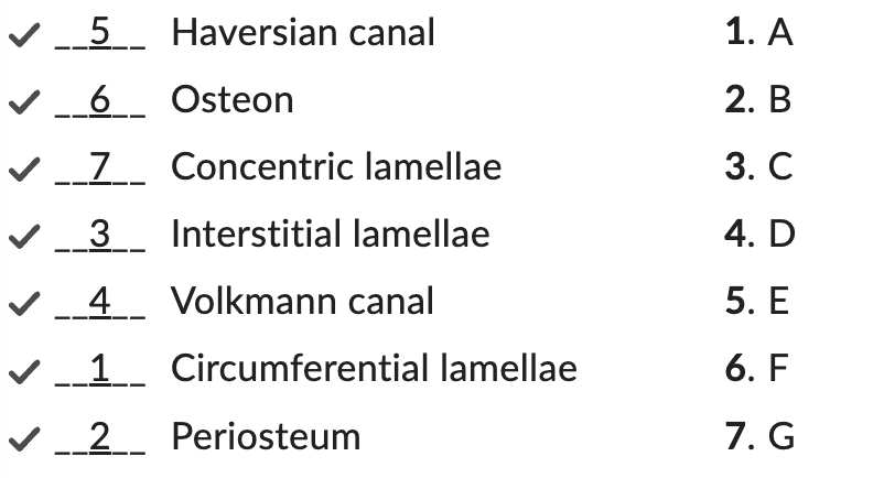
Label this diagram.
Terms: Haversian canal, osteon, concentric lamellae, interstitial lamellae, volkmann canal, circumferential lamellae, periosteum
Assignment 1

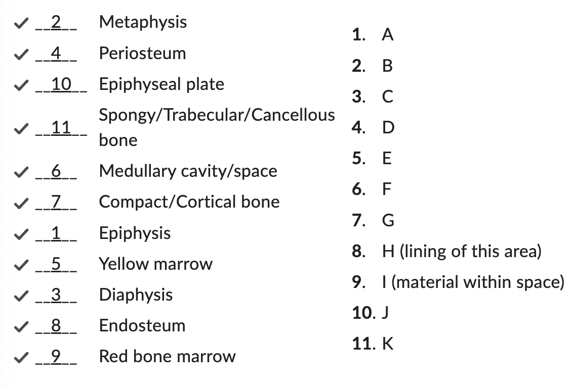
Label this diagram.
Terms: metaphysis, periosteum, epiphyseal plate, spongy/trabecular bone, medullary cavity/space, compact/cortical bone, epiphysis, yellow marrow, diaphysis, endosteum, red bone marrow
Assignment 1

Label this diagram.
Terms: lamellae, lacuna, canaliculi, osteoblast, haversian canal, osteoblast, osteocyte
Assignment 1

What is the role of Vitamin D hormone of Ca2+ homeostasis?
increases efficiency of Ca2+ and phosphorus absorption in intestine
Quiz 1
What is the difference between red and yellow bone marrow?
red bone marrow is found in long bones and most flat bone, and it is the location of hematopoietic cell production
yellow bone marrow is found primarily in long bones, and it is a storage place for adipose tissue
Quiz 1
What are centers of ossification?
the locations in the hyaline cartilage model that ossify first
Quiz 1
Explain the process of collagen matrix formation.
first, osteoblasts synthesize procollagen
then, there is hydroxylation of proline and lysine groups with the help of vitamin C
this process helps stabilize procollagen for the development of helices
3 procollagen are combined to form the tropocollagen
5 of these combine to create a microfibril
finally, staggered microfibrils end to end then make up the collage matrix
Quiz 1
What is the purpose of the epiphyseal plate?
allow long bones room to grow in length as an animal matures
Quiz 1

State the function of osteoprogenitor cells, osteoclasts, osteoblasts, and osteocytes. Identify them in the picture.
A is osteoclasts
large multi nucleated cells at bone surface and actively resorb bone
B is osteoblasts
mononuclear cells that for new bone (ossification) and originate from mesenchymal cells in bone marrow
C is osteocytes
inactive & entrapped osteoblasts within bone matrix
Osteoprogenitor cells
germ cell that turns into other cells in bone
Quiz 1
What causes hyena disease?
vitamin A toxicosis in calves
Quiz 1
What is the difference between epiphyseal and diaphyseal ossification?
epiphyseal ossification won’t replace articular cartilage
diaphyseal ossification replaces all cartilage
Quiz 1
What is the difference between Haversian canals and Volkmann canals?
Haversian canals run parallel with the bone and are at the center of osteons
Volkmann canals run perpendicular to bone and feed the periosteum and medullary cavity
Quiz 1
Osteocalcin is produced by osteoblasts in the presence of vitamin K. What does it do?
Inhibits formation of hydroxyapatite precipitates and mineralization
Attracts osteoclasts to areas in need of resorption and remodeling
Quiz 1
Explain the process of growth plate closure.
first, diaphyseal side chondrocytes experience apoptosis
Then, blood vessels and osteoprogenitor cells invade into lacunae of marrow spaces in primary ossification zone
After invasion, osteoblasts then differentiate along calcified cartilage
They deposit collagen matrix on calcified cartilage
In favorable conditions, this matrix quickly calcifies and forms spongy bone in the metaphysis
As plate finishes its closure, osteoclasts move into metaphysis to resorb woven bone
At the same time of woven bone resorption, osteoblasts put down new bone matrix
By the end, the metaphysis has extended farther from diaphysis and growth plate has closed
Quiz 1
In what location does intramembranous ossification happen most often in fetal development?
skull
Quiz 1
Describe the anatomy of cartilage.
Cells
chondrocytes are cells responsible for cartilage formation
chondrocytes secrete a amorphous gel-like matrix: contains collagens, proteoglycans, and non-collagenous proteins
contains polysaccharides (chondroitin sulfates) complex
young chondrocytes can divide after they come isolated in lacunae → daughter cells secrete new matrix between them & move apart in separate lacunae
Layers
Perichondrium: dense fibrous layer lined by cells that have the capacity to secrete hyaline matrix
outer fibrous layer: provides protection, mechanical support, and attaches cartilage to other structures
inner cellular: involved in growth & maintenance of cartilage
lacks nerves & blood vessels
overall resistant to compression
L3

What are the different types of cartilages?
hyaline: bulk of cartilage found in animals
found on ventral ends of ribs, in tracheal rings & larynx, articulating surfaces of bones, and in growth plates in growing animals
randomly oriented but little elastin
elastic: relatively scare but found in external ear & epiglottis
has a ton of elastic fibers embedded in it
more flexible due to elastin fibers in ground substance in matrix
fibrocartilage: found at site of insertion of many larger ligaments onto bone, intervertebral disks, and symphysis of pubis
proportion of dense collagen bundles oriented parallel
cells occupy lacunae arranged in rows between coarse bundles of collagen
collagen content of matrix greatly increased & collagen fibrils from thick though bundles aligned in 1 direction to improve tensile strength
L3
Describe the growth of cartilage.
chondrocytes come from mesenchymal cells
undergo mitosis and spread out
continue to surround themselves with specialized mix of proteins to form interstitial matrix
fine fibrils of collagen exist within matrix
originally secreted into matrix as tropocollagen by cells → aggregate & assemble themselves into collagen fibrils
L3
What is the difference between interstitial and appositional growth?
interstitial: process of growth that occurs through the addition of new cells within existing tissue
involves mitosis
increases tissue size since add new cells
occurs throughout life
appositional: process of growth that involves the addition of new cells to surface of the tissue
involves differentiation of stem cells
increases tissue size by increase in new layers
occurs during growth
L3

What are the types of collagen in cartilage?
type II: main collagen form in cartilage
only found in cartilage
type IX: on surface of type II & doesn’t form fibrils
functions to regulate spatial arrangement of type II
type X: found in chondrocytes within growth plates in long bones
L3
Define joints.
aka articulation
junction between any 2 bones
attaches bone to another → allows bones to move freely with little resistance
movement controlled & limited by action of muscles & by ligaments and tendons
types of joints: immovable & synovial
L3
Describe synovial joints.
contained in a capsule
outer fibrous layer of collagen fibers that run from periosteum of 1 to bone another
lends side to side stability to joint
ligaments are special extension of fibrous capsule
location on inside or outside of joint capsule
L3

Describe synovial fluid.
inner surface of capsule composed of synovial membranes
involved in production of synovial fluid (viscous liquid with consistency similar to egg white)
provides lubrication to joint
reduces wear on opposing surfaces
L3
Define ligaments.
tough, elastic bands of connective tissue that surround joint to give support & limit movement
connects bone together
L3
Define tendons.
another type of tough connective tissue thats on each side of a joint & attaches on each side of a joint
attaches to muscles that control movement of joint
connect muscles to bones
L3
Define bursas.
fluid-filled sacs between bones, ligaments, or other nearby structures
help cushion the friction in a joint
L3
Define menisci.
special areas of dense fibrocartilage that exist between articulating cartilage surfaces on some bones
most common in areas bearing weight
cushions compressive forces
L3
Name the different types of joints.
pivot
hinge
ball & socket
condyloid
plane
L3
Describe pivot joints.
freely moveable joints that allows only rotary movement around a single axis
moves in a rotation pattern within ring formed by 2nd bone & ligament
allows for turning of head
radius & ulna at elbow and carpus
L3
Describe hinge joints.
primarily only allows movement in 1 plane, but can in small movements in other directions
up/down or side to side but can’t do both well
toes, elbows, stifles, tarsi
L3
Describe ball and socket joints.
joint where rounded surface of a bone moves within a depression on another bone
allows greater freedom of movement
shoulders and hips
L3