Ch. 14 - Brain and Cranial Nerves
1/101
There's no tags or description
Looks like no tags are added yet.
Name | Mastery | Learn | Test | Matching | Spaced |
|---|
No study sessions yet.
102 Terms
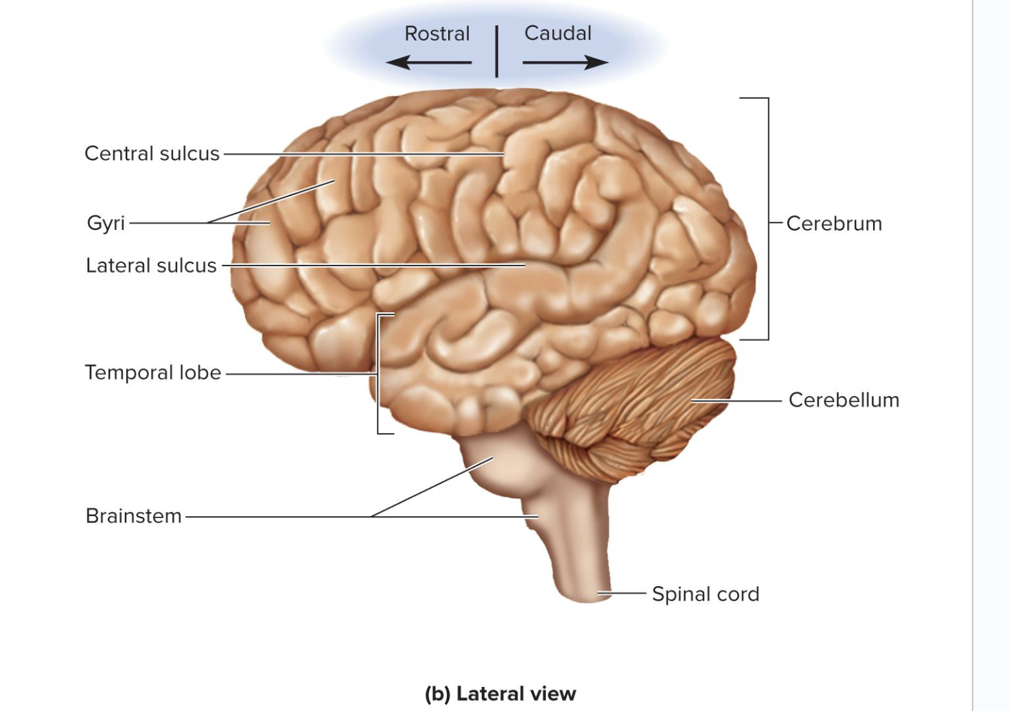
Rostral Vs. Caudal
rostral - toward the frontal brain
caudal - towards the brain stem
Gray Matter
unmyelinated
“thinking matter”
have neurosomas, dendrites, and synapses
Dull color due to little myelin
Forms surface layer (cortex) over cerebrum and cerebellum
Forms nuclei deep within brain
White Matter
highly myelinated
“transmission matter”
bundles of axons
Lies below the cortical gray matter
Pearly white color from myelin around nerve fibers
made of bundles of axons that connect one part of the brain to another, and to the spinal cord
Ventricles
4 internal chambers within brain:
two lateral ventricles
third ventricle
fourth ventricle
there is a high concentration of ependymal cells in the ventricles, specifically the lateral ventricles
CSF is pressurized in the lateral ventricles
also circulates through subarachnoid space
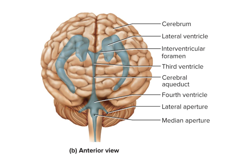
Interventricular Foramen
a tiny pore that connects to the third ventricle
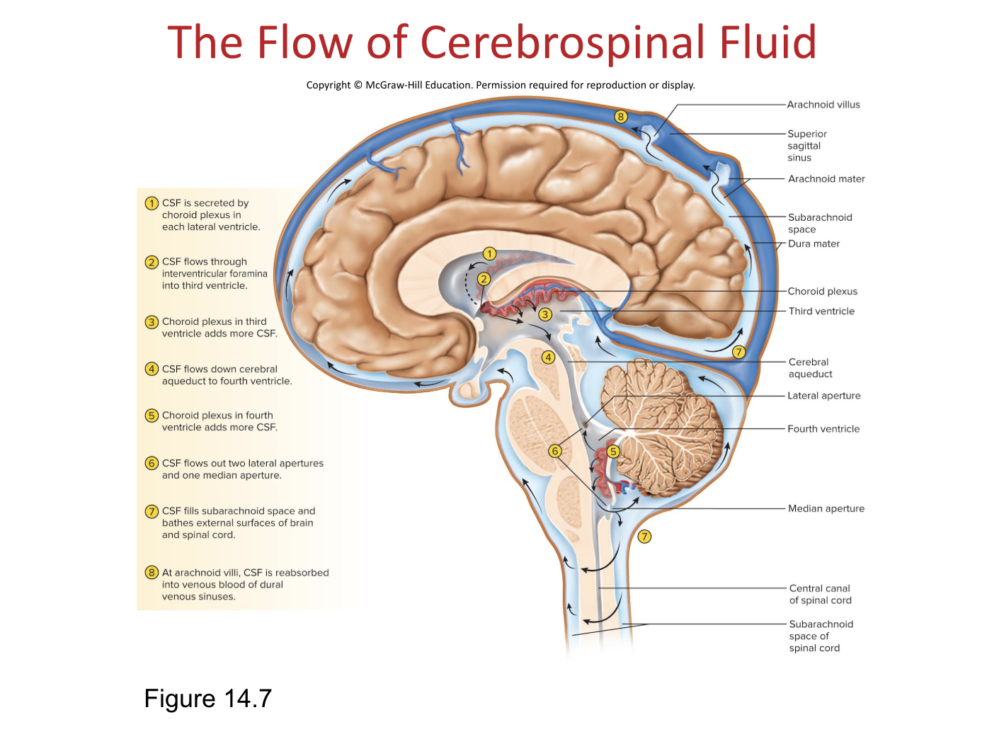
Choroid Plexus
a spongy mass of blood capillaries on the floor of each ventricle
covered by ependymal cells which produce CSF
Cerebrospinal Fluid (CSF)
clear, colorless liquid that fills the ventricles and canals of CNS and bathes their surfaces
basically blood plasma without the proteins
more lactate than glucose will be floating around in it compared to blood
most CSF is produced in the meninges
Brain produces and absorbs 500 mL/day
Production of CSF begins with filtration of blood plasma through capillaries of the brain, then ependymal cells modify it
Functions of CSF:
Buoyancy
Allows brain to gain mass without being impaired by its own weight. If it rested heavily on the floor of the cranium, the pressure would kill the nervous tissue.
Protection - shock absorber
Protects the brain from striking the cranium when the head is jolted.
Chemical stability
Flow of CSF rinses away metabolic wastes from nervous tissue and regulates its chemical environment.
The Flow of Cerebral Spinal Fluid
CSF gets produced in the ventricles
CSF then goes through the ventricles to the central canal
Then it fills the subarachnoid space and bathes the outer surfaces of the spinal cord and brain
there are holes in the dura mater for excess CSF, called arachnoid villi
Blood Supply to the Brain
The brain is only 2% of adult body weight, but receives 15% of our blood
Neurons have a high demand for ATP so a constant supply of blood is required
10s interruption of blood flow may cause loss of consciousness
1 to 2 min interruption can cause significant impairment of neural function
4 minutes without blood causes irreversible brain damage
Brain Barrier System
regulates what substances can get from the bloodstream into the tissue fluid of the brain
Although blood is crucial, it can also contain harmful agents
Two points of entry must be guarded
Blood capillaries throughout the brain tissue
Capillaries of the choroid plexus
transcytosis is one of the only ways to get to the tissues of the CNS
Blood-Brain Barrier
protects blood capillaries throughout brain tissue
Astrocytes build the blood brain barrier by forming tight junctions with the capillaries
Anything leaving the blood must pass through the cells, and not between them
Endothelial cells in the capillary walls can exclude harmful substances from passing to the brain tissue while allowing necessary ones to pass
Blood-CSF Barrier
protects brain at the choroid plexus
Forms tight junctions between the ependymal cells
Tight junctions are absent from ependymal cells elsewhere
Important to allow exchange between brain tissue and CSF
Brain barrier system is highly permeable to water, glucose, and lipid-soluble substances such as oxygen, carbon dioxide, alcohol, caffeine, nicotine, and anesthetics (narcotics)
Slightly permeable to sodium, potassium, chloride, and the waste products urea and creatinine
What are some issues with the brain barrier system?
The brain barrier system (BBS) can be an obstacle for delivering medications such as antibiotics and cancer drugs
Trauma and inflammation can damage the BBS and allow pathogens to enter brain tissue
Ex) Circumventricular organs
Circumventricular Organs (CVOs)
places in the third and fourth ventricles where the barrier is absent
Blood has direct access to the brain
allows the brain to monitor and respond to fluctuations in blood glucose, pH, osmolarity, and other variables
CVOs allow for invasion of HIV
any intracellular pathogen can do this (like viruses)
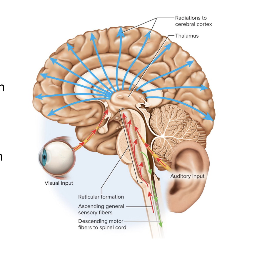
Reticular Formation
A loose web of gray matter that runs up through all levels of the brainstem
Occupies space between white fiber tracts and brainstem nuclei
Has connections with many areas of the cerebrum
Has more than 100 small neural networks without distinct boundaries
Includes:
somatic motor control
cardiovascular control
pain modulation
sleep and consciousness
habituation
Role of Reticular Formation in Somatic (Voluntary) Motor Control
Adjusts muscle tension to maintain tone, balance, and posture, especially during body movements
Relays signals from eyes and ears to the cerebellum
Integrates visual, auditory, balance and motion stimuli into motor coordination
includes gaze centers and central pattern generators
Gaze Centers
allow eyes to track and fixate on objects
Central Pattern Generators
neural pools that produce rhythmic signals to the muscles of breathing and swallowing
Role of Reticular Formation in Cardiovascular Control
monitors contraction strength and frequency of the heart
Role of Reticular Formation in Pain Modulation
Some pain signals ascend through the reticular formation
reticular formation downplays pain
ex) not remembering how you got a bruise
Some descending analgesic pathways begin in the reticular formation
They end in the spinal cord where they block transmission of pain signals
Role of Reticular Formation in Sleep and Consciousness
Reticular formation plays a central role in consciousness, alertness and sleep
Injury here can result in irreversible coma
Role of Reticular Formation in Habituation
the reticular activating system changs activity in the cerebral cortex so that it ignores repetitive, inconsequential stimuli
training your central pattern generators
ex) wearing glasses and learning to ignore that they are there
Cerebellum
the little brain
mostly gray matter
little branching white matter is the arbor vitae
contains most of our neurons in our brain
largest part of the hindbrain and second largest part of the brain as a whole
has right and left cerebellar hemispheres
important for motor coordination and movement
Nonmotor Functions of the Cerebellum
a lot this is subconscious work
Comparing textures of objects
Perceiving space
Recognizing objects from different views
Keeping time and maintaining tapping rhythm
Helping direct eye movements that compensate for head movements (so that gaze stays on a fixed object)
Judging the pitch of tones and distinguishing between similar spoken words
Helping in verbal association tasks
Planning, scheduling, and emotion control
The Forebrain
consists of two parts
Diencephalon
Telencephalon (the cerebrum)
Diencephalon
Encloses the third ventricle
Most rostral part of the brainstem
has 3 parts
thalamus
hypothalamus
epithalamus
Thalamus
an oval shaped structure/mass on each side of the brain on the top of the brainstem
is like the mail room/ receptionist
makes up 4/5 of the diencephalon
Two thalami are joined in the middle by a narrow intermediate mass
Made of 23 nuclei within five major functional groups
Functions of the Thalamus
takes info from brain stem and determines what part of the cerebrum will process this info
“Gateway to the cerebral cortex”: the thalamus filters nearly all info to the cerebrum as it passes through synapses in the thalamus
key role in motor control by relaying signals from cerebellum to cerebrum and providing feedback loops between the cerebral cortex and the basal nuclei
involved in the memory and emotional functions of the limbic system
Hypothalamus
forms part of the walls and floor of the third ventricle
Extends anteriorly to optic chiasm and posteriorly to mammillary bodies
Each mammillary body has 3 or 4 mammillary nuclei
Relay signals from the limbic system to the thalamus
Performs regulatory functions
bypasses the blood-brain barrier
control center of autonomic nervous system and endocrine system
maintains homeostasis across the body
Functions of Hypothalamus
Thermoregulation
is like a thermostat and monitors body temperature
Food and water intake
Regulates hunger and satiety
responds to hunger, energy expenditure, and long-term control of body mass
Thirst center monitors osmolarity of blood and can stimulate production of antidiuretic hormone
Sleep and circadian rhythms
Suprachiasmatic nucleus sits above optic chiasm
Memory
Mammillary nuclei receive signals from hippocampus
short term to long term memory during sleep in the hypothalamus
Emotional behavior and sexual response
Anger, aggression, fear, pleasure, contentment, sexual drive - aspects of limbic system
Functions of the Nuclei of the Hypothalamus
Hormone secretion
Controls pituitary gland so it regulates growth, metabolism, reproduction, and stress responses
Produces pituitary hormones for labor contractions, lactation, and water conservation
Autonomic effects
Major integrating center for autonomic nervous system
Influences/regulates heart rate, blood pressure, gastrointestinal secretions, motility, etc
Infundibulum
the stalk attaching the pituitary gland to the hypothalamus
Epithalamus
very small mass of tissue composed of:
Pineal gland - endocrine gland
produces melatonin
Habenula
Habenula
functions as a relay/connection from the limbic system to the midbrain
Thin roof over the third ventricle
Cerebrum
largest, most conspicuous part of human brain
processes sensory perception, memory, thought, judgment, and voluntary motor actions
the two hemispheres are connected by the corpus callosum
has gryi and sulci that increase surface area to increase processing
Made mostly of white matter
5 Lobes of the Cerebrum
Frontal
Parietal
Temporal
Occipital
Insula
there is an overlap in function of the lobes
3 Types of Tracts in the Cerebrum
Projection tracts
Association tracts
Commissural tracts
Projection Tracts
Extend vertically between higher and lower brain and spinal cord centers
Example: corticospinal tracts
Association Tracts
Connect different regions within the same cerebral hemisphere
Long fibers connect different lobes
short fibers connect gyri within a lobe
ex) connect one lobe to another lobe or one gyri to another gyri
Commissural Tracts
Cross from one cerebral hemisphere to the other, allowing communication between two sides of the cerebrum
Largest example: corpus callusum
Other crossing tracts: anterior and posterior commissures
Where is neural integration carried out?
in the gray matter of the cerebrum
Cerebral gray matter found in 3 places
Cerebral cortex
Limbic system
Basal nuclei
Cerebral Cortex
gray matter that covers surface of the cerebral hemispheres
Only 2 to 3 mm thick
has a high surface area and has lots of neurons and connections
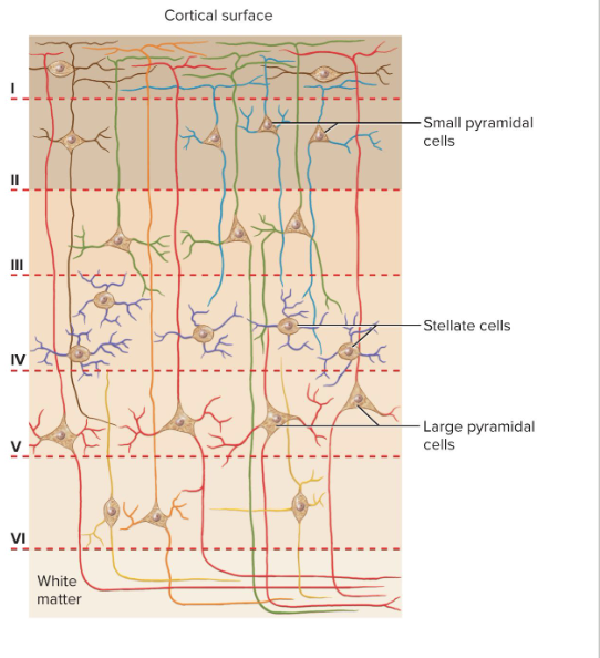
90% of human cerebral cortex is neocortex—six-layered tissue
Contains primarily 2 types of neurons:
stellate cells
pyramidal cells
Stellate Cells
Have sphere-shaped neurosomas with dendrites pointing in all directions
Receives sensory input and processes information on a local level

Pyramidal Cells
Tall and cone-shaped, with apex toward the brain surface
has a thick dendrite with many branches with small knobs
Includes the output neurons of the cerebrum
are the only neurons that leave the cortex and connect with other parts of the CNS
Limbic System
important center of emotion and learning
components:
Cingulate gyrus: arches over corpus callosum in frontal and parietal lobes
Hippocampus
Amygdala: immediately rostral to hippocampus (emotion functions)
There is a limbic system in each cerebral hemisphere
Limbic system components are connected through a loop of fiber tracts
allows for circular patterns of feedback
Limbic system structures have centers for both gratification and aversion
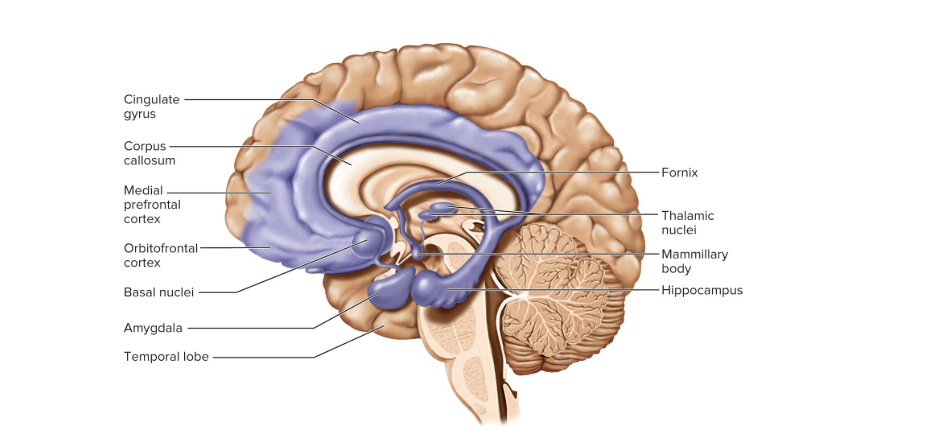
Hippocampus
in medial temporal lobe (memory functions)
performs short term to long term memory
is completely enclosed by the limbic system
Basal Nuclei
masses of cerebral gray matter buried deep in the white matter, lateral to the thalamus
Receive input from the substantia nigra of the midbrain and the motor areas of the cortex
Send signals back to both of these locations
Involved in motor control*
Formed by 3 different brain centers (are collectively called the corpus striatum)
Caudate nucleus
Putamen
Globus pallidus
Lentiform nucleus
putamen and globus pallidus together
Higher Brain Functions:
sleep, memory, cognition, emotion, sensation, motor control, and language
Involve interactions between the cerebral cortex and basal nuclei, brainstem, and cerebellum
Functions of the brain do not have easily defined anatomical boundaries
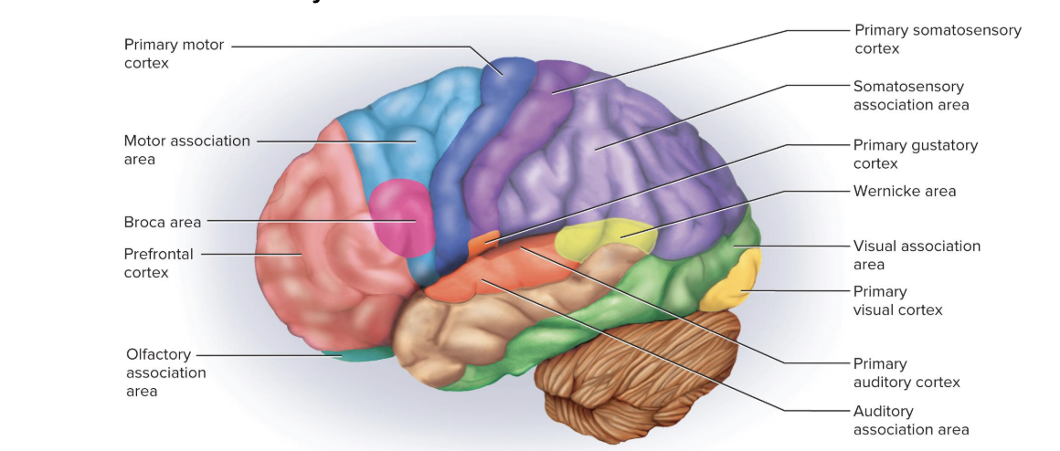
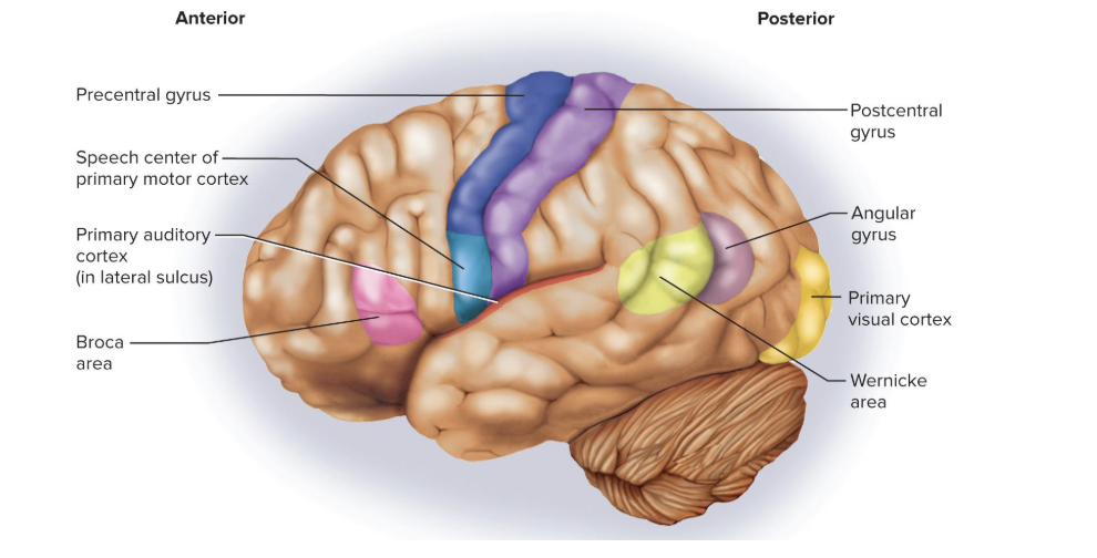
Primary Sensory Cortex
sites where sensory input is first received and you becomes conscious of the stimulus
Association areas near the primary sensory areas process and interpret that sensory information
ex) primary visual cortex, multimodal association areas
located on post-central gyrus
process things like heat, temperature, pressure, not the special senses
Primary Visual Cortex
makes cognitive sense of visual stimuli
is bordered by visual association areas
Multimodal Association Areas
receives input from multiple senses and integrates this into an overall perception of our surroundings
Special Senses
senses limited to the head and employ complex sense organs
vision
hearing
equilibrium
taste and smell
Vision
Visual primary cortex is in the posterior of the occipital lobe
Visual association area: takes up the rest of the occipital lobe
Much of the inferior temporal lobe deals with recognizing faces and familiar objects
Hearing
Primary auditory cortex is in the superior region of the temporal lobe and insula
Auditory association : temporal lobe deep and inferior to the primary auditory cortex
Recognizes spoken words, a familiar piece of music, or a voice on the phone
Equilibrium
Signals for balance and sense of motion are mainly sent to the cerebellum and several brainstem nuclei involved in head and eye movements and visceral functions
Association cortex: in the roof of the lateral sulcus near the lower end of the central sulcus
consciousness of our body movements and orientation in space is processed here
Taste and Smell
Gustatory (taste) signals:
received by primary gustatory cortex in inferior end of the postcentral gyrus of the parietal lobe and anterior region of the insula
Olfactory (smell) signals:
received by the primary olfactory cortex in the medial surface of the temporal lobe and inferior surface of the frontal lobe
sense of smell skips the thalamus, goes straight to cerebrum
General (somesthetic, somatosensory, or somatic) Senses
distributed over entire body and employ simple receptors
Includes touch, pressure, stretch, movement, heat, cold, and pain
For the head, cranial nerves carry general sensory information
For the rest of the body, ascending tracts bring general sensory information to the brain
How are general senses passed along/processed?
The thalamus processes the input from the contralateral side
it then selectively relays signals to the postcentral gyrus of the parietal lobe
Functionally known as the primary somesthetic cortex
Provides awareness of stimulus
processed on postcentral gyrus
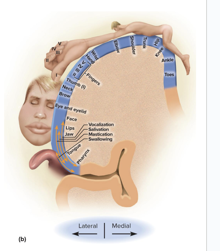
Sensory Homunculus
diagram of the primary somesthetic cortex which resembles an upside-down sensory map of the contralateral side of the body
Shows receptors in lower limbs projecting to superior and medial parts of the gyrus
Shows receptors from face projecting to the inferior and lateral parts of the gyrus
Somatotopy
point-to-point correspondence between an area of the body and an area of the CNS
how we were able to map out the homunculi on the postcentral gyrus
The intention to contract a muscle begins in…
The motor association (premotor) area of the frontal lobes
this is where we plan our behavior
also where neurons make a “program” for the degree and sequence of a muscle contraction required for a certain action
Where does the motor “program” get sent to after being created?
the program is transmitted to neurons of the precentral gyrus (primary motor area)
These neurons send signals to the brainstem and spinal cord leading ultimately to muscle contractions
The Pre-Central Gyrus in Motor Control
exhibits somatotopy
Neurons for toe movement are deep in the longitudinal fissure of the medial side of the gyrus
The summit of the gyrus controls the trunk, shoulder, and arm
The inferolateral region controls the facial muscles
Motor Homunculus
diagram of the motor cortex
has a distorted look because the amount of cortex devoted to a given body region is proportional to the number of muscles and motor units of that body region (not body region size)
voluntary movements only
no abdominal viscera or genitals in this diagram because they are not voluntary
if more fine motor control is needed, the organ takes more surface area on the homunculus
Pyramidal Cells of the Precentral Gyrus
are called upper motor neurons
Their fibers project caudally
lots of their fibers end in nuclei of the brainstem
their fibers also form the corticospinal tracts
Most fibers decussate in the lower medulla oblongata
In the brainstem/spinal cord, the fibers from upper motor neurons connect with lower motor neurons whose axons innervate skeletal muscles
Basal Nuclei in Motor Control
Important motor functions include helping to control:
start and stop of intentional movements
Repetitive hip and shoulder movements in walking
Highly practiced, learned behaviors such as writing, typing, driving a car
- Lie in a feedback circuit from the cerebrum, to the basal nuclei, to the thalamus, and back to the cerebrum
Dyskinesias
movement disorders caused by lesions in the basal nuclei involving abnormal movement initiation
Cerebellum in Motor Control
Highly important in motor coordination
Aids in learning motor skills
Maintains muscle tone and posture
Smooths muscle contraction
Coordinates eye and body movements
Coordinates motions of different joints with each other
Lesions can cause ataxia
Ataxia
clumsy, awkward gait
alcoholics can have lesions in their cerebellum, causing ataxia
causes them lose ability to regulate motor movements
causes constant tremors
Language includes:
reading, writing, speaking, and understanding words
Wernicke’s Area
Posterior to lateral sulcus usually in left hemisphere
Permits recognition of spoken and written language
When we want to speak, Wernicke’s area makes phrases and transmits a plan of speech to Broca’s area
controls comprehension of language

Broca’s Area
Inferior to the prefrontal cortex, usually in left hemisphere
Generates a motor program for the muscles of the larynx, tongue, cheeks, and lips for speaking and for hands when signing
Transmits the program to the primary motor cortex for commands to the lower motor neurons that lead to muscles
controls movement of mouth for speech
Affective Language Area
usually in right hemisphere
Lesions produce aprosody
Aprosody
flat emotionless speech
Aphasia
a language deficit from lesions to the hemisphere with Wernicke and Broca areas
Nonfluent (Broca) Aphasia
caused from a lesion in Broca’s area
Slow speech
difficulty in choosing words
using words that only approximate the correct word
Fluent (Wernicke) Aphasia
Caused from a lesion in Wernicke’s area
Speech is normal and excessive, but uses senseless sentences
Cannot comprehend written and spoken words
Anomic Aphasia
Can speak normally and understand speech, but cannot identify written words or pictures
Cerebral Lateralization
the difference in the structure and function of the cerebral hemispheres
Left Hemisphere
usually the categorical hemisphere
Specialized for spoken and written language
Sequential and analytical reasoning (math and science)
Breaks information into fragments and analyzes it
Right Hemisphere
usually the representational hemisphere
Perceives information in a more integrated way
Involves imagination and insight
Musical and artistic skill
Perception of patterns and spatial relationships
Comparison of sights, sounds, smells, and taste
Differences in lateralization between genders and right vs. left handed:
Right handed:
left hemisphere is typically the categorical one
Left-handed:
left hemisphere is typically the categorical one, but less than righties
some lefties have neither hemisphere specialized
Lateralization differs with age and sex
Children are more resilient to lesions on one side
Males exhibit more lateralization than females and suffer more functional loss when one hemisphere is damaged
Cranial Nerves
12 pairs of cranial nerves
arise from the base of the brain
Exit the cranium through foramina
Lead to muscles and sense organs located mainly in the head and neck
allows us to still have coordination even if we have a spinal cord injury
Cranial Nerve Pathways (sensory vs motor)
motor fibers of the cranial nerves begin in the brainstem and lead to glands and muscles
Sensory fibers begin in receptors located mainly in head and neck and lead mainly to the brainstem
Most cranial nerves carry fibers between brainstem and ipsilateral receptors and effectors
so a lesion in the brainstem causes a deficit on the same side
Exceptions:
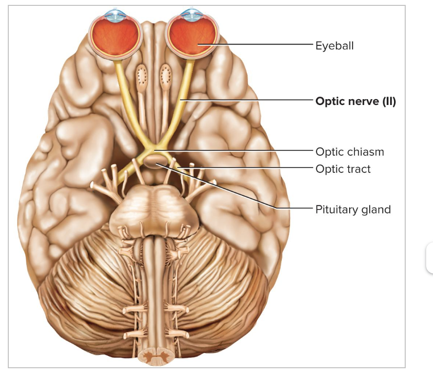
optic nerve: half the fibers decussate
trochlear nerve: all efferent fibers lead to a muscle of the contralateral eye
Sensory Cranial Nerves:
l, ll, Vlll
Motor Cranial Nerves:
III, IV, VI, XI, and XII
Stimulate muscle but also contain fibers of proprioception
Mixed Cranial Nerves:
V, VII, IX, X
Sensory functions may be quite unrelated to their motor function
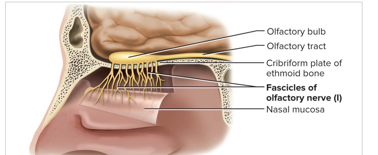
Olfactory Nerve (l)
sends the sense of smell to the brain
has rootlets into the nasal cavity
Optic Nerve (ll)
Used to send visual signals to the brain using the optic chiasma
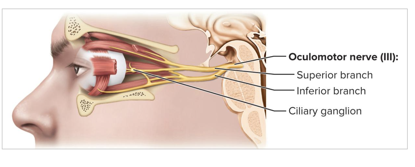
Oculomotor Nerve (lll)
Control the size of the pupil and movement of eye
Controls:
Superior Rectus
Inferior Rectus
Medial Rectus
Inferior Oblique
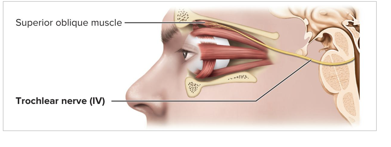
Trochlear Nerve (lV)
Controls movement of eye
controls the superior oblique muscle
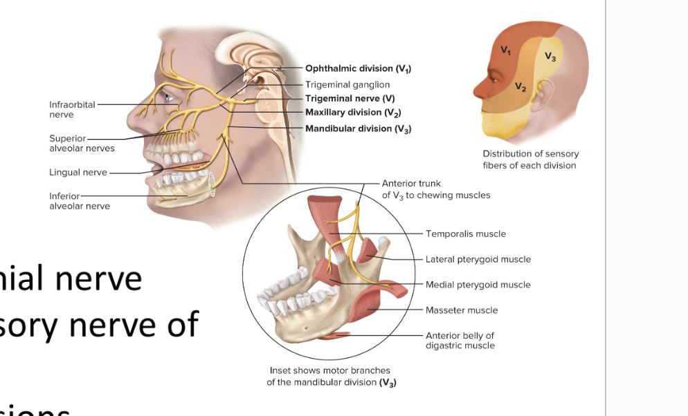
Trigeminal Nerve (V)
largest cranial nerve
most important sensory nerve of the face
forks into 3 divisions
Ophthalmic Division (V1)
sensory
Maxillary Division (V2)
sensory
Mandibular Division (V3)
mixed
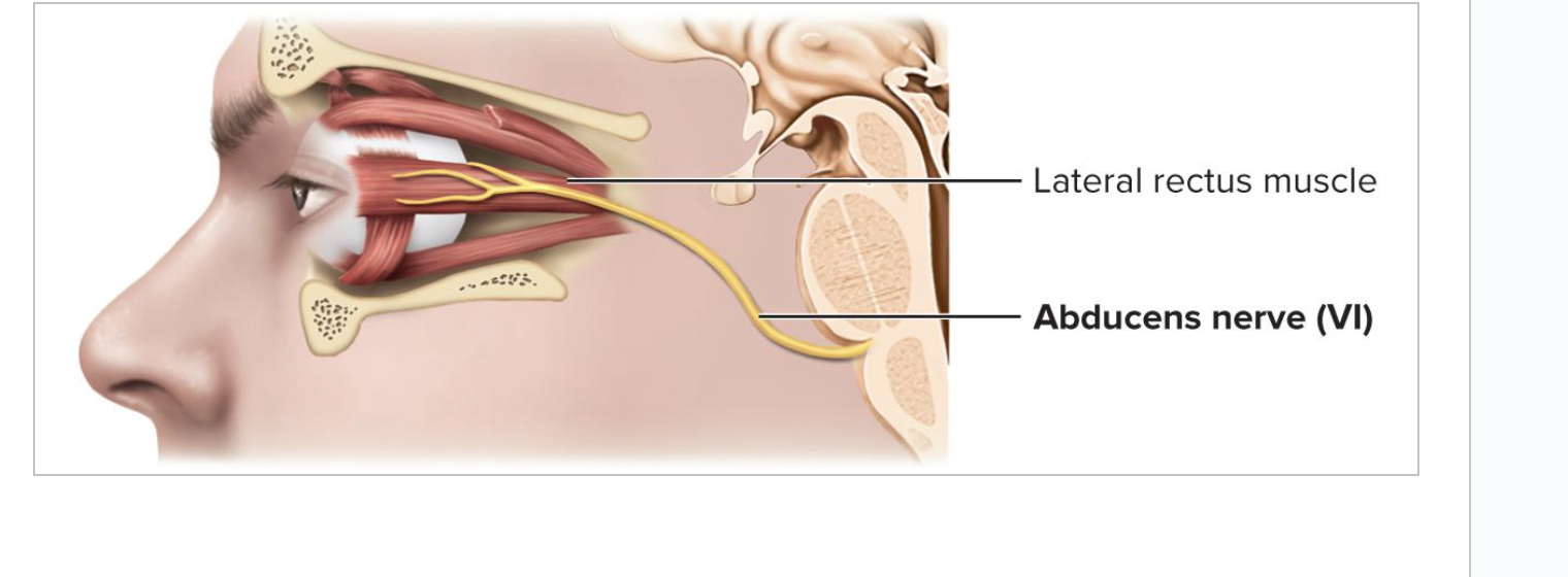
Abducens Nerve (Vl)
Movement of eyeball
controls lateral rectus muscle
allows us to move eyes laterally
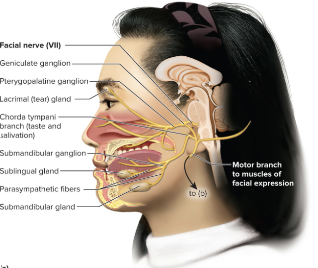
Facial Nerve (Vll)
Motor function:
is the major motor nerve of facial muscles:
facial expressions, salivary glands, tear, nasal, and palatine glands
Sensory function:
controls taste on anterior two-thirds of tongue
Damage:
Damage produces sagging facial muscles and disturbed sense of taste
Clinical test:
test anterior two-thirds of tongue with sugar, salt, vinegar, and quinine
test response of tear glands to ammonia fumes
test motor functions by asking subject to close eyes, smile, whistle, etc.
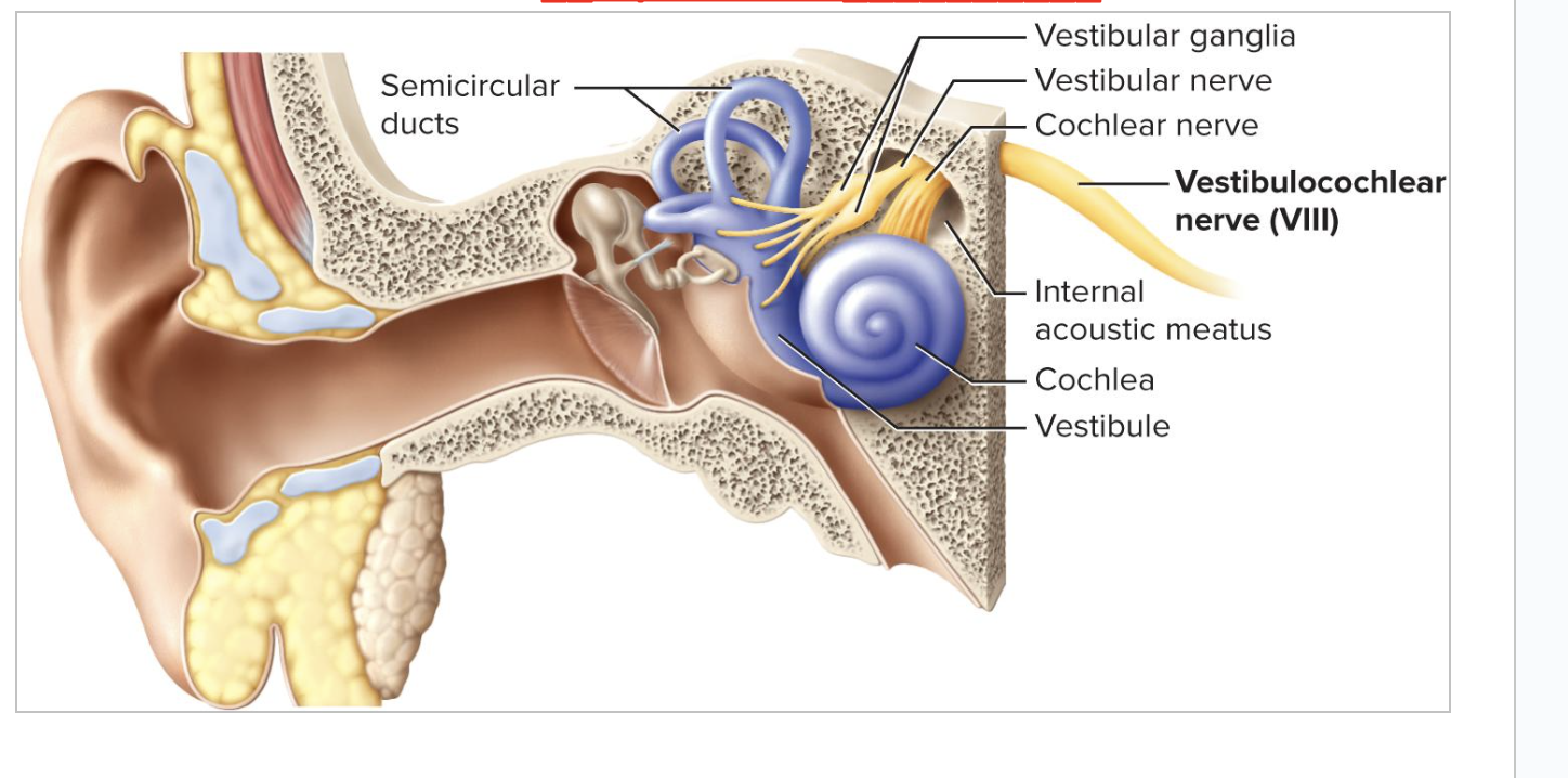
Vestibulocochlear Nerve (Vlll)
Controls sense of equilibrium and sound
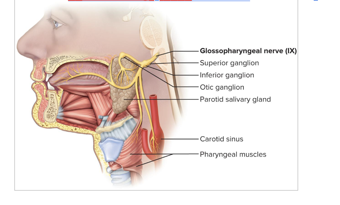
Glossopharyngeal Nerve (lX)
Used to control tongue, salivary glands, and swallowing muscles
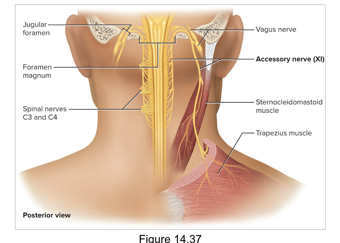
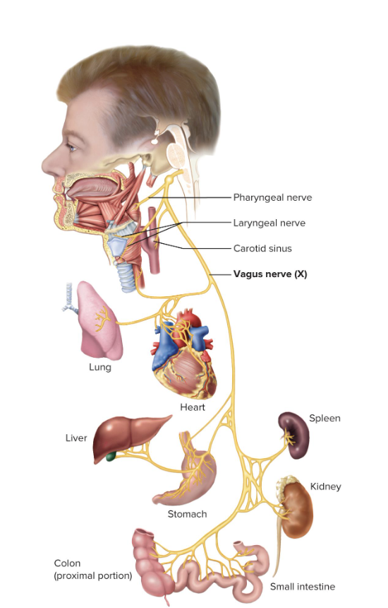
Vagus Nerve (X)
The wandering nerve
Most extensive distribution of any cranial nerve
Major role in the control of cardiac, pulmonary, digestive, and urinary function
Controls swallowing, speech, and regulation of viscera
Damage causes hoarseness or loss of voice, impaired swallowing
fatal if both are cut
Accessory Nerve (Xl)
Movement of sternocleidomastoid and trapezius