GBS709 Week 1
 Call Kai
 Learn
Learn Practice Test
 Spaced Repetition
 Match
Match Flashcards
 Knowt Play
1/108

There's no tags or description
Looks like no tags are added yet.
Name | Mastery | Learn | Test | Matching | Spaced | Call with Kai | Chat |
|---|
No analytics yet
Send a link to your students to track their progress
109 Terms
list membrane bound organelles and non-membrane bound***
membrane bound: plasma membrane, nucleus, endoplasmic reticulum, golgi apparatus, lysosomes, endosomes, peroxisomes, mitochondria
non-membrane bound: ribosomes, proteasomes, cytoskeleton elements, centrioles, basal bodies, cilia, flagella

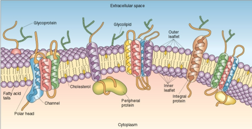
what is dis?
fluid mosaic model, contains lipid rafts which serve as “signaling platforms”
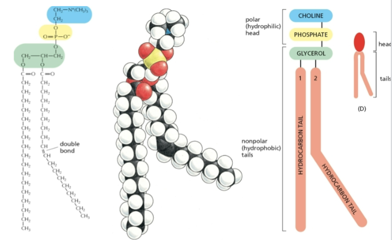
glycerophospholipid
main phospholipid in most animal cell membranes
lipid composition
polar, hydrophilic head: choline, phosphate
nonpolar hydrophobic tails: 3C glycerol backbone, hydrocarbon tails with double bonds
Lipids that form a bimolecular leaflet are ______?
amphipathic meaning they contain hydrophobic and hydrophilic components
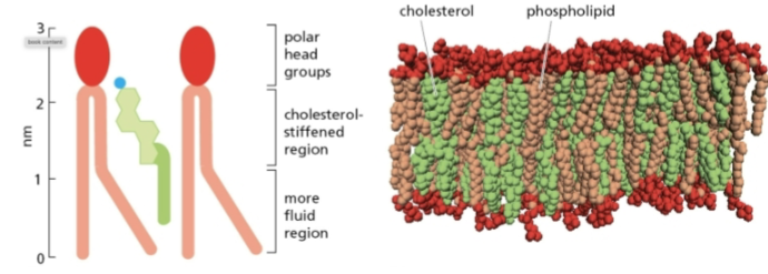
cholesterol
rigid, in lipid bilayer
cell membranes are composed of
proteins and lipids
3 major lipids in cell membrane
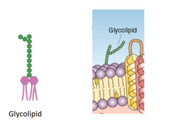
phospholipids, glycolipids, cholesterol
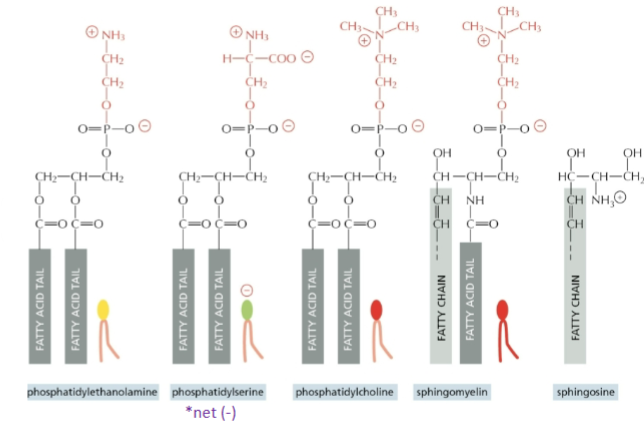
4 major phospholipids
phosphatidylethanolamine, phosphatidylserine (net negative), phosphatidylcholine, sphingomyelin, sphingosine
2 major protein groups in cell membrane
Integral: transmembrane, multipass
Peripheral: extracellular, cytosolic
6 functions of cell membranes
maintains structural integrity
controls movement of substances in and out
regulates cell-cell interactions
serves as recognition board via receptors
establishes transport system for specific molecules
transduces extracellular physical and chemical signals into intracellular events
4 kinds of membrane proteins
channel proteins, pump + carrier proteins, surface receptors, linkers + structural prteins
channel proteins + 3 examples
allow passage of ion or charge; voltage-gated, ligand-gated, mechanically-gated
pumps and carrier proteins + 3 examples
bind and transport ions and molecules; Na+- K+ pump, Ca++ pumps, glucose transporter
surface receptors + 2 examples
bind to ligand to elicit cellular response
involved in receptor-mediated endocytosis
signaling molecules which are or are associated with kinases (enzymes that phosphorylate)
coupled to G-proteins
ex) cytokine and steroid receptors
linkers and structural proteins
cell-cell attachment; cell-matrix attachment, scaffolds for cytoskeleton
Histogenesis***
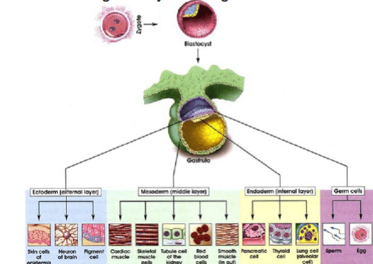
formation or development of tissues, begins at gastrulation (day 15/16 in early embryogenesis)
Gastrulation***
process where 3 germ layers are generated and placed in proper position in embryo:
ectoderm externally
mesoderm in between
endoderm internally
4 essential processes for generation of multicellular organism***
cell proliferation, specialization, interaction, and movement

4 tissue types
epithelial, connective, muscle, nerve
epithelial tissue/epithelium + 2 types
sheet covering free surfaces, creates border between environment and organ, controls movement of substances
cells collectively bound and sit on basement membrane
basis for gland formation
avascular; no blood vessels
polarized structure and function
derived from all 3 germ layers (ectoderm, mesoderm, endoderm)
endothelium vs epidermis***
endothelium - epithelium lining blood vessels, epidermis - epithelium of skin
basement membrane
attaches epithelium to underlying connective tissue by specialized cell junctions, PAS-positive layer is visible by light microscopy
7 functions of epithelium ***
protection: epidermis
absorption: digestive tract
secretion: secretory cells within glands are derived from epithelial tissue
excretory: kidneys
lubrication: pleura, pericardium and peritoneum; allows movement of organs within cavities.
sensory: many specialized sensory cells are derived from epithelium such as taste buds, retinal pigmented epithelia
reproduction: sperm are modified epithelial cells transformed into spermatozoa
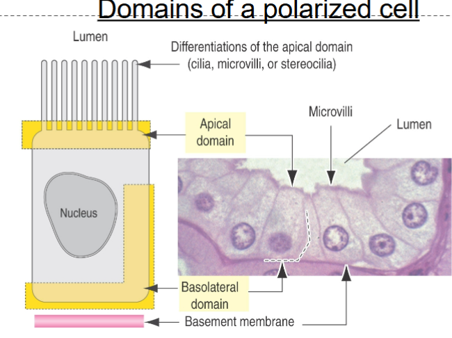
2 domains of polarized cell
apical and basolateral, results in polarization of cell in epithelium
Basal Lamina
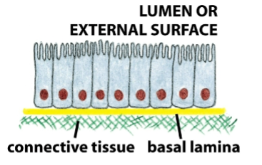
secreted by epithelial cells, interface between epithelia and underlying supportive tissues
permits movement of nutrients to epithelial cells from blood, and movement of metabolites towards blood
binds and concentrates growth factors in cell growth and proliferation
influences cell metabolism and cell-to-cell interactions/signaling
part of basement membrane
2 types of epithelial glands
Exocrine: organized into specific units that secrete into a tubular network of ducts
Endocrine: cells that secrete directly into blood stream
all glands are derived from epithelial tissue

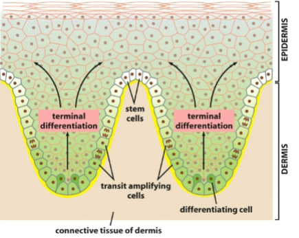
Epithelium Renewal
variable; fast (intestinal epithelium replaced weekly) or slow (large glands)
in stratified layers, mitosis only occurs in cells at basal layer
rapid repair and replacement of damaged cells
carcinomas
tumor of epithelial origin. adenocarcinomas are tumors of glandular epithelial
Connective Tissue + know diagram
mesodermal origin, tissue-specific ECM consisting of fibers and ground substance (clear, colorless gel-like fluid) with blood vessels and nerves (except cartilage)
forms a continuum with epithelial, muscle and nervous tissues
stores fat as adipose tissue
facilitates signaling; cell membrane receptors bind matrix molecules to control adhesion, differentiation, cell cycle
defense, protection, wound healing, tissue repair
ex: connective tissue proper (loose or dense), cartilage, bone, adipose tissue, blood, mesenchymal connective tissue (embryonic, undifferentiated)
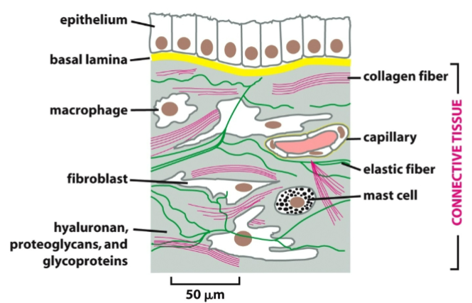
2 examples of connective tissue
parenchyma: perform major function of that organ (hepatocytes in liver, alveolar cells in lung)
stroma: connects/supports other elements of tissue
2 types of connective tissues ***
connective tissue proper (loose and dense w/ irregular and regular) and specialized connective tissue (cartilage, bone. blood, adipose)
2 types of cells in connective tissue
resident (fibroblasts, adipocytes, pericytes, mast, tissue macrophages, stem cells) and transient (white blood cells)
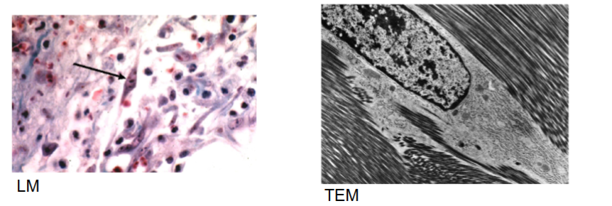
Fibroblasts
principal cell of connective tissue, synthesis of ECM fibers (collagen, elastic, reticular) and ground substance, rarely divide but mitosis resumes during wound healing, not terminally differentiated
Adipocytes
energy storage, insulation, cushioning of organs and secretion of hormones
large cells, up to ~100um
lipid mass is not membrane bound
white (unilocular) or brown (multilocular)
hormones involved in weight control
leptin and insulin
Leptin
produced exclusively by adipocytes, reduce appetite (obese people have high levels and are resistant to leptin activity)
insulin
stimulates uptake and synthesis of triglycerides
blood
specialized connective tissue, develop in bone marrow and enter circulation fully formed
adults have ~5-6 liters
red blood cells and platelets generally remain in vascular system
white blood cells can leave circulation and enter interstitial compartments, only lymphocytes can return to vascular compartment
6 functions of blood ***
transports: gases (O2, CO2), nutrients, hormones, chemical signals
buffer system: maintains pH at 7.4
temperature control: conjunction with blood vessels that dilate or constrict to release or conserve heat
removal: cellular and metabolic wastes
immune functions: defense against infection
coagulation functions: prevent massive blood loss
hematocrit of human blood
volume of packed erythrocytes in a sample of blood (cells 45%, plasma 55%, leukocytes and platelets 1%). a normal hematocrit is 39-50% in males and 35-45% in females

plasma
fluid of blood, 55% of total blood volume, liquid extracellular material (ECM). Plasma w/o coagulation is called serum
7 key components of blood (may not be important, idk)
albumin (most abundant protein in plasma)
immunoglobulins (IgA, IgG, IgM, IgE, IgD), Complement system
lipoproteins; chylomicrons, VLDL, IDL,LDL (‘bad cholesterol”), HDL
haptoglobin, plasminogen, transferrin, and transport proteins.
fibrinogen and other clotting molecules
hormones (soluble or protein bound)
electrolytes, nutrients, gases, etc.

blood composition ***
plasma (water, protein, solutes)
erythrocytes
buffy coat (platelets and leukocytes)
Cartilage
specialized connective tissue, part of skeletal system
provide flexible support (bone rigid template for bone formation)
in respiratory system, joints, external ear
avascular, no nerves or lymphatics
composed of cells (chondroblasts, chondrocytes) + matrix (fibers: collagen II, elastic fibers and ground substance: proteoglycans and glycosaminoglycans/GAGs)
shock absorber: w/ pressure, water forced out of tissue to absorb pressure, w/o pressure, water rebinds PG aggregate and tissue returns to original size
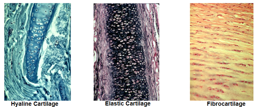
3 types of cartilage
Hyaline: most common arrangement – nasal septum, joint surface, ribs
Elastic: like hyaline but enriched with elastic fibers – ear, larynx
Fibrocartilage: rows of chondrocytes with increased fibrous matrix – intervertebral disks, tendon/ligament attachment
bone
calcium store (also Mg++ and Na+), hematopoiesis (marrow)
alive, dynamic and continually remodeling
highly vascularized (compared to cartilage)
composed of cells and matrix (inorganic and organic)
organic is fibers: type I collagen, highly organized and ground substance: little, some PG as cartilage
inorganic: 50% of matrix is calcium phosphate complexes, rigid

3 types primary cells in bone ***
Osteoblasts: immature, synthesize and secrete osteoid which becomes mineralized to give bone; do not divide.
Osteocytes: surrounded by and maintain matrix; do not divide.
Osteoclasts: large multinucleated macrophages, from monocyte lineage, remodel bone by resorbing bone matrix.

2 types mature bone ***
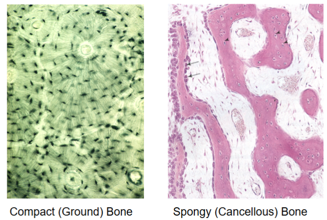
Cancellous (spongy): trabeculae (fine irregular plates), inside long bones (marrow), gives strength without weight
Compact: highly ordered, outer and inner circumferential lamellae, contains Haversian systems (osteons)
muscle
contractile cells that are responsible for movement.
contraction is dependent on interactions between actin and myosin (rearrangement of fibers will differ)
voluntary/involuntary; striated or nonstriated
3 types muscles
Skeletal: striated/voluntary, attached to bone (bicep, oblique), few places not bundled (tongue, esophagus)
Cardiac: striated/involuntary, wall of heart and base of veins that empty into heart.
Smooth: non-striated/involuntary, lining blood vessels, visceral organs

Muscle Tissue
derived from mesoderm
sarcolemma (cell membrane) vs sarcoplasm (cytoplasm)
sarcoplasmic reticulum (SR) is modified ER.
contraction of muscle depends on interaction of actin and myosin and is regulated by cellular calcium levels
muscle tissue cells
length up to ~30 cm; diameter 10-100 um
multinucleated syncytium (muscle fiber)
continuous with tendon or other dense connective tissue
highly vascularized
skeletal muscle tissue
bundles of long, cylindrical multinucleated cells w/ cross striations
contraction thro thin actin and thick myosin filaments
controlled by somatic nervous system
attached to bone
visceral striated muscle (oral cavity, larynx, pharynx, upper esophagus, anal region)
extraocular muscles in eye
3 types of connective tissue associated with muscle tissue ****
epimysium: dense connective tissue surrounding entire muscle.
perimysium: connective tissue surrounding bundles of muscle fibers
endomysium: delicate connective tissue surrounding each muscle fiber
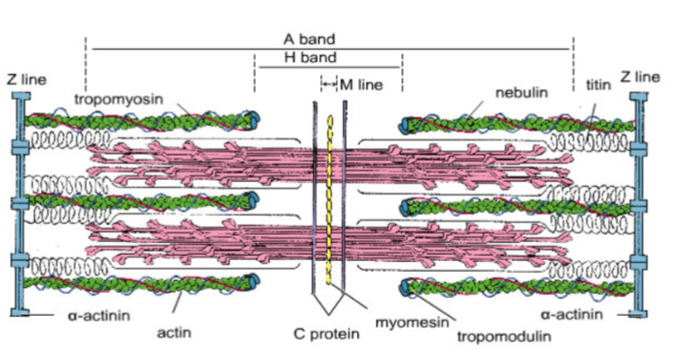
Sarcomere
extends from Z-line to Z-line, smallest repetitive subunit of contractile unit
Arrangement of Thick and Thin Filaments
have an appreciate for this image
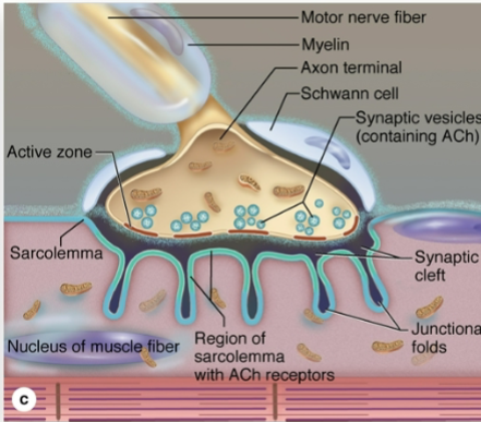
Neuromuscular Junction
this type of junction is only found in voluntary muscle
Cardiac Muscle Tissue
tightly knit bundles of cells, produces a characteristic wave of contraction that leads to a wringing out of heart ventricles
Cardiac Muscle Cells ***
15 um in diameter, 85-100 um long
cross-striated banding pattern and sarcomere arrangement/function same as skeletal muscle
each cell has 1-2 central nuclei, covered by sheath of endomysial connective tissue w/ capillary network w/ no perimysium or epimysium
instead of forming syncytial cells like skeletal muscles, they branch and form complex junctions between their processes and neighboring cells
intercalated disks: dark-staining transverse lines that cross chains of cardiac cells at irregular intervals.
cardiac muscle contraction passed from cell to cell via communication through gap junctions.
smooth muscle tissue
elongated, nonstriated, fusiform cells (largest at midpoint and tapered at ends)
cells enclosed by basal lamina and network of reticular fibers; both combine forces generated by smooth muscle fiber into concerted action
smooth muscle contraction is passed from cell to cell via communication through gap junctions.
found lining GI tract (esophagus, stomach, intestines), respiratory tract, uterus, bladder and urethra, lining blood vessels, dermis of skin (arrector pili muscles)
tight packaging of tissue achieved by lining up cells with narrow parts of some cells against broad parts of neighboring cells
contain dense bodies for attachment of filaments and propagation of contraction
nervous system
receives, transmits and integrates information from outside and inside body to control and coordinate activities in body
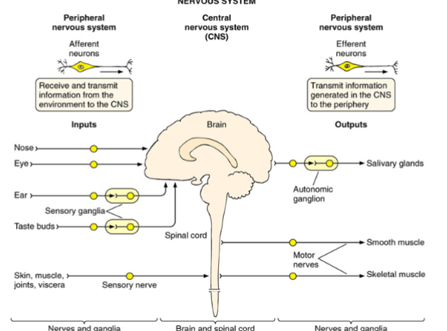
Central Nervous System (CNS) and Peripheral Nervous System (PNS)
Central Nervous System (CNS)
brain, spinal cord, eyeballs
Peripheral Nervous System (PNS)
peripheral (cranial and spinal) nerves and ganglia (aggregations of nerve cell bodies, somatic/sensory and autonomic)
somatic and autonomic (parasympathetic division (rest and digest), sympathetic division (fight or flight), enteric division (GI function))
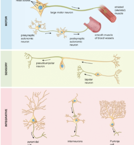
Neurons (nerve cells)
nerve cells specialized to transmit electrical impulses, made of axon, cell body and dendrites; morphology (uni-, bi-, multi-polar), function (integrative, sensory, motor)
11 organ systems
gastrointestinal (digestive)
musculoskeletal
integumentary (skin)
blood and lymphatic
endocrine
nervous
skeletal
reproductive
pulmonary
urinary
cardiovascular
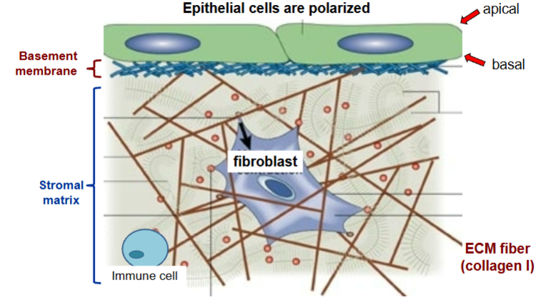
epithelial cells
polarized, apical and basal, consist of basement membrane and stromal matrix
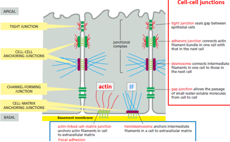
cell-cell junctions
tight, adherens, desmosome, gap
tight junction
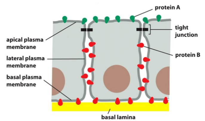
seals gap between epithelial cells
restricts flow of solutes, water, and movement of plasma membrane proteins
this creates apical and basolateral domains with different surface proteins
mediated by claudins and occludins/tricellulins
tight epithelium vs endothelium
tight epithelium has a lot of tight junctions such as the collecting duct in kidney. tight endothelium (blood-brain barrier)
apical vs basal surfaces
apical: lumen of intestine. basal: supportive connecting tissue and muscle
adherens junction
connects actin filament bundle in cells, mediated by cadherins (E-cadherin in epithelial cells)
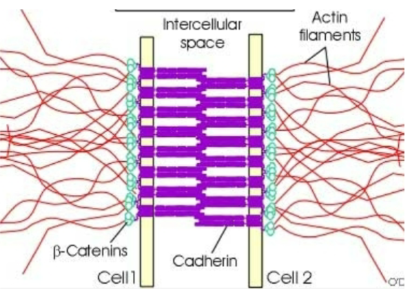
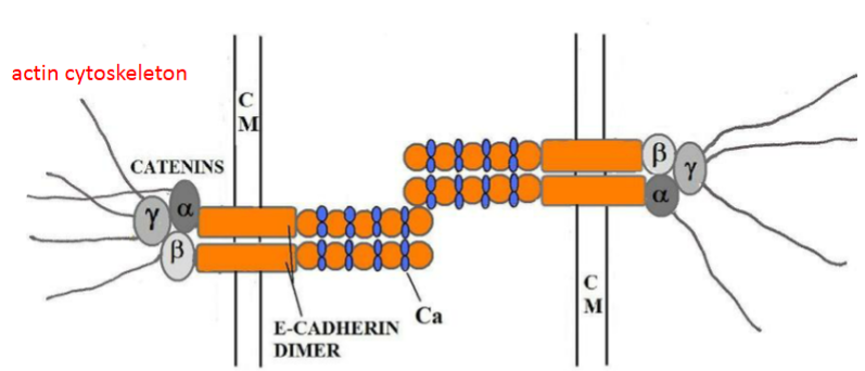
E-cadherins
homodimers, mediates epithelial cells in adheren junctions
E-cadherins and cell association regulated by Ca++ binding
this causes E-cadherin to extend out like a rod, enabling contact with other cell
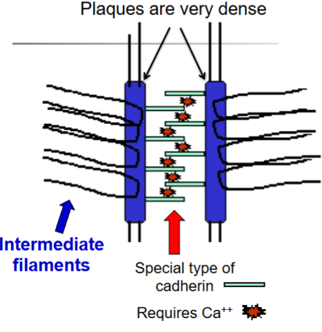
desmosome
dense plaques, connects intermediate filaments in cells, mediated by desmoglein (a type of cadherin), requires Ca++
gap junction
allows passage of small water-soluble molecules from cell to cell

cell-matrix junctions
actin-linked cell-matrix, hemidesmosome
actin-linked cell-matrix junctions
anchors actin filaments in cell to extracellular matrix, focal adhesion
hemidesmosome
anchors intermediate filaments in a cell to extracellular matrix
Cell-cell and cell-matrix junctions are critical for_____?
maintaining cell polarity!
why is apical-basal polarity important?
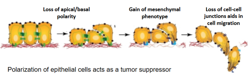
critical for normal cell function, locomotion and formation of immune synapse, polarization of epithelial cells also acts as a tumor suppressor
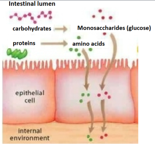
what does loss of polarity (loss of cell-cell or cell-matrix adhesion) cause?
de-differentiation
epithelial to mesenchymal transition
increased migration/invasion
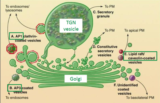
How do you get different proteins on the apical surface versus the basal surface?
cargo sorting by the trans-golgi network (TGN) - specific coat proteins direct TGN vesicles to targets
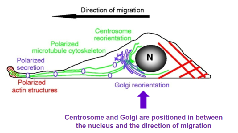
6 steps as to how polarity causes cell locomotion
direction of movement
front protrusion: protrusion forms at leading edge w/ F-actin polymerization by Rac, Cdc42, Rho (G-proteins that organize cytoskeleton)
front adhesion: formation of new adhesion
cell-body translocation: actomyosin contraction allows cell body to translate
tail deadhesion: adhesion at trailing end disintegrates and F-actin depolymerizes
tail retraction
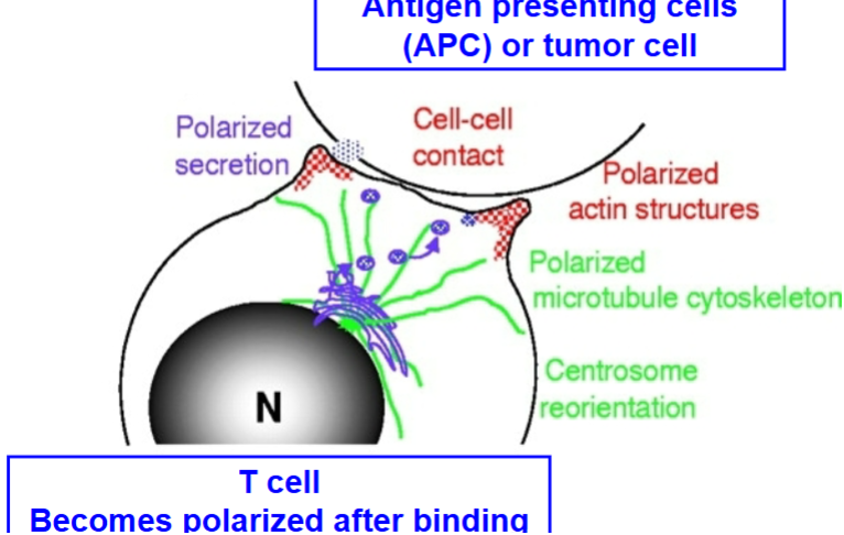
how does polarity form the immune synapse?
Antigen presenting cells (APC) or tumor cell causes T cell to become polarized after binding
cytoskeletal filaments + 3 types
polymers composed of non-covalently-bound monomers
interaction between two monomers is weak and noncovalent, stabilized by multiple bonds
weak bonds between individual monomers allows filament polymerization to be dynamic
actin filaments (microfilaments), intermediate filaments, microtubules
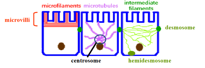
actin filaments
determine cell shape, and regulate cell locomotion
2 protofilaments that twist around in a right-handed helix, POLAR
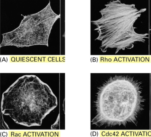
can combine to form actin bundles (stress fibers), requires accessory G-proteins
4 G-proteins involved in actin bundle formation
quiescent cells, Rho activation, Rac activation, Cdc42 activation
actin monomer
globular structure, ATP cap
have ATP bound because more ATP than ADP in cell
can hydrolyze ATP
intermediate filaments
rope-like, provide mechanical stability and serve as scaffolds for signaling molecules
stabilize epithelial and endothelial cell monolayers in the presence of shear stresses
NO POLARITY (ends are symmetrical)
regulation of polymerization appears to involve phosphorylation
can withstand greater deformation and force without breaking compared to other cytoskeleton filaments (MTs or actin)
intermediate filaments composition
Keratins (variant) – epithelial cells
Vimentin – fibroblasts, endothelial cells, leukocytes, contribute to mechanical stability of lymphocytes
Desmin – muscle cells
Glial fibrillary acidic protein – glia
Lamins – ubiquitously expressed, localized to nucleus
how are intermediate filaments made?
a-helical region in monomer
coiled-coil dimer
staggered tetramer of 2 coiled-coil dimers
2 tetramers packed together
8 tetramers twisted into rope filament
microtubules
determine positions of membrane-enclosed organelles, and direct chromosomal separation during mitosis
made of α/β tubulin dimers w/ GTP cap, have ring of 13 protofilaments w/ + and - ends
originate in centrosome, lie just under the plasma membrane to provide support

how does the polar structure of actin and microtubules contribute to polymerization? also what happens if the GTP cap is lost?
head and tail ends of polymer are different w/ ATP (actin) or GTP (microtubules) on one end, and ADP/GDP at the other
+ end: fastest polymerization
- end: slowest polymerization
ATP or GTP “caps” promote assembly
ADP/GDP ends tend to disassemble
GTP cap forces GDP-bound monomers to stay in a linear conformation
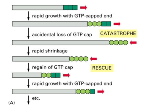
filament disassembles if cap is lost
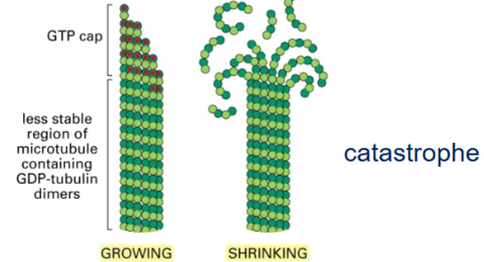
DYNAMIC INSTABILITY
minor, temporary fluctuations in rates of assembly/disassembly, characteristic of microtubule polymerization.
TREADMILLING
rate of assembly at plus end exactly matches the rate of disassembly at the minus end
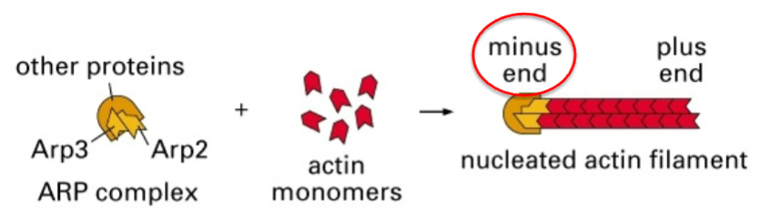
ARP-mediated nucleation of actin filaments
on inner leaflet of plasma membrane – cell locomotion
on side of actin filament – creates web-like structure
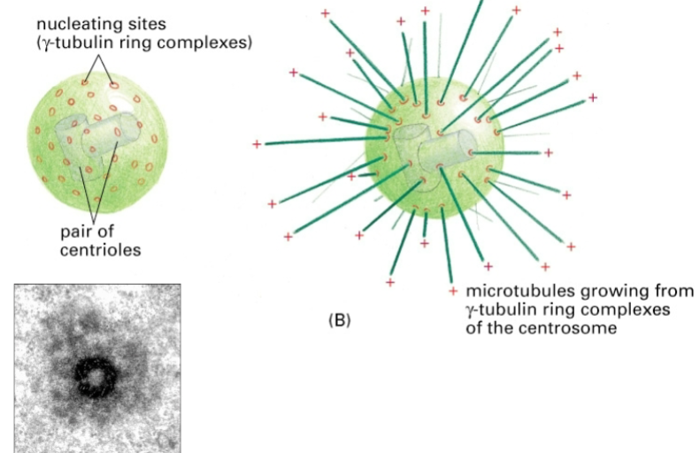
Nucleation of microtubules
γ-tubulin forms a ring-like structure that serves as nucleating site for microtubules
what do accessory proteins that modulate filament polymerization or stability do exactly? + 3 examples
regulate nucleation (proteins that help/inhibit recruitment of monomers to nucleation site)
stabilize/destabilize pre-existing filaments by binding to sides/ends of polymer
filament-cleaving/bundling proteins, actin-bundling, microtubule-bundling
Filament bundling proteins
Actin bundling proteins, can link actin with α-actin, fimbrin, and filam
actin filaments linked to α-actin
creates a contractile bundle: loose packaging allows myosin-II to enter bundle, stress fibers
stress fibers
actin filaments linked to fimbrin
causes parallel bundle: tight packaging prevents mysoin-II from entering bundle
gel-like network, cell cortex
bundles within microvilli