ANATOMY & PHYSIOLOGY EXAM 1
0.0(0)
Card Sorting
1/118
There's no tags or description
Looks like no tags are added yet.
Study Analytics
Name | Mastery | Learn | Test | Matching | Spaced |
|---|
No study sessions yet.
119 Terms
1
New cards

Which one is the Pacinian corpuscle/mechanoreceptor:
E
2
New cards

Which one is the Sebaceous gland:
C
3
New cards

Which one is the Papillary layer
I
4
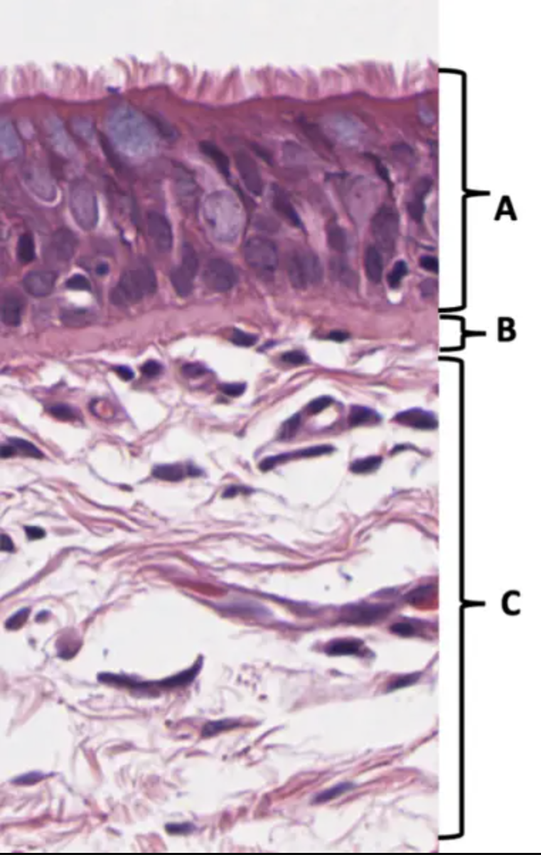
New cards
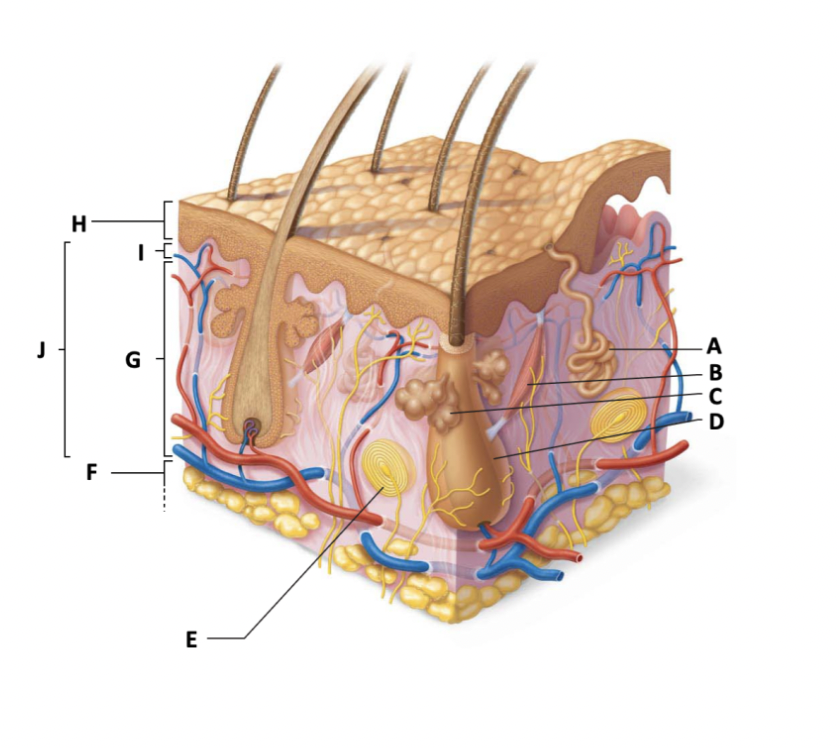
Which one is the Arrector pili muscle:
B
5
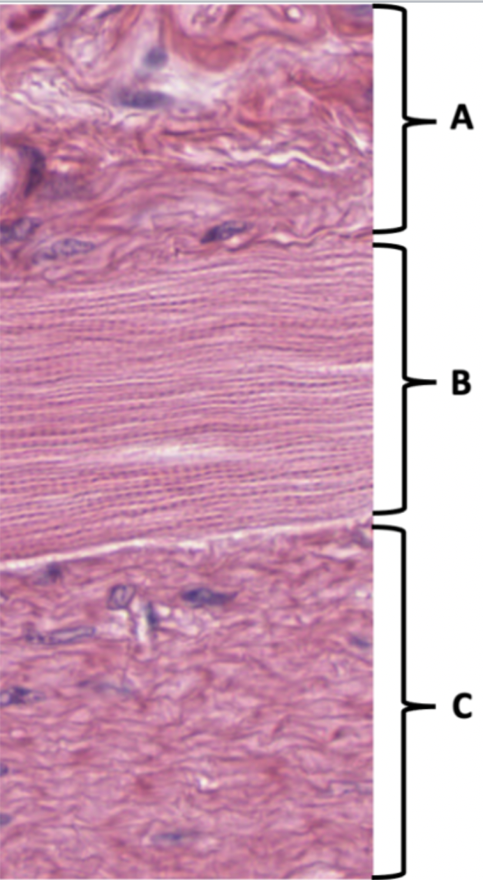
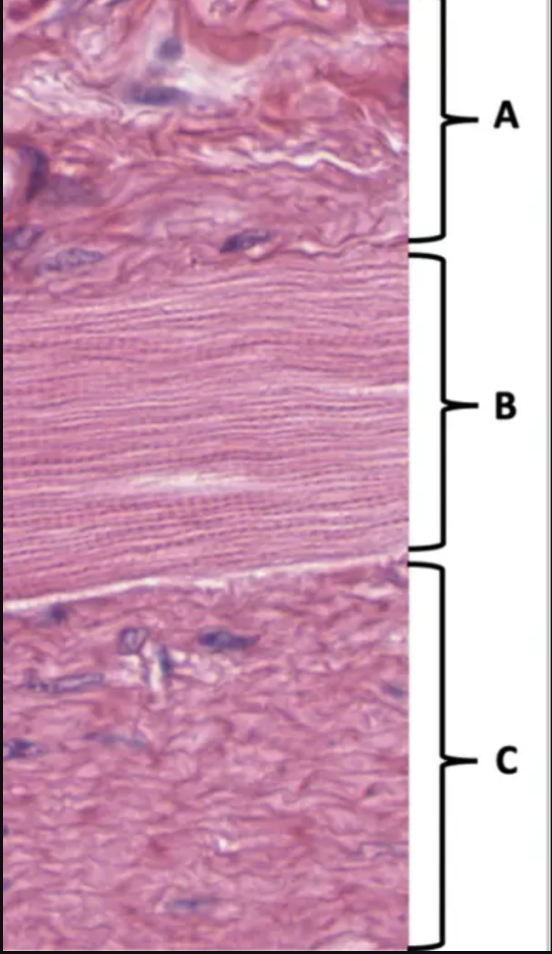
New cards

Which one is the Reticular layer:
G
6
New cards

Which one is the Hair follicle
D
7
New cards

Which one is the Sweat/eccrine gland
A
8
New cards

Which one is the Epidermis
H
9
New cards

Which one is the Dermis
J
10
New cards

Which one is the Hypodermis
F
11
New cards

What organ is this?
This organ is from the digestive system (small intestine)
12
New cards

Name the tissue at D:
__**Name the tissue at D:**__ loose irregular connective tissue, areolar connective tissue
13
New cards

Which letter identifies the structure that increases the surface area for absorption:
__**Which letter identifies the structure that increases the surface area for absorption**__: A
14
New cards
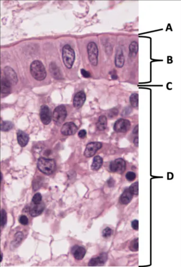
Name the tissue at B
__**Name the tissue at B:**__ simple columnar epithelia
15
New cards

Name the structure at C:
__**Name the structure at C:**__ basal lamina, basement membrane
16
New cards
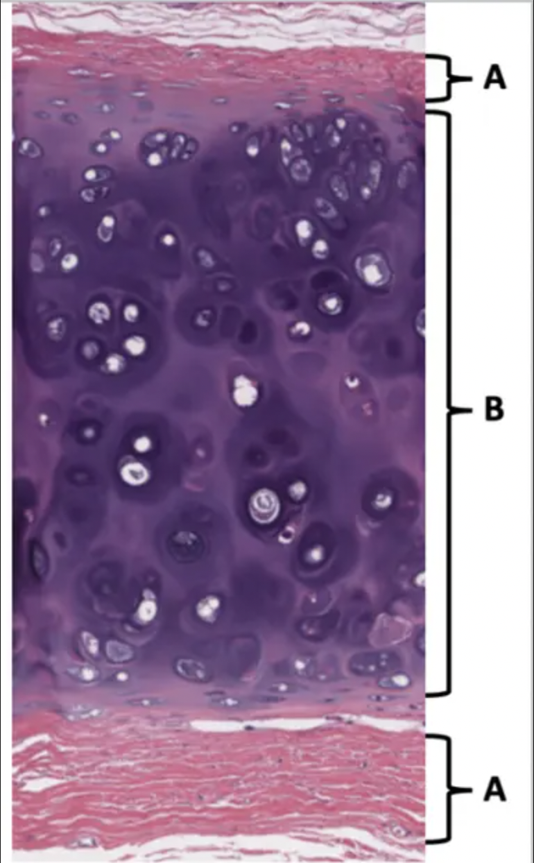
Name the tissue at A:
dense regular connective tissue
17
New cards

Name the tissue at B:
cartilage, hyaline cartilage
18
New cards

Name the tissue at A:
stratified squamous epithelium
19
New cards
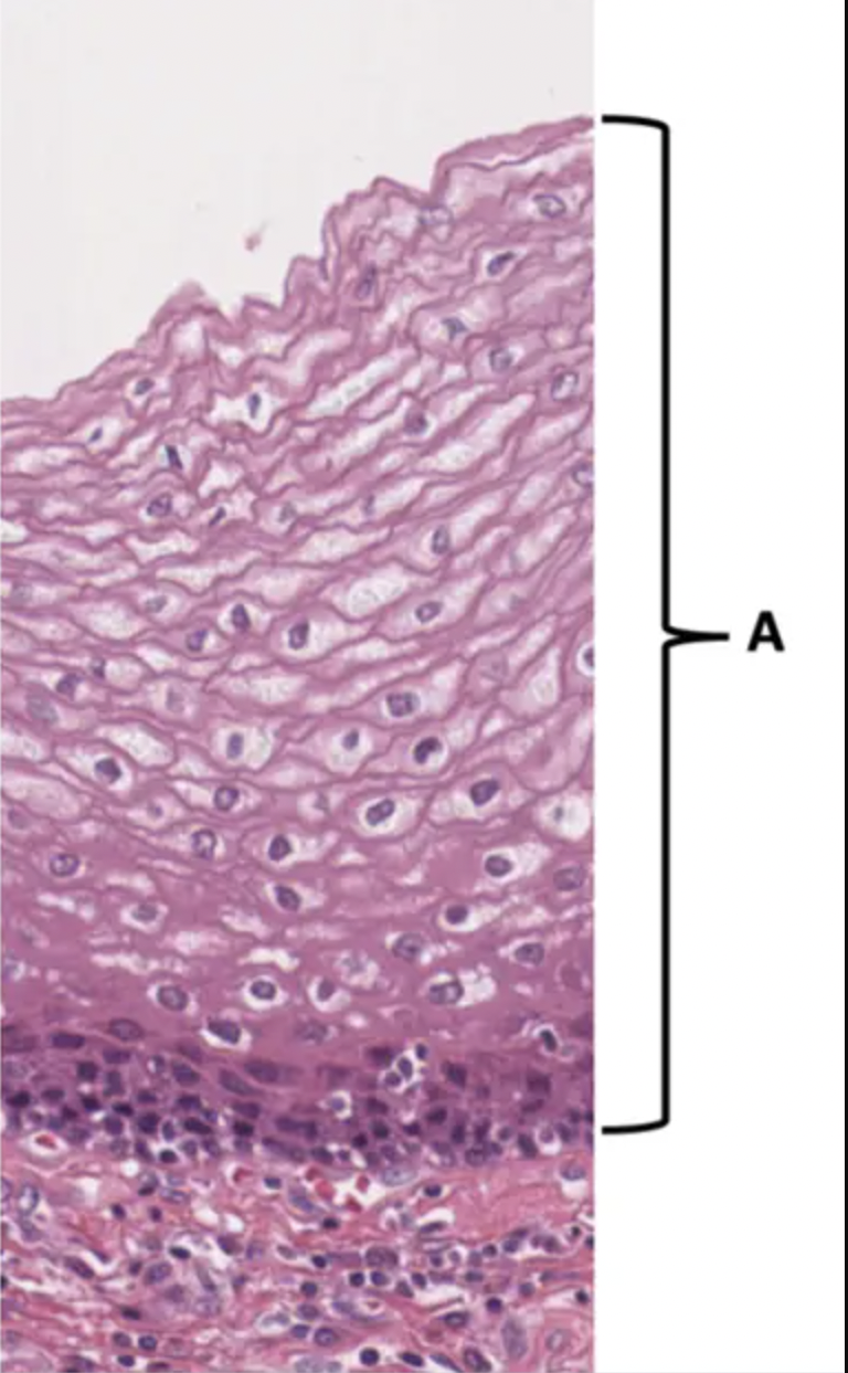
Is this a cutaneous or mucous membrane?:
Mucous
20
New cards
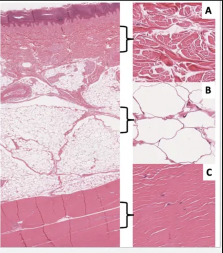
Name the tissue in image A:
Dense irregular connective tissue
21
New cards

Name the tissue in image B:
Adipose tissue
22
New cards

Name the tissue in image C:
Dense regular connective tissue
23
New cards

Which letter identifies a tissue that is mostly cytoplasm?
Adipose (B)
24
New cards

Name the tissue at A:
Pseudostratified columnar epithelia, respiratory epithelia
25
New cards

Name the tissue at C:
Loose connective tissue, areolar connective tissue
26
New cards
Name the structure at B:
Basal lamina, basement membrane
27
New cards

Does the tissue in A contain unicellular exocrine glands (yes or no)?
yes
28
New cards

Name the tissue at A:
dense irregular connective tissue
29
New cards
Name the tissue at B:
skeletal muscle
30
New cards
Name the tissue at C:
smooth muscle
31
New cards

The pink-staining material in this section is protein. Which letter identifies a tissue where the protein is mostly extracellular (outside the cell)?:
A
32
New cards

Name the layer at A:
Epidermis
33
New cards
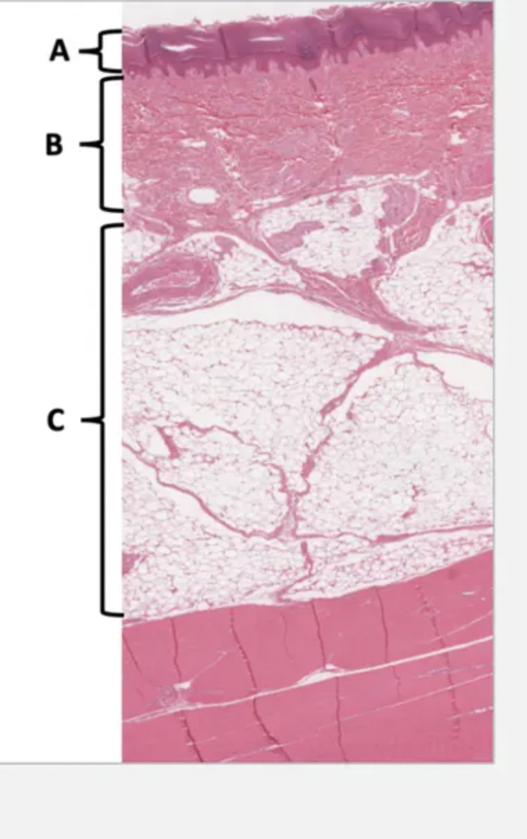
Name the layer at B:
Dermis
34
New cards

Name the layer at C:
Hypodermis
35
New cards

Which layer contains Merkel cells?
A, Epidermis
36
New cards

Which layer contains Meisner’s corpuscles
B, Dermis
37
New cards

Is this skin glabrous (yes or no)?
yes
38
New cards

Which is the Stratum lucidum:
C
39
New cards
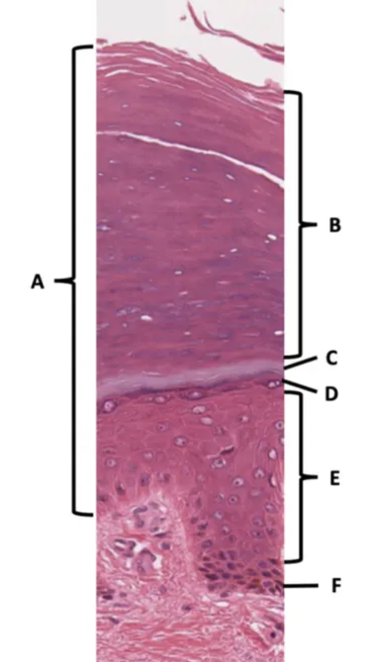
Which is the Stratum spinosum
E
40
New cards

Which is the Epidermis of thick skin:
A
41
New cards

Which is the Stratum corneum
B
42
New cards

Which is the Stratum basale/germinativum
F
43
New cards

Which is the Stratum granulosum
D
44
New cards

Which is a melanoma
C
45
New cards
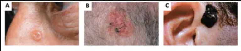
Which is a Basal Cell Carcinoma?
A
46
New cards

Which is a Squamous Cell carcinoma
B
47
New cards
Define anatomy as a science
Anatomy is the oldest discipline of medicine. It studies the structure involved in the function. Physiology studies how it works via chemical and physical properties
48
New cards
What are the subdivisions of anatomy?
Surface Anatomy
Gross Anatomy
Regional Anatomy
System (organ) Anatomy
Histology
Cell Biology
\
SALLYS - Surface anatomy
GOING- gross anatomy
RIGHT- regional anatomy
SINCE- system (organ) anatomy
HENRYS- histology
COMING- Cell Biology
Gross Anatomy
Regional Anatomy
System (organ) Anatomy
Histology
Cell Biology
\
SALLYS - Surface anatomy
GOING- gross anatomy
RIGHT- regional anatomy
SINCE- system (organ) anatomy
HENRYS- histology
COMING- Cell Biology
49
New cards
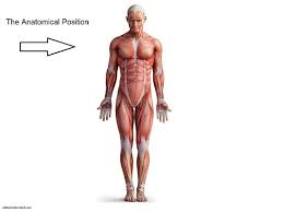
Describe the human body in anatomical position & how to use right and left in anatomical reference
Anatomical position is when the body is standing up right, flat footet with feet slightly apart and parallel, arms down with palms facing forward. When referring to the use of right and left in anatomical reference, we are referring to the body we are observing not our own. (If you are facing the body and the body is facing you, your left is there right and vice versa)
50
New cards
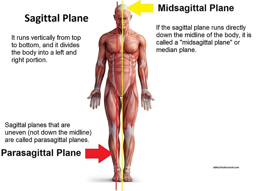
What planes can be used to view the body?
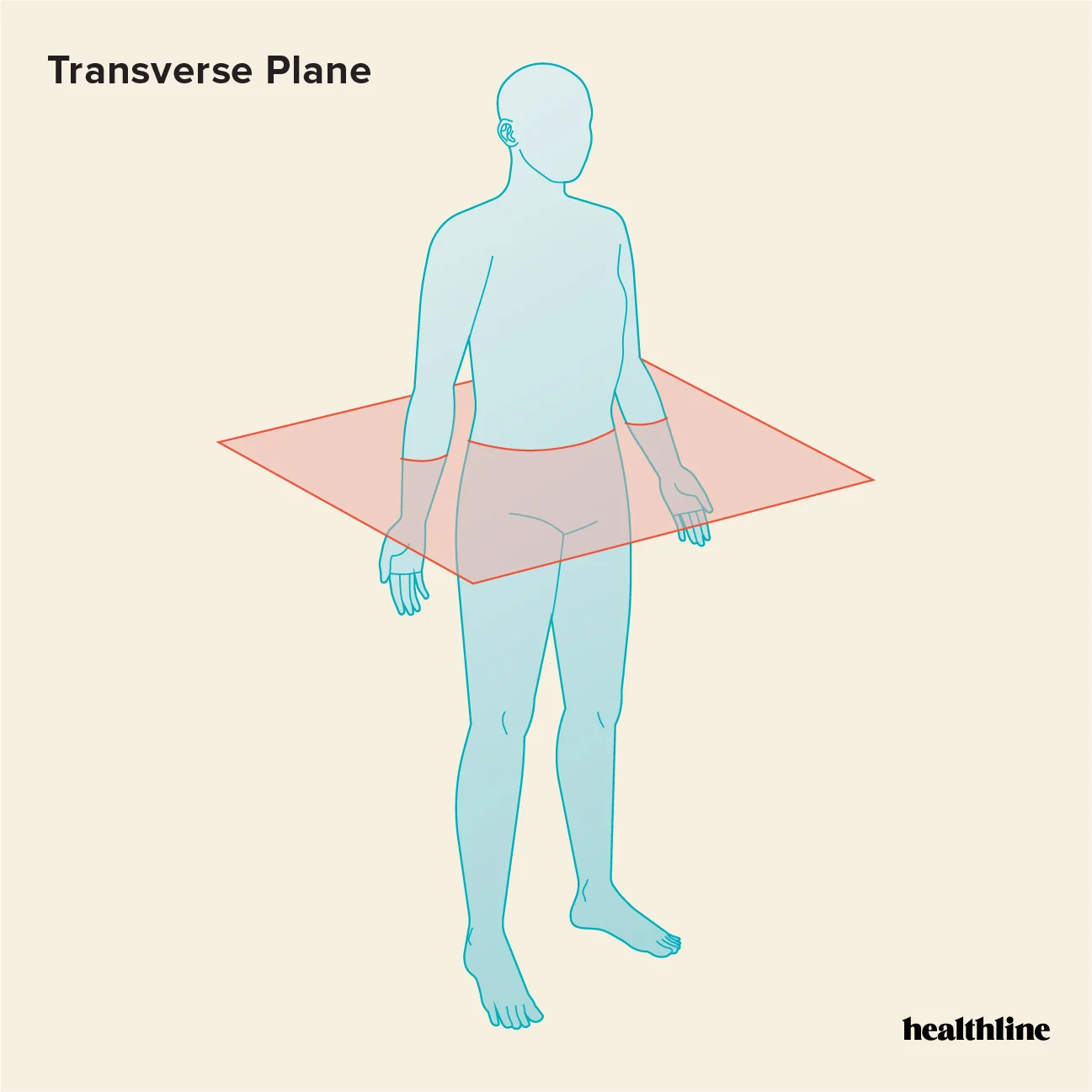
Transverse
Tangential
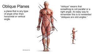
Oblique
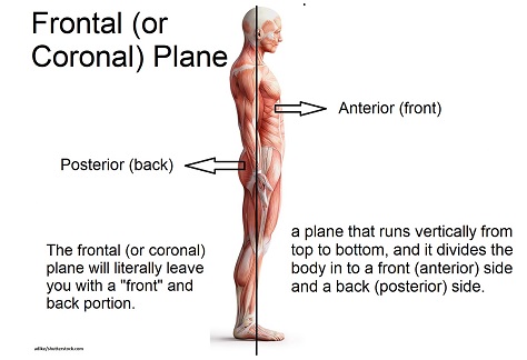
Frontal
Coronal
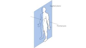
Sagital
Midsagital
Parasagittal
\
TOMMY - transverse
TAKES - tangential
ORANGES - oblique
FROM - frontal
CASEY - coronal
SINCE - sagittal
MONEYS - midsagittal
POOR - parasagittal
Tangential
Oblique
Frontal
Coronal
Sagital
Midsagital
Parasagittal
\
TOMMY - transverse
TAKES - tangential
ORANGES - oblique
FROM - frontal
CASEY - coronal
SINCE - sagittal
MONEYS - midsagittal
POOR - parasagittal
51
New cards
Whats a transverse plane?
section across the body 90 degrees to the superior/inferior axis
52
New cards
What is the tangential/oblique plane?
section across the body < or > 90 degrees to the superior/inferior axis
53
New cards
What is the frontal/coronal plane?
section that divides the anterior/posterior body (coronal more commonly used for the brain.)
54
New cards
What is the sagittal plane?
section that divides the right and left sides of the body along the superior/inferior axis
55
New cards
What is the mid/midial sagittal plane?
cut along the midline

56
New cards
What is the parasagittal plane?
Splits into right and left sides away from the midline
57
New cards
What is negative feedback and give an example
Negative feedback is when there are stimuli that change the environment, triggering a response for the system to maintain near the system’s set point.
An example of negative feedback is temperature
An example of negative feedback is temperature
58
New cards
What is positive feedback and give an example
\n Positive feedback is when there is a stimuli the system enhances it up until the event of it. The response of the stimuli may get amplified.
An example of positive feedback is childbirth
An example of positive feedback is childbirth
59
New cards
Explain why negative feedback is the most common control mechanism used to maintain homeostasis.
Negative feedback is the most common because its job is to reduce excessive response to stimuli and keeps things within their normal range.
60
New cards
Name the three tissue layers of the embryo and the tissue types that develop from these layers.
1. Ectoderm - nervous tissue
2. Mesoderm - muscle and connective tissue
3. Endoderm - inner lining of digestive system
*epithelium from all three germ layers*
61
New cards
What is the formation of tissues and organs?
epithelial tissue
connective tissue
nervous tissue
muscle tissue
membranes
\
EPIC - epithelial
CATS- connective
NEVER- nervous
MURDERED- muscle
MICE- membranes
connective tissue
nervous tissue
muscle tissue
membranes
\
EPIC - epithelial
CATS- connective
NEVER- nervous
MURDERED- muscle
MICE- membranes
62
New cards
Define Tissue
A tissue consists of similar types of cell
63
New cards
Define Organ
an organ is made up of different types of tissues
64
New cards
List the four major tissue types
Epithelial
Connective
Muscle
Nerve
\
EXCITED - epithelial
CHIEFS - connective
MAKE - muscle
NUTELLA - nerve
Connective
Muscle
Nerve
\
EXCITED - epithelial
CHIEFS - connective
MAKE - muscle
NUTELLA - nerve
65
New cards
What does epithelial tissue do?
Covers
66
New cards
What does connective tissue do?
Connects and supports
67
New cards
What does muscle tissue do?
Moves you and things within you
68
New cards
What does nerve tissue do?
Communicates between the external and internal environments
69
New cards
What are the functions of epithelial tissue?
\-Form barriers (membranes) on the inside and outside of the body
\-Protection mostly from surface epithelia (epidermis)
\-Absorption - uptakes nutrients and water
secretion - mucous, sweat, and milk
\-Excretion - removal of waste
\-Filtration - blood via the kidneys
\-Sensory functions - taste, smell (common germ layer origin)
\
FANCY- form barriers
ANIMALS- Absorption
FAKE- filtration
PROTECTING- Protection
ELEPHANTS- Excretion
SADLY- Sensory functions
70
New cards
What are the structural characteristics of Epithelial Tissue?
1. Historically polarized
2. Have cell-to-cell junctions that result in coordinated activity > function as a syncytium of cells
3. Form sheets of cells to give macroscopic/functional membrane
4. Have cell-to-matrix (basement membrane) connection on the basal side
5. Junctions and polarization allow for absorption and secretion
6. Cover external and internal surfaces of the body
7. Invaginations of epithelia into underlying tissue forms glands
8. Avascular > lack a blood supply
9. Highly mitotic relative to other tissues
10. Can be single to multiple layers of cells
11. Highest number of all tissue cell types by cell number (nuclei) not by volume
71
New cards
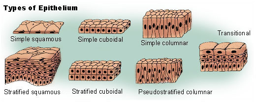
What are the different types of Epithelial tissue?
Squamous- flat
cuboidal- round/cube
columnar - rectangular/tall
Simple - 1 layer
stratified - 2 or more layers
pseudostratified - looks stratified but is only one layer, all cells connect to the basement membrane
Transitional - can change the number of layers (a form of stratified)
cuboidal- round/cube
columnar - rectangular/tall
Simple - 1 layer
stratified - 2 or more layers
pseudostratified - looks stratified but is only one layer, all cells connect to the basement membrane
Transitional - can change the number of layers (a form of stratified)
72
New cards
Describe the structural characteristics common to all types of epithelia
Epithelial cells lie on the basement membrane. Epithelial cells have two different “sides”—apical and basolateral. The apical side always faces out of the body (outside or into a lumen). There is a small amount, or an absence of, extracellular matrix.
73
New cards
Differentiate between exocrine and endocrine gland
Exocrine is onto the surface
Endocrine is into the blood
Endocrine is into the blood
74
New cards
What are Unicellular Glands?
Unicellular glands are just one cell and are dispersed amongst other epithelial cells. Multicellular is more than one.
Unicellular glands mostly secrete a protecting mucous
Unicellular glands mostly secrete a protecting mucous
75
New cards
What are multicellular glands?
Multicellular glands form by invagination of epithelia and most retain connection to surface epithelia
76
New cards
\
Describe mesenchyme and explain its role in the classification of all types of connective tissue.
Describe mesenchyme and explain its role in the classification of all types of connective tissue.
Mesenchyme is a type of loosely organized embryonic connective tissue of undifferentiated cells. Its role in the classification of connective tissues is it directly gives rise to most of the body's connective tissues, from bones and cartilage to the lymphatic and circulatory systems.
77
New cards
\
What are the shared characteristics of connective tissues?
What are the shared characteristics of connective tissues?
1. Develops from mesenchyme via mesoderm
2. Developing/dividing/active cells called “blasts”
3. Cartilage -→ chondroblasts
4. Adult/nondividing/matrix maintaining cells called “cytes”
5. Cartilage -→ Chondrocytes
78
New cards
Describe the structural characteristics common to all types of connective tissue
1. Connective tissue is composed primarily of an extracellular matrix and a limited number of cells
2. large amounts of amorphous ground substance, and protein fibers.
79
New cards
What are the functions of nervous tissue?
1. Involved in short-term and long-term homeostasis via its integrator function
2. Communicates with the outside and inside environments
3. Sensory receptors
4. Interprets/processes sensory information
5. Integration center > central nervous system
6. Sends signals to effector organs
7. Response to environmental
8. Stores information
80
New cards
Describe the characteristics of nervous tissue cells and where this tissue is found
1. Develops from ectoderm
2. Specialized for membrane depolarization and cell-to-cell communication via synapses
3. The main cell type (neuron) has long processes involved in receiving and sending information via membrane depolarization.
4. The main cell type is enriched in cytoskeletal proteins for structural support and transport.
5. Protected by connective tissues
6. Mostly found in the brain, spinal cord, and nerves
81
New cards
Describe the structure and function of mucous membranes, and where is it found?
1. membranes line the digestive, respiratory, urinary, and reproductive tracts. They are coated with mucous gland secretions (contains glands)
2. The mucous membrane lubricates and protects these organs and cavities from abrasive particles and bodily fluids, as well as invasive pathogens
82
New cards
Describe the structure and function of serous membranes, and where it can be found
Serous membranes line body cavities closed to the exterior of the body: the pericardial, peritoneal and pleural cavities. These membranes are thin and help to reduce friction. (secrete fluids)
83
New cards
What is the structure and function of cutaneous membranes and where can it be found.
an upper layer called the epidermis, and underlying or basement layer called the dermis, and connective tissue called the subcutaneous or hypocutaneous. It helps protect the rest of the body's tissues and organs from physical damage such as abrasions, chemical damage such as detergents, and biological damage from microorganisms (makes up the skin)
84
New cards
What is the structure and function of synovial membranes and where can they be found
1. A distinct intimal lining layer of 1-2 cells thickness and a synovial sublining layer. It protects the joints they surround
2. Synovial membranes are located between the bones, muscles, tendons, and ligaments of synovial joints
85
New cards
What are the functions of Muscle tissue?
1. Moves you via skeletal muscle
2. Moves contents within you via smooth and cardiac muscle
3. Smooth muscle lines the hallow organs of the body
4. Moves digesta, urine, gametes, and regulates blood and airflow
5. Forms sphincters to regulate movement between organs of organ systems
6. Cardiac muscle moves/pumps blood
7. Amino acid storage from muscle proteins
8. Major target/effector organ of the nervous system involved in homeostasis
86
New cards
Describe the structural characteristics common to all types of muscle tissue.
excitability. contractility. Extensibility - they can be stretched.
87
New cards
Describe the functions of the epidermis.
protecting your body from the outside world, keeping your skin hydrated, producing new skin cells and determining your skin color
88
New cards
Identify and describe the tissue type making up the epidermis.
The epidermis is made up of stratified squamous epithelial cells called keratinocytes. It functions primarily as a protective barrier and also provides touch sensation.
89
New cards
Identify and describe the layers of the epidermis, indicating which are found in thin skin and which are found in thick skin.
four layers in thin skin
stratum corneum
stratum granulosum
stratum spinosum
stratum basale
five layers in thick skin... all of the above plus stratum lucidem
\
CORRUPT - corneum
GANGS - granulosum
SPIN - spinosum
BLOCKS - basale
^^THIN SKIN
\
Lucidem- thick skin
stratum corneum
stratum granulosum
stratum spinosum
stratum basale
five layers in thick skin... all of the above plus stratum lucidem
\
CORRUPT - corneum
GANGS - granulosum
SPIN - spinosum
BLOCKS - basale
^^THIN SKIN
\
Lucidem- thick skin
90
New cards
Describe the processes of growth and keratinization of the epidermis.
New cells formed in Stratum Basale are pushed upwards away from source of nutrients & oxygen, accumulating more keratin. They undergo apoptosis and when they reach Stratum Corneum they slough off and are replaced by underlying cells going through keratinization.
91
New cards
Explain how Stem cells of the stratum basale contribute to the functions of the epidermis
single layer of columnar cells, and are the only cells to undergo mitosis in the epidermis; as a result, cells transfer or migrate outward from the basal to the other layers until they are shed from the skin surface
92
New cards
Explain how keratinocytes contribute to the functions of the epidermis
filled with keratin and are arranged in distinct layers, and are the most distinct layer in the epidermis
93
New cards
Explain how melanocytes contribute to the functions of the epidermis?
contribute colored pigments to the skin and serve to decrease the amount of Ultraviolet light that can penetrate into the deeper layers of the skin
94
New cards
Explain how the epidermal dendritic (Langerhans) cells contribute to the function of the epidermis
branched cells that play a role in immunity; each DC finds markers (antigens) on bacteria and other invaders and presents them to other immune system cells for recognition and destruction
95
New cards
Explain how tactile (Merkel) cells contribute to the function of the epidermis
Located in the deepest layer of the epidermis; they connect to sensory nerve endings to form structures that serve as light touch receptors
96
New cards
Explain how Discs contribute to the function of the epidermis
Merkel discs respond to tactile stimuli by generating slowly adapting type 1 (SA1) impulses
97
New cards
Compare and contrast thin and thick skin with respect to location and function.
1. Thin Skin: covers most of the body; the number of cell layers in each epidermal stratum is fewer; stratum lucidum is absent; raised parallel ridges are not present
2. Thick Skin: hairless; covers palms, soles, and other areas subject to friction; all 5 strata are present; dermal papillae are raised in curving parallel epidermal ridges (friction ridges) to form finger prints
98
New cards
Identify and describe the dermis and its layers, including the tissue types making up each dermal layer
1. Dermis: Deep primary layer of the skin; made up of fibrous tissue; also includes some blood vessels, muscles, and nerves; derived from the mesoderm
2. Papillary Layer: Loose fibrous tissue with collagenous and elastic fibers; forms nipplelike bumps called papillae; includes tactile corpuscles, which are touch receptors, and other sensory receptors
3. Reticular Layer: Touch network (reticulum) of collagenous dense irregular fibrous tissue with some elastic fibers; forms most of the dermis
99
New cards
Identify and describe the subcutaneous layer, including the tissue types.
Hypodermis (superficial fascia): Loose fibrous (areolar) connective tissue and adipose tissue; under the skin (not part of the skin); includes fibrous bands or skin ligaments that connect the skin strongly to underlying structures; includes lamellar corpuscles, which are pressure receptors, and other sensory receptors
100
New cards
Describe the functions of the subcutaneous layer.
1. It forms a connection between the skin and the underlying structures of the body.
2. The density and arrangement of fat cells and collagen fibers in this area determine the relative mobility of the skin.
3. The rich blood supply and loose spongy texture of this area make it an ideal site for the rapid and relatively pain-free absorption of injected material.