Embryology of the Central Nervous System
1/57
There's no tags or description
Looks like no tags are added yet.
Name | Mastery | Learn | Test | Matching | Spaced | Call with Kai |
|---|
No analytics yet
Send a link to your students to track their progress
58 Terms
Reminder** How does a gamete form and what are the subsequent stages of development?
Fecundation: Union of 2 gametes = zygote
Segmentation: Zygote → Morula → Blastula
Blastulation: Blastula → Blastocyst
Gastrulation: New layer formation (3 layers - ectoderm, mesoderm, endoderm) - the three layer embryo
The nervous system starts to form during the _______ part of embryonation.
Gastrulation
Reminder, what organs does each layer of the trilaminar embryo give rise to?
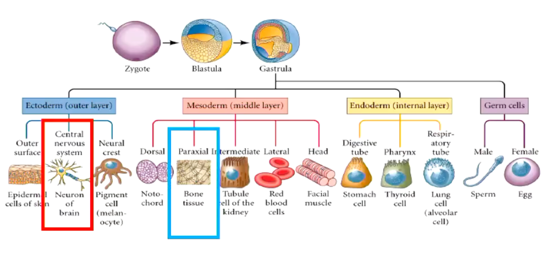
What were the three parts of the trilaminar embryo previously?
Ectoderm = previously epiblast
Mesoderm = new layer
Endoderm = previously hypoblast
The notochord appears within the mesoderm of the embryo, what function does this have?
Functions:
• Define the embryonic midline (as a reference)
• Cellular inductor (role in neural plate) → marks center of body and directs the formation of the other organs
• Vertebral precursor
• Signalling
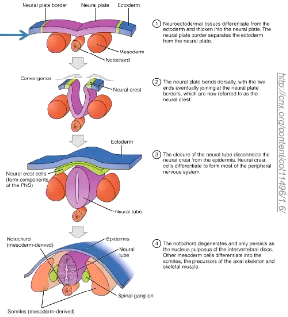
The notochord will disappear in the adult (only persist into the nucleus pulposus).
After the three germinal layers have become established, the development of the nervous system begins, what is this process called?
Neurulation
What is the region of the ectoderm forming the CNS called?
What is the rest of the ectoderm called?
The ectoderm which will form the CNS = Neuro-Ectoderm
The rest of the ectoderm which will form other tissues (skin) = Non- neural Ectoderm
The neuro-ectoderm has three components, what are they?
• Neural plate= thicker central part of the ectoderm
• Neural folds (or neural plate border) = margins of the neural plate
Depends on if open or closing
• Neural groove = the sulcus of the neural plate
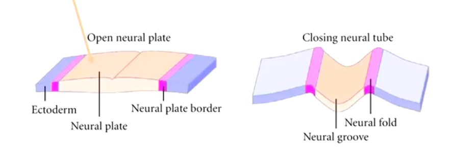
How does the neurulation process occur?
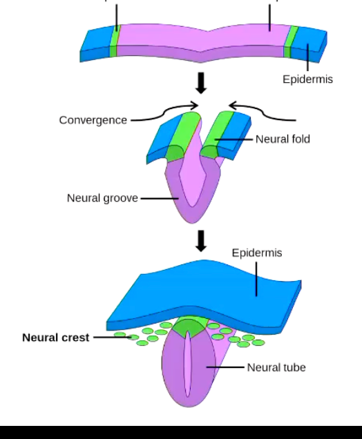
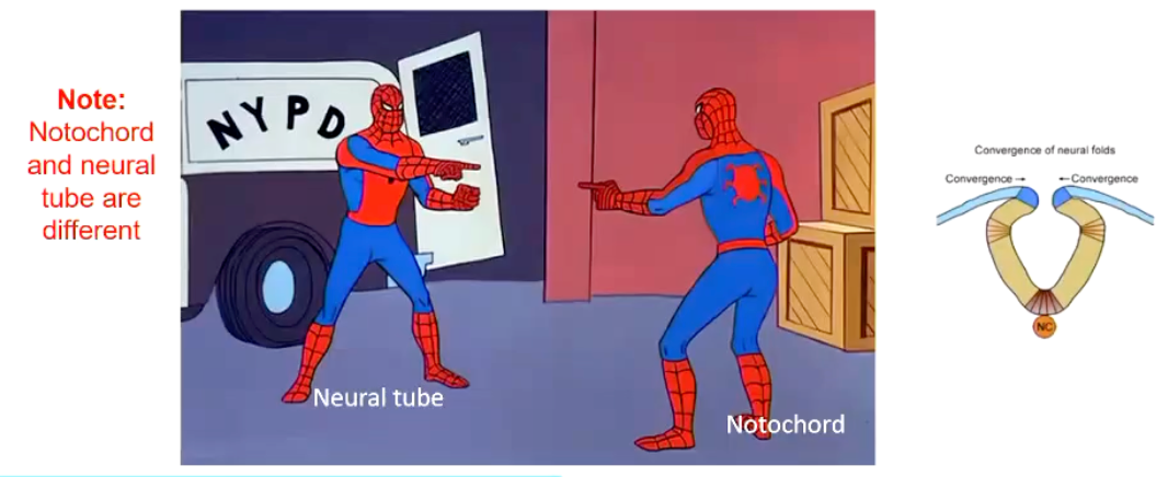
Neurulation starts with a folding process which includes the transformation of the neural plate into the neural tube by the joint of the neural fold.
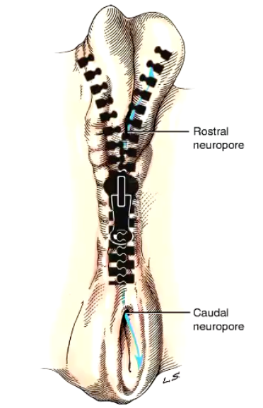
The formation of the neural tube does not close ______, usually it starts in what part of the body?
homogenously
The neural tube forms (closing) gradually from the neural plate by "zippering" in caudal and cephalic directions from the cervical region.
The last portions of the neural tube to close are the rostral and caudal neuropores.
Rostral Neuropore and Caudal Neuropore
In the dog, when does the neural tube begin to close?
In the dog, neural tube closure starts around day 16 of gestation and is complete by around day 18.
During the formation of the neural tube, it will separate from what structures?
Separates from the non-neural ectoderm which grows over the dorsum of the tube to fuse along the midline
While this fusion and separation of ectodermal layers occurs, a longitudinal column of ectodermal epithelial cells arises from the neural folds (junction of non-neural and neural ectoderm) and separates from these two structures when the neural tube is formed (ectodermal cells are induced to develop mesenchymal features).
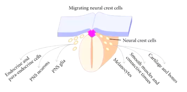
These two bilateral columns, situated dorsolateral to the neural tube throughout its length, are the neural crest.
What is the neural crest?
What region of the nervous system does it give rise to?
The neural crest is a transitory structure exclusive to the vertebrates' embryo.
Cells in the neural crest initially lie between the dorsal ectoderm and the neural tube but, then the cells migrate to different locations, settle, and differentiate, giving rise the spinal ganglia but also the rest of the peripheral nervous system and a remarkably wide array of cell types:
What is the hollow structure located in the neural tube?
What does it become in the adult animal?
Neural Canal
Becomes the central canal in the spinal cord and the ventricular system in the brain
Are the neural tube and the notochord the same thing?
NO
The notochord appears within the mesoderm (invagination from the endoderm into the mesoderm)
Functions:
Define the embryonic midline
Cellular inductor (role in neural plate).
Vertebral precursor
Signalling
Neural tube appears from the neuro-ectoderm
Precursor to the central nervous system (brain and spinal cord)
How does the neural tube develop into the nervous system?
Neuroectodermal cells = neuroepithelial cells (proliferation)
• It is a multistep process controlled by various genes and environmental factors.
• Initially monolayer (1 cell layer) of neurectodermal cells* organized in a pseudostratified arrangement. (Then proliferates, 1 layer → 2 layers → 3 layers, etc.)
Cells mitotically active = in a short time the wall of the tube will increase in thickness, and a new layer of differentiating cells appears on the external surface of the actively dividing layer.
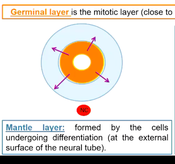
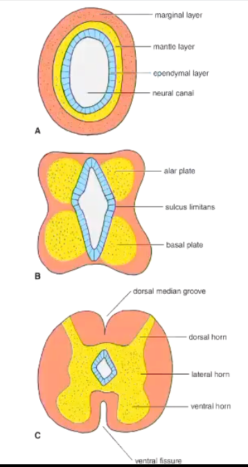
Describe the layers of the neural tube as it proliferates.
Germinal layer is the mitotic layer (close to the neural canal)
Mantle layer: formed by the cells undergoing differentiation (at the external surface of the neural tube).
The cells differentiating from the germinal layers are of two types, what are they?
The cells that are differentiated are of 2 types which start to migrate to the external layer.
• Immature neurons: progenitors of the neurons
• Spongioblasts: are the progenitors of the neuroglia. Astrocytes and oligodendrocytes are derived from these spongioblasts.
**The microglial cell is mesodermal in origin, as it is a monocyte that enters the nervous system from the blood supply.
Describe the makeup of the three layered neural tube.
Germinal layer is the mitotic layer (proliferation of neuroepithelial cells) → which will be exhausted reducing this layer into a single layer of cells = ependymal cells (ependyma)
Mantle layer: will ultimately become the gray matter of the definitive spinal cord and brain
Marginal layer (the external layer): is composed primarily of the growing axonal processes of the neuronal cell bodies in the mantle layer. These axons will form tracts in the white matter
Describe the sequence of events in the different layers in the process of neuronal migration.
Induction (one group of cells, the inducing tissue, directs the development of another group of cells)
Proliferation (germinal layer)
Cellular migration (mantel and marginal layer)
Differentiation (glia vs neurons)
Establishment of connections (among cells)
Apoptosis (if required)
The three layered relationship of cells persists in what structure of the body?
The developing spinal cord
In the telencephalon, the neurons migrate from the mantle layer to the external surface of the neural tube. For that reason the grey matter is located externally in the forebrain.
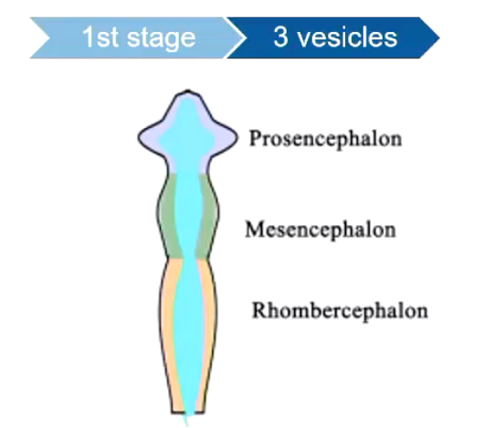
What will occur in the brain, at the level of the cranial neuropore during brain development?
• At the level of the neuropore: the rostral end of the neural tube develops rapidly and produces three vesicles, from rostral to caudal:
Cranial Part - prosencephalon
Middle - mesencephalon
Caudal - rhombercephalon
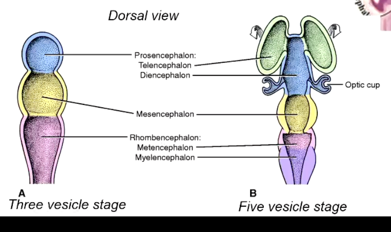
After the three vesicles have been formed in the brain, they will progress to produce what?
5 vesicles total, becoming:
Prosencephalon → Telencephalon (2 hemispheres), Diencephalon (optic nerves, eyes form around?
2 lateral enlargements growing laterally overlying by skin
ectoderm = optic vesicles
Mesencephalon → remains mesencephalon
Rhombencephalon → metencephalon, myelencephalon
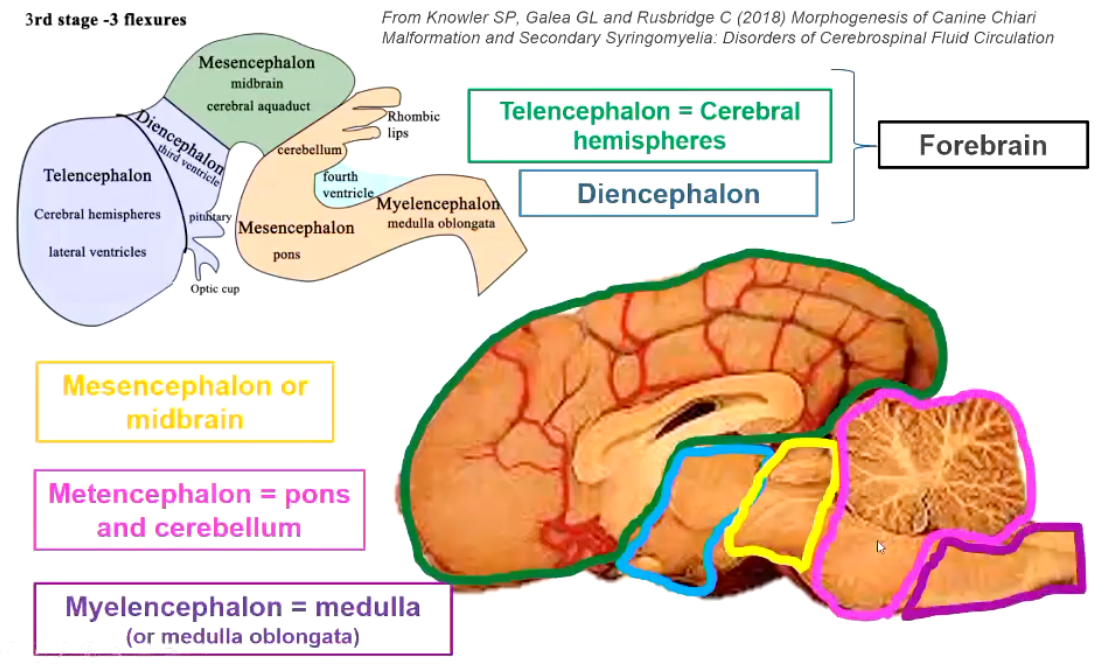
During the 3rd stage of brain development, ______ form.
flexures
What structures produce the cerebral hemispheres?
telencephalon
What do the telencephalon, diencephalon, mesencephalon, myelencephalon and metencephalon produce in the adult?
Forebrain
Telencephalon = Cerebral hemispheres
Diencephalon
Midbrain
Mesencephalon
Pons & Cerebellum
Metencephalon
Medulla (Or medulla oblongata)
Myelencephalon
Is the pattern of malformation in the brain linked more severely to the aetiology or gestational age of the insult?
Gestational age of insult
More dependent on when in gestation
Therefore, the same malformation may be seen with a number of different aetiologies.
Sometime there is a very brief period of susceptibility (as short as 8 hours) → which makes difficult to tract the primary cause
In general, the earlier the disruption occurs, the more severe the resultant malformation.
When can malformations of CNS occur?
• Can occur at any time of CNS development:
Malformations of the brain may or may not be associated with malformations of the neurocranium
Malformations of the CNS are relatively common in dogs & cats, but it is important to note what?
• Relatively common in dogs & cats BUT not all diagnosed malformations are of clinical relevance ~ incidental finding
Importance of correlating diagnostic imaging features with results of neurological examination
What do malformations of the brain & neurocranium cause?
What do malformations of the spinal cord typically cause?
• Malformations of the brain & neurocranium can cause abnormal behaviour, seizures & vestibular or cerebellar signs
• Malformations of the spinal cord can cause gait abnormalities & urinary and/or faecal incontinence
What are the main causes of malformations of the CNS?
• Some breeds overrepresented ~ genetic predisposition
• In utero exposure to infectious agents and other teratogens
What are the main types of malformations that can occur in the CNS?
1. Neural tube defects
A. Failure of neural tube closure:
Cerebral aplasia (Anencephaly)
Meningocele / Meningoencephalocele / Myelomeningocele
Spina bifida
B. Other neural tube defects
Hydrocephalus
2. Neuronal migration disorders
Lissencephaly & polymicrogyria
3. Destructive process
Hydranencephaly / Porencephaly
4. Other complex malformations
Cerebellar aplasia/hypoplasia
Chiari-like malformation & syringohydromyelia
What is the most common type of neural tube defects?
Most common NTD is the failure of complete closure of the neural groove to form a neural tube:
May occurs in different localizations (brain / spinal cord)
NTDs may be associated to altered induction of mesodermal tissue which is forming the bones surrounding the CNS; this will result in an affection of the bone (skull (=craniochischisis- skull cannot close) and or vertebrae (=spina bifida & rachischisis - vertebrae cannot close).
Cranial NTD: anencephaly, meningoencephalocele
Caudal NTD: meningocele, myelomeningocele
There is a range of phenotypes observed in NTDs, why is this?
What are the related causes?
Due to complex nature of neural tube closure, non-neural ectoderm (skin) may be also affected
Leading to OPEN: exposed neural tissue and leaking CSF
OR, closed, not exposed neural tissue, not leaking CSF out of body but covered by skin
• Related cause: (multiple)
• Hyperthermia
• Genetic
• Teratogenic substances
• Folic acid deficiency (mainly in people)
• Most of time, cause unknown
• Nutritional deficiencies
• Multifactorial
What is cerebral aplasia and how does it occur?
Type of NTDs
• Failure of the closure of the cranial neuropore + failure in development of the telencephalic vesicles = absent cerebral hemispheres → forebrain
Skull may be partially open = cranioschisis
Presence caudal structures such as brainstem and cerebellum and eyes (although may be abnormal too).
• Commonly referred as anencephaly but incorrect (as some parts of the brain are present, ancephaly technically means absence of brain)
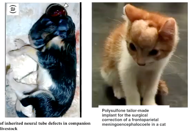
What is meningocele / meningoencephalococele and how does it occur?
Type of NTDs
• Disturbance in separation of surface ectoderm and neuroectoderm during final phase of neural tube formation = Protrusion of brain and/or meninges through an opening in the calvaria.
Since neuroectoderm and non-neural ectoderm cannot separate
This can be disturbed at any of the sites of neural tube closure.
Meningocele: protrusion of meninges, covered by skin through the skull
Meningoencephalocele: as above but contains brain parenchyma in the protrusion (Far more severe)
Size variation
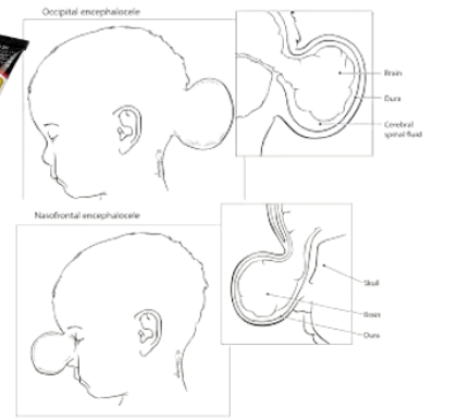
Where can Meningocele / Meningoencephalocele be observed within the skull?
What are some signs?
What are frequent causes?
• Not always is externally observed (nasal, ethmoidal, orbital, sphenoidal localization)
• Signs in dogs:
Depending on the localization
Most common presenting complaints: Seizures and/or behavioural abnormalities
• Causes:
Griseofulvin administration in cats.
Inherited malformation in Burmese cats (related to select shortened faces).
How does Meningocele / Meningoencephalocele often appear in cats?
The abnormality is characterized by agenesis of all derivatives of the medial nasal prominence; lateral duplication of most derivatives of the maxillary process; including the canine teeth and whiskers fields; telencephalic meningoencephalocele; and secondary ocular degeneration (Fig. 1c and d)
Genes associated with Burmese Cats due to trying to make faces flatter
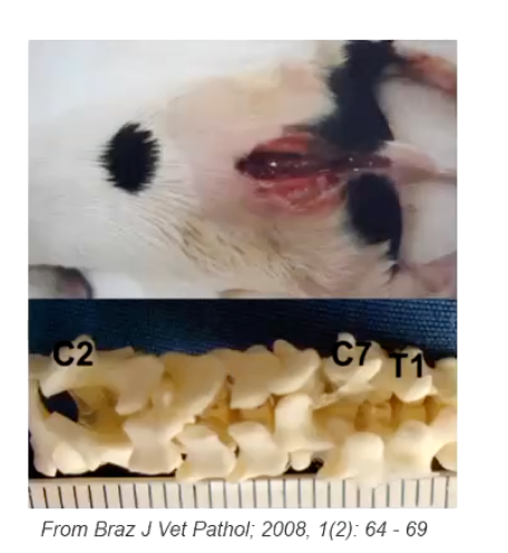
What is Meningocele / Meningomyelocele and how does it occur?
• If the failure occurs at the vertebral column = Protrusion of spinal cord and meninges through an opening in the vertebrae (spina bifida or rachischisis).
• Meningocele: protrusion of meninges, covered by skin through the vertebrae. Associated to accumulation of CSF.
• Meningomyelocele: as above but also contains spinal cord parenchyma in the protrusion.
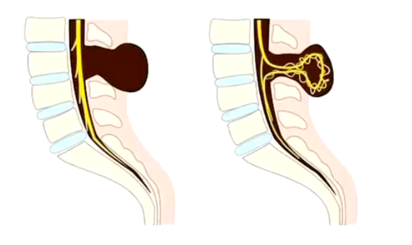
Where can Meningocele / Meningomyelocele be observed within the spinal cord?
What are some signs?
What are frequent causes?
• Not always is externally observed or just associated to small concavity of the skin or coat-whirl.
• Signs (based on the localization, but commonly at sacrocaudal area since birth:
Urinary and/or faecal incontinence.
Paraphimosis in males
Paraparesis (weak gait in pelvic limbs)
• Causes:
Genetic factors in French bulldogs (Screw-Tail Dog) and Manx cat (absent tail)
What is Spina bifida and how does it occur?
How does it differ form rachischisis?
How are these abnormalities observed?
• Failure to the closure of the dorsal aspect of the vertebra (one or few vertebrae) (AKA split vertebra)
• If many adjacent vertebrae are involved = named rachischisis Different process = due failure of the vertebral arches to develop
• Not always is externally observed
Signs in dogs:
No signs if only spina bifida
Variable clinicals signs if spina bifida is associated to other anomalies (such as myelomeningocele/meningocele).
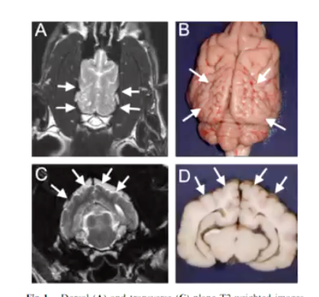
What is the most common brain malformation and how does this abnormality occur?
Congenital Hydrocephalus
• Abnormal flow or absorption of CSF = accumulation of CSF w/in cranial cavity = reduce thickness of cerebral cortex.
The most common brain malformation.
Excess CSF due: stenosis (absent) or small size of mesencephalic aqueduct, increased CS production from choroid plexus, abnormal CSF absorption due malformed/lack of arachnoid villi.
**** Although there also exist acquired causes of hydrocephallus, these are typically congenital
What animal are most susceptible to congenital hydrocephalus?
What are the signs and when do they become evident?
• Toy & brachycephalic breed dogs most common
• Signs (may not be evident until several months of age):
• Dome-shaped head
• Persistent fontanelle
• Ventrolateral strabismus ('setting sun sign')
• Abnormal behaviour
• Cognitive dysfunction
• Blindness
• Ataxia
• Death
**Note: there is expansion of the skull; different from meningoencephalocele!
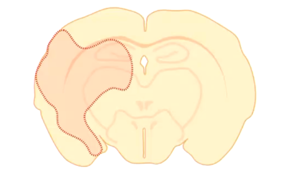
What are the characteristics of Lissencephaly?
• Slow or failed neuronal migration of brain neurons = Lack of development of the surface folding of the cerebrum
Characterized by a small, smooth-appearing cerebrum with rudimentary or absent gyri (agyria) and sulci.
What are the signs and causes of lissencephaly?
• Signs:
• Cognitive dysfunction
• Abnormal behavior
• Seizures
• Causes:
• Suspected inherited condition in Lhasa Apso (common)
• Intrauterine hypoxia (human)
What are the characteristics of polymicrogyria?
Also note the signs.
• Abnormal neuronal migration of brain neurons (specifically of abnormal cortical lamination) = excessive cortical folding
Resulting in increase in numbers of smaller irregularly convoluted cortical gyri
• Signs:
Blindness with or without other neurological deficits.
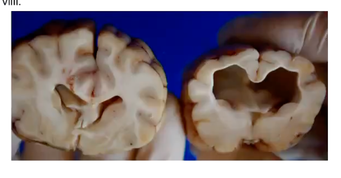
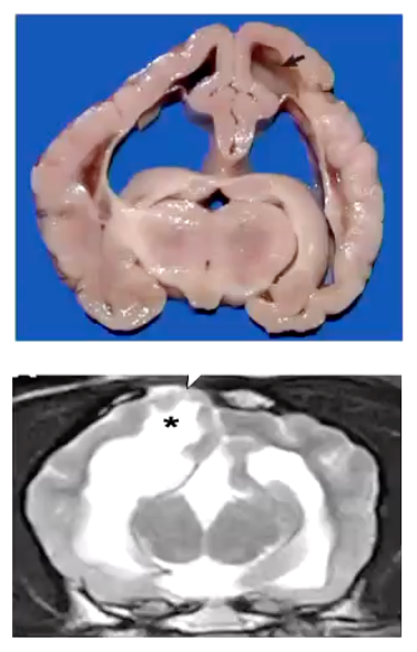
What are the characteristics of hydranencephaly?
• Almost complete loss of one or both cerebral hemispheres which is replaced by CSF (=cerebral cavity)
• It was initially well-formed but is destroyed.
• Almost complete absence of cerebral cortex, reduced to a thin layer.
• The adjacent skull may be abnormal too.
What are some of the main causes of hydranencephaly?
Perinatal stroke = lack of perfusion of cells = necrosis.
Viral infection = destruction of germinal layer
(Cattle: Bovine Viral Diarrhoea, virus, Blue tongue virus, etc...; Cat: Panleukopenia virus).
Genetic transmission in sheep
What are the signs of hydranencephaly?
• Signs:
• Seizures - most commonly + Mild deficits: unilateral/bilateral blindness.
• Cognitive disfunction
What are the characteristics of porencephaly?
• Same physiopathology than hydranencephaly but the cavity formed is smaller
• The insult occurs later on the developmental stage.
E.g.: fetal lamb with bluetongue infection from 50-55 days = hydranencephaly. Compared with porencephaly which occurs when infection at 75-78 days. And infection after 100days of pregnancy cause mild encephalitis in the fetus, with no malformation.
The insult affects to a smaller region of the brain.
• Usually clinically normal or just seizures in adulthood (<3y of age in dogs)
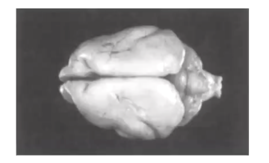
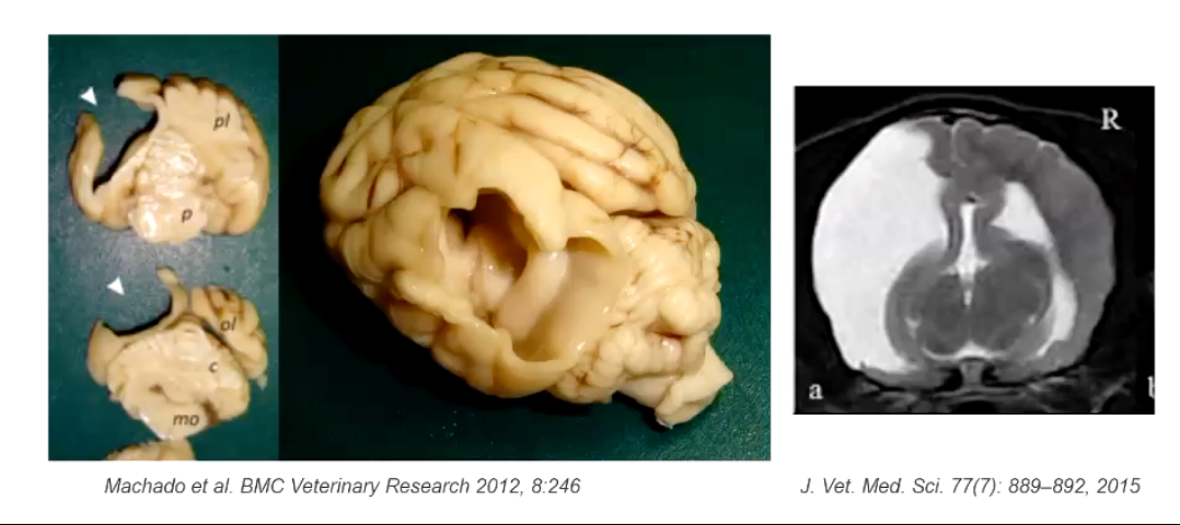
What are the key features of cerebellar aplasia?
• Cerebellar aplasia
Complete or almost complete absence of cerebellar tissue
Cerebellum is replaced by CSF
Most common in calves
Clinical signs: unable to stand up after birth or even to right themselves in a sternal position.
What are the key features of cerebellar hypoplasia?
• Cerebellar hypoplasia
Uniform paucity of cerebellar tissue
Kittens: most common following in utero or perinatal infection w panleukopenia virus
Piglets: treated with an organophosphate insecticide during the second half of gestation (= cerebellar hypoplasia and hypomyelination).
Clinical signs: cerebellar ataxia from birth or shortly thereafter that do not progress
Wobbly cats!
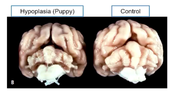
Cerebellar hypoplasia, not to be confused with…
cerebellar abiotrophy → caused by cells degeneration = slow progressive signs (normal development of cerebellum but progressive destruction of cells). which is a degenerative condition, but hypoplasia is a stable condition that will not worsen
What are the key features of chiari-like malformations?
• Complex skull malformation
Reduced development of the occipital bone = impacted and/or herniated cerebellum through the foramen magnum and compromises CSF flow.
Flat faced dogs or cats have a thicker bone in the occipital area, in caudal fossa region, which decreases space for in the occipital region, pushing the brain stem and deforming the cerebellum
Causes accumulation of CNF
In what breeds is chiari-like malformation most common in?
How does it present secondarily?
What signs may be present?
• Very common in small and brachycephalic breed dogs
• Secondarily: may be associated to dilation of the central canal (named hydromyelia) or a cavitation of the spinal cord parenchyma (named syringomyelia).
Most common signs: spinal pain, neck scratching
Why do we not see cancer as often derived from neurons?
In the adult animal with fully developed tissues, where might certain cancers arise in CNS tissue?
In general, mature neurons will not divide → no cancer from neurons
Glioma (astrocytomas, oligodendrogliomas)
Meningioma
Choroid plexus tumour
Ependimoma
Nerve sheath tumour
How do embryonal cell tumours develop and what are a few examples?
Embryonal cells tumours: develop from residual cells from the embryo that have remained in the brain/spinal cord after birth
Medullloblastoma
Nefrobrastoma
Chordoma
Primitive neuroectodermal tumors
Germ cell tumors
What is the nucleus pulposus and what is it surrounded by?
The nucleus pulposus is the gelatinous, jelly-like inner core of an intervertebral disc, located between the vertebrae. Its primary function is to act as a shock absorber for the spine, absorbing compressive forces and helping to maintain the spine's flexibility and stability.
Surrounded by anulus fibrosus