🆑Carlton Ch. 33 Fluoroscopy
1/52
There's no tags or description
Looks like no tags are added yet.
Name | Mastery | Learn | Test | Matching | Spaced | Call with Kai |
|---|
No analytics yet
Send a link to your students to track their progress
53 Terms
1. Fluoroscopy is a ____ x-ray examination.
a. dynamic, real-time
b. digital, static
c. therapeutic
d. nonionizing
a. dynamic, real-time
2. Fluoroscopy is primarily the domain of the
a. radiographer.
b. radiologist.
c. radiation physicist.
d. radiology manager.
b. radiologist.
3. Fluoroscopy is commonly used for observation of
a. healing bone fractures.
b. growing breast cysts.
c. small bowel disorders.
d. central nervous system tumors.
c. small bowel disorders.
4. The fluoroscopic x-ray tube and image receptor are mounted
a. on a carriage assembly.
b. coincident to each other.
c. from the ceiling.
d. orthogonal to each other.
b. coincident to each other.
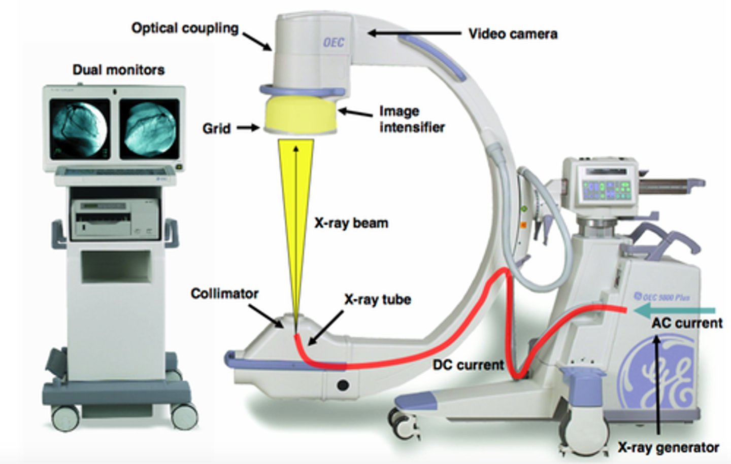
5. The fluoroscopic carriage commonly supports the
a. image receptor.
b. power-assist drive controls.
c. fluoroscopic and digital controls.
d. all of the above.
d. all of the above.
6. The fluoroscopic mA range is commonly ____ mA.
a. 0.5 to 5.0
b. 5.0 to 10.0
c. 10.0 to 100
d. 100 to 500
a. 0.5 to 5.0
7. During fluoroscopy, the SOD on a fixed unit cannot be less than ____ inches.
a. 12
b. 15
c. 20
d. 40
b. 15
8. During fluoroscopic image intensification, the primary x-ray beam exits the patient and strikes the ____ of the image intensifier.
a. input screen
b. electrostatic lenses
c. photocathode
d. output screen
a. input screen
9. The input screen absorbs ____ and emits ____.
a. x-ray photons; light photons
b. x-ray photons; electrons
c. light photons; x-ray photons
d. electrons; x-ray photons
a. x-ray photons; light photons
10. The photocathode absorbs ____ and emits ____.
a. x-ray photons; light photons
b. light photons; electrons
c. light photons; x-ray photons
d. electrons; light photons
b. light photons; electrons
11. Electrostatic lenses are used to accelerate and focus
a. light photons.
b. x-ray photons.
c. electrons.
d. scattered photons.
c. electrons.
12. The primary ____ occurs from the acceleration and focusing of the electron beam.
a. phosphorescence
b. minification
c. magnification
d. brightness gain
d. brightness gain
13. The output screen absorbs ____ and emits ____.
a. electrons; light photons
b. electrons; x-ray photons
c. light photons; electrons
d. light photons; x-ray photons
a. electrons; light photons
14. The input screen of an image intensifier is ____ in shape.
a. oval
b. rectilinear
c. concave
d. convex
c. concave
15. The shape of the image intensifier input screen helps to control
a. image distortion.
b. pixel binning.
c. electron scattering.
d. minification.
a. image distortion.
16. The photocathode of an image intensifier is composed of ____ metals.
a. TFT
b. photoabsorptive
c. photoemissive
d. electrostatic
c. photoemissive
17. Photoemissive materials absorb ____ and emit ____.
a. x-ray photons; light photons
b. light photons; electrons
c. light photons; x-ray photons
d. x-ray photons; electrons
b. light photons; electrons
18. Electrostatic focusing lenses are actually
a. concave optical lenses.
b. convex optical lenses.
c. planar lenses.
d. charged electrodes.
d. charged electrodes.
19. The greater the voltage supplied to the electrostatic lenses, the ____ the acceleration and the ____ the focal point to
the input screen.
a. greater; farther
b. greater; closer
c. lesser; smaller
d. greater; larger
b. greater; closer
20. Total brightness gain in an image intensifier is
a. a measure of the increase in image intensity.
b. determined by minification gain.
c. determined by flux gain.
d. all of the above.
d. all of the above.
21. Fluoroscopic resolution with image intensification will vary according to the
a. minification gain.
b. electrostatic focal point.
c. input and output screen diameter.
d. all of the above.
d. all of the above.
22. The edge distortion problem in image intensification tubes is called
a. minification.
b. magnification.
c. vignetting.
d. blooming.
c. vignetting.
23. The most common solution for quantum mottle is to
a. increase the fluorotube mA.
b. increase the distance between the patient and the primary barrier.
c. decrease the efficiency of the input screen.
d. decrease the efficiency of the flux gain.
a. increase the fluorotube mA.
24. A fluoroscopic image that appears dark during fluoroscopy indicates
a. too much radiation is striking the output phosphor.
b. too little radiation is hitting the input phosphor.
c. the fluoroscopic filed size exceeds the dimension of the input phosphor.
d. the fluoroscopist has exceeded the limits of the fluoroscopic generator.
b. too little radiation is hitting the input phosphor.
25. With a flat panel, digital fluoroscopic detector, image noise can be reduced by
a. pixel binning.
b. selecting smaller DEL dimensions.
c. using detectors with a lower quantum detection efficiency (DQE).
d. selecting the magnification mode.
d. selecting the magnification mode.
26. Digitization of the fluoroscopic image permits
a. image postprocessing.
b. transfer via PACS.
c. electronic archival.
d. all of the above.
d. all of the above.
27. Digital fluoro pixels are between ____ μm.
a. 50 and 75
b. 100 and 200
c. 200 and 400
d. 500 and 600
c. 200 and 400
28. Flat panel digital detectors used for fluoroscopy use
a. amorphous silicon as the digital detector.
b. a cesium iodide scintillator.
c. TFT technology.
d. all of the above.
d. all of the above.
29. All of the following are true of flat panel, digital fluoroscopic systems EXCEPT
a. there is no dose increase in the magnification mode
b. digital detectors offer less peripheral image blooming
c. the photoconductive layer in a digital fluoroscopic detector is amorphous selenium
d. there is no peripheral image falloff as the detector is a flat surface
c. the photoconductive layer in a digital fluoroscopic detector is amorphous selenium
30. All of the following are common post-processing features with digital fluoroscopy EXCEPT
a. last image hold (LIH).
b. digital subtraction.
c. window level and width.
d. edge enhancement.
a. last image hold (LIH).
31. During fluoroscopy, the principal source of radiation exposure to personnel in the room is the
a. fluoroscopy x-ray tube.
b. patient.
c. radiologist.
d. table top surface.
b. patient.
32. In a fluoroscopy system, the x-ray tube and image receptor must be interlocked in order to enable fluoroscopy. This
classifies the image receptor as the
a. secondary barrier.
b. primary barrier.
c. scatter barrier.
d. principal barrier.
b. primary barrier.
33. All of the following are true of the fluoroscopic lead apron drape EXCEPT:
a. It should be 0.25 mm Pb equivalent.
b. It is designed to absorb a large percentage of patient scatter.
c. It covers the Bucky slot in the table.
d. It may be detached for specific cases.
c. It covers the Bucky slot in the table.
34. Methods to reduce image intensifier fluoroscopic radiation exposure include:
1. automatic brightness control
2. Bucky tray slot cover
3. switching to magnification imaging
4. fluoroscopic lead apron drape
5. dead-man type fluoroscopy switch
6. LIH
7. increase the SID
8. decrease the SOD
9. decrease the IOD
a. 1, 2, 4, 7 and 9
b. 1, 2, 4, 5, 6 and 9
c. 2, 3, 4, 6 and 7
d. 1, 4, 5, 6, 8 and 9
b. 1, 2, 4, 5, 6 and 9
35. Digital fluoroscopy is typically
a. a progressive, pulsed fluoroscopic exposure.
b. capable of producing higher contrast images.
c. capable of 256 shades of gray through 8-bit processing.
d. all of the above.
d. all of the above.
36. The fluoroscopic x-ray tube is different from a radiographic tube in that the fluoro tube
a. has high heat-loading potential, as it operates at very high milliamperes.
b. cannot have an SOD less than 15 inches.
c. does not employ a cathode focusing cup assembly.
d. emits predominantly characteristic photons.
b. cannot have an SOD less than 15 inches.
37. Flux gain
a. improves image resolution due to the small penumbra created by the output phosphor crystals.
b. deals with the gain resulting from the conversion of light at the output phosphor.
c. is directly related to the input phosphor conversion efficiency.
d. all of the above.
b. deals with the gain resulting from the conversion of light at the output phosphor.
38. Mobile fluoroscopic systems are different from fixed systems in that they
a. do not have a primary barrier.
b. do not use video displays.
c. have separate fluoroscopy and radiographic tubes.
d. have a minimum SOD of 12 inches.
d. have a minimum SOD of 12 inches.
39. When using automatic brightness control (ABC), it is important to remember that
a. image brightness is not dose related.
b. a dark video image indicates photon starvation to the receptor.
c. collimation and part placement do not impact video image quality.
d. none of the above.
b. a dark video image indicates photon starvation to the receptor.
40. Digital fluoroscopy typically
a. uses pulsed x-ray exposures timed with the detector.
b. uses indirect detector technology with thin film transistors (TFT).
c. provides resolution of 1-2 lp/mm.
d. all of the above.
d. all of the above.
41. A digital flat panel detector with a pixel pitch of 141 microns would typically yield
a. 3.54 lp/mm of resolution.
b. a magnified image with increased dose.
c. 7.0 lp/mm of resolution.
d. increased noise due to photon starvation.
a. 3.54 lp/mm of resolution.
42. A digital R/F system that uses a single detector for all examinations would
a. have the x-ray tube positioned above the patient.
b. offer lower fluoroscopic radiation dose to the operator's head and neck.
c. require the fluoroscopist to stand behind the control booth for all procedures.
d. not be capable of chest radiography at 72".
a. have the x-ray tube positioned above the patient.
43. Prior to beginning a fluoroscopic procedure using a mobile fluoroscopic system, your responsibility as a
radiographer is to
a. ensure all personnel are using personal monitoring devices
b. use the highest fluoroscopic kVp and lowest mA permissible, to reduce absorbed dose
c. ensure the fluoroscopic lead curtain is attached to the front of the image receptor
d. make sure that all involved personnel are wearing lead protection
d. make sure that all involved personnel are wearing lead protection
44.With newer, digital flat panel fluoroscopic systems, visualization of small guide wires and catheters in
patients is best achieved due to the
a. increased ergonomic design of these systems.
b. improved low-contrast detectability of the detector.
c. lower degree of image blooming or white-out on the edge of the image.
d. both b and c.
d. both b and c.
45.With digital, flat panel fluoroscopic receptors, pixel binning will
a. improve spatial resolution.
b. decrease image "smearing" or lag.
c. increase image file size.
d. improve low-contrast resolution.
b. decrease image "smearing" or lag.
Match the following choices with the correct statement as each relates to fluoroscopic technology.
a. input phosphor
b. 1-3 R/min
c. 10 R/mn
d. fluoroscopic carriage
e. brightness gain
f. primary barrier
g. output phosphor
h. CsI
i. photocathode
j. ZnS-CdS:Ag
k. vignetting
l. TFT resolution
46. image intensifier or digital detector
47. input phosphor material
48. mechanical attachment for detector and fluoroscopic controls
49. uniform across the detector surface array
50. a function of flux and image minification
51. loss of brightness and resolution on the edge of the image
52. output phosphor material
53. emits electrons via the photoelectric effect
54. maximum exposure rate
55. converts electrons to light
46. image intensifier or digital detector = f. primary barrier
47. input phosphor material = h. CsI
48. mechanical attachment for detector and fluoroscopic controls = d. fluoroscopic carriage
49. uniform across the detector surface array = l. TFT resolution
50. a function of flux and image minification = e. brightness gain
51. loss of brightness and resolution on the edge of the image = k. vignetting
52. output phosphor material = j. ZnS-CdS:Ag
53. emits electrons via the photoelectric effect = i. photocathode
54. maximum exposure rate = c. 10 R/mn
55. converts electrons to light = g. output phosphor
56. What is the magnification factor for an image viewed with an image intensification tube with an input screen diameter
of 23 cm that is using a 13 cm diameter area during magnification?
1.8
57. What is the magnification factor for an image viewed with an image intensification tube with an input screen diameter
of 23 in. that is using a 10 in. diameter area during magnification?
2.3
58. What is the minification gain for an image intensification tube with an input screen diameter of 5 in. and an output
diameter of 1 in.?
25
59. What is the minification gain for an image intensification tube with an input screen diameter of 10 in. and an output
diameter of 2 in.?
25
60. What is the total brightness gain for an image intensification tube with a minification gain of 25 and a flux gain of 50?
1,250
61. What is the lp/mm resolution from a flat panel detector with a pixel pitch of 175 microns?
2.85 lp/mm
62. What is the lp/mm resolution from a flat panel detector with a pixel pitch of 125 microns?
4.00 lp/mm