Digestive system - lecture
1/54
There's no tags or description
Looks like no tags are added yet.
Name | Mastery | Learn | Test | Matching | Spaced |
|---|
No study sessions yet.
55 Terms
Gastrointestinal (GI) Tract
What is it also called?
What is it lined with?
what are the parts?
Gastrointestinal (GI) Tract – also called the alimentary canal
A continuous tube that food passes through.
Lined with mucous membrane to protect and aid digestion.
Includes:
Oral cavity (mouth) and pharynx (throat)
Esophagus
Stomach
Small intestine
Large intestine
Anus
Inside space = lumen, where food is broken down and absorbed
Accessory Digestive Organs
What do they do? What are they?
Accessory Digestive Organs – help with digestion, but food doesn’t pass through them directly.
Salivary glands – make saliva and enzymes
Teeth & tongue – help with chewing and swallowing
Liver – produces bile to break down fats
Gallbladder – stores and concentrates bile from the liver
Pancreas – releases digestive enzymes
6 main functions of the digestive system
describe them
Ingestion – taking food/drink into the mouth.
Motility – muscular movements that mix and move food along.
Can be voluntary (chewing/swallowing) or involuntary (peristalsis).
Secretion – release of digestive fluids (enzymes, acids, bile, mucus).
Digestion – breaking food down:
Mechanical digestion – physical breakdown (chewing, churning).
Chemical digestion – enzymes breaking down large molecules.
Absorption – nutrients, water, and electrolytes move from the GI tract into blood or lymph.
Elimination – removal of indigestible waste (feces).
how many layers of tunica are in the GI track? from inner most to outer.
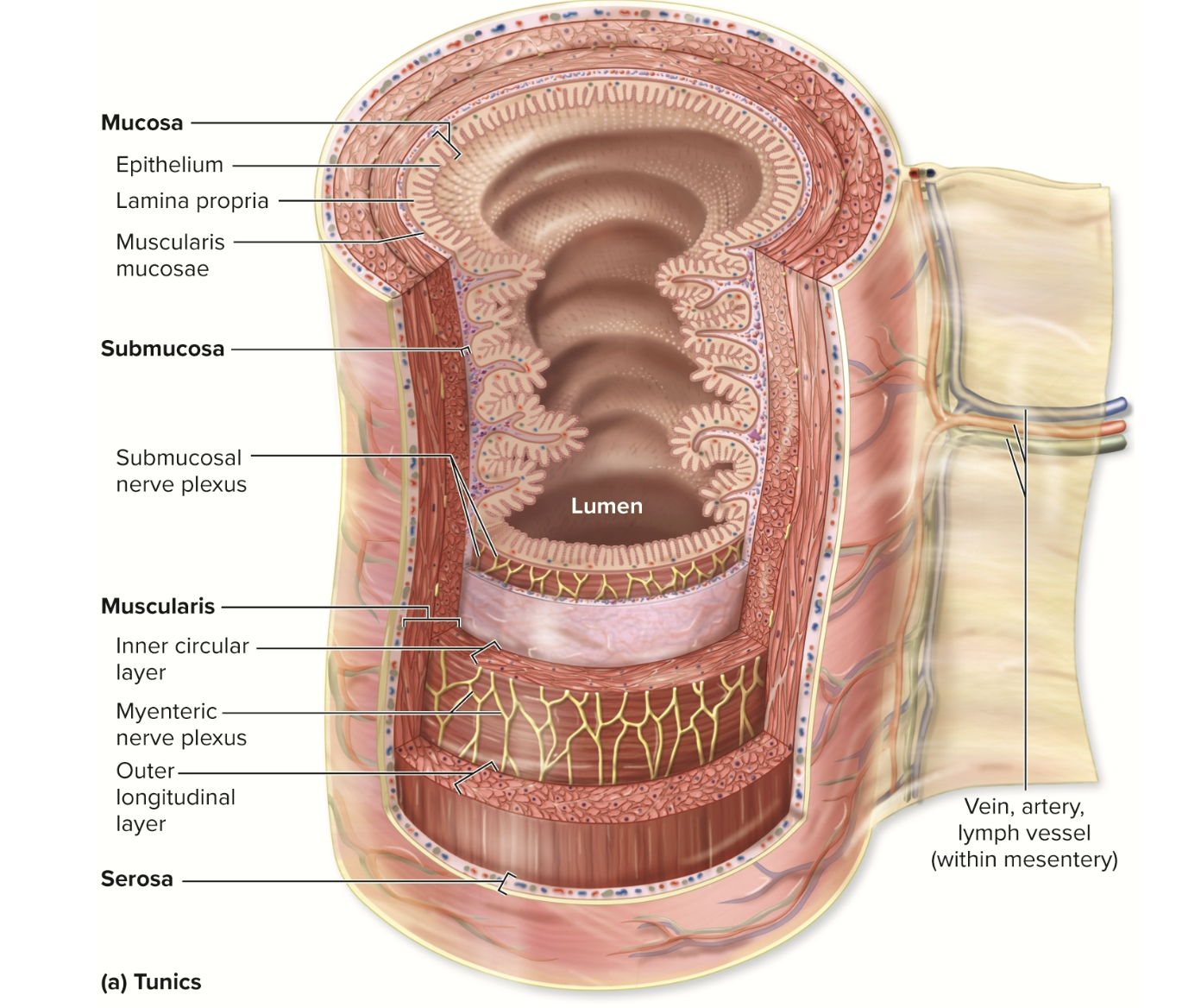
The GI tract wall has 4 main layers (tunics)
Mucosa, Submucosa, Muscularis, Adventitia / Serosa
Mucosa layer
Made of 3 things ?
Mucosa
Innermost layer that touches the food (lumen).
Made of:
Epithelium – secretes, absorbs, protects.
Simple columnar = for absorption/secretion (stomach, intestines)
Stratified squamous = for protection (mouth, esophagus, anus)
Lamina propria – connective tissue with blood & lymph vessels.
Muscularis mucosae – thin muscle layer that helps move contents.
Submucosa
Connective tissue with …..
Contains …….
what helps protect from microbes?
? large clusters of lymph tissue in small intestine.
Submucosa
Connective tissue with blood vessels, lymph vessels, and nerves.
Contains submucosal nerve plexus (controls glands & smooth muscle).
Includes MALT (mucosa-associated lymphatic tissue) to protect from microbes.
Peyer patches = large clusters of lymph tissue in small intestine.
Muscularis
Two layers of smooth muscle?
Between them?
Forms ?
Responsible for …. and …
Muscularis
Two layers of smooth muscle:
Inner circular – squeezes (narrows) the lumen.
Outer longitudinal – shortens the tube.
Between them is the myenteric nerve plexus, which controls contractions.
Forms sphincters to control food movement between regions.
Responsible for peristalsis (wave-like movement) and segmentation (mixing).
Adventitia / Serosa
Adventitia ?
Serosa ?
Adventitia / Serosa
Outermost layer:
Adventitia = areolar connective tissue (outside peritoneal cavity).
Serosa = same but covered by visceral peritoneum (inside peritoneal cavity).
Regulation
Explain Enteric Nervous System (ENS)
Enteric Nervous System (ENS)
Enteric Nervous System (ENS)
“Brain of the gut” — local network of sensory & motor neurons in the GI wall.
Controls reflexes like mixing and peristalsis.
2. Autonomic Nervous System (ANS)
Parasympathetic = increases digestion (rest & digest).
Sympathetic = slows digestion (fight or flight).
Regulation
Explain Reflexes
Hormones
Receptors
Reflexes
Short reflexes – controlled locally by ENS.
Long reflexes – involve the brain/CNS (e.g., smell of food triggering digestion).
4. Hormones
Examples: Gastrin, Secretin, Cholecystokinin (CCK), Motilin – regulate digestion and movement.
5. Receptors
Baroreceptors – sense stretching in the GI wall.
Chemoreceptors – detect chemical changes (pH, nutrients, etc.).
Peritoneum :
What are the 2 peritoneums and cavities of the abdomen?
Peritoneum = thin, slippery membrane that lines and covers abdominal organs.
Parietal peritoneum – lines inner abdominal wall.
Visceral peritoneum – covers surface of organs.
Peritoneal cavity – space between them, filled with fluid to reduce friction.
What is the difference between intraperitoneal organs and retroperitoneal organs?
Intraperitoneal organs – completely covered by visceral peritoneum
(stomach, most of small intestine, parts of large intestine)Retroperitoneal organs – behind peritoneum, only front covered
(duodenum, pancreas, ascending & descending colon, rectum)
Mesenteries?
Describe each ?
Greater omentum
Lesser omentum
Falciform ligament
Mesentery proper
Mesocolon
Mesenteries – double layers of peritoneum that support and anchor organs:
Greater omentum – “fatty apron,” hangs from stomach.
Lesser omentum – connects stomach/duodenum to liver.
Falciform ligament – attaches liver to front wall of abdomen.
Mesentery proper – holds small intestine in place.
Mesocolon – anchors large intestine.
Name the organs of the upper respitory track?
Organs Included:
Oral cavity & salivary glands
Pharynx
Esophagus
Stomach
Duodenum (beginning of small intestine)
oral cavity
main function/ mechanism ?
contains ?
Organ | Main Function / Mechanism | Location / Notes |
|---|---|---|
Oral cavity (mouth) | Start of digestion. Mechanical digestion by chewing; small amount of chemical digestion by saliva. | Entry to GI tract. Contains teeth, tongue, and openings of salivary glands. |
saliva glands
main function / mechanism?
contains ?
Salivary glands |
Secrete saliva containing salivary amylase (starts starch digestion). Lubricates food to form bolus. chemical. |
3 pairs: parotid (by ear), submandibular (under jaw), sublingual (under tongue). |
Pharynx
main function?
contains?
Pharynx (throat) |
Moves bolus from mouth to esophagus during swallowing (deglutition). Mucus helps food slide down. |
Behind oral cavity, connects to esophagus. |
Esophagus
main function?
contains?
Esophagus |
Tube transporting bolus to stomach via peristalsis (muscle contractions). |
Extends from pharynx → through diaphragm → into stomach. |
Stomach
main function/mechanism?
contains?
Stomach |
Mixes bolus with gastric juices to form chyme. Starts protein digestion with acid and enzymes. mechanical / chemical |
Upper left abdomen; connects to esophagus above and duodenum below. |
Duodenum
main function/mechanism?
contains?
Duodenum |
First part of small intestine; continues digestion using bile and pancreatic enzymes. |
C-shaped section just after stomach. |
oral cavity
Vestibule =
Lined with
Oral Cavity
Mouth = entrance to GI tract.
Vestibule = space between lips/cheeks and teeth.
Lined with stratified squamous epithelium → protection from friction.
Tongue
Things it helps with:
Tongue
Location: Floor of mouth.
Mechanism/Function:
Moves and mixes food during chewing (mechanical digestion).
Helps form bolus.
Has taste buds (papillae) and lingual tonsils for immune defense.
Assists in speech and swallowing.
Name the 3 saliva glands, thier location and what they produce
Gland | Location | % of Saliva | Main Function |
|---|
Parotid | In front of ear | 25–30% | Secretes watery saliva with amylase; can be infected (mumps). |
Submandibular | Under jaw | 60–70% | Produces most saliva. |
Sublingual | Under tongue | 3–5% | Adds mucus-rich saliva. |
Saliva contents:
Regulation:
Controlled by
Parasympathetic →
Sympathetic →
Saliva contents:
99.5% water
Salivary amylase (digests starch)
Mucus (lubricates)
Lysozyme & IgA (antibacterial)
Regulation:
Controlled by salivary nuclei in brainstem.
Parasympathetic → watery saliva (normal digestion).
Sympathetic → thick saliva (stress).
What do lingual glands help with?
Lingual Glands
Small glands at the base of the tongue.
Continuously release lingual lipase → begins fat digestion (even before stomach).
What is mastication?
Mastication
Purpose:
Controlled by:
Structures involved:
Mastication (Chewing)
Purpose: Mechanical digestion to increase surface area and mix food with saliva.
Controlled by: Brainstem (pons & medulla, mastication center).
Structures involved: Teeth, tongue, lips, cheeks, and jaw.
Structure | Location | Function / Mechanism | Extra Info |
|---|
Pharynx | ? | Moves? ? helps it slide down. | 3 parts? |
Esophagus | Extends from what to what? | Uses ? to push bolus down. Has ? to prevent air in or reflux. ?to help keep food out of respitory track | ? closes during breathing. |
Structure | Location | Function / Mechanism | Extra Info |
|---|
Pharynx | Posterior to oral cavity | Moves bolus into esophagus; mucus helps it slide down. | 3 parts: naso-, oro-, laryngopharynx (only oro- and laryngo- used for food). |
Esophagus | From pharynx → through diaphragm → to stomach | Uses peristalsis to push bolus down. Has upper and lower sphincters to prevent air in or reflux. Epiglottis to help keep food out of respitory track | Upper sphincter: closes during breathing. |
Histology of esophagus : | ?epithelium | ? lubrication. | Top ⅓ = bottom ⅓ = |
Swallowing (Deglutition): | 3 phases | Voluntary (mouth) → ? → ?. | Controlled by |
Histology: | Nonkeratinized stratified squamous epithelium (for protection) | Mucus glands in submucosa for lubrication. | Top ⅓ = skeletal muscle; bottom ⅓ = smooth muscle. |
Swallowing (Deglutition): | 3 phases | Voluntary (mouth) → Pharyngeal (involuntary, food to esophagus) → Esophageal (peristalsis to stomach). | Controlled by brainstem. |
Stomach:
Location:
Function:
Mechanism?
Starts ? digestion
Acts as ? — food can stay here for 2–6 hours.
Absorbs ?
Location: Upper left abdomen, just below the diaphragm.
Function:
Continues digestion (both chemical & mechanical).
Starts protein digestion (first place proteins are broken down).
Acts as a storage bag — food can stay here for 2–6 hours.
Absorbs small, nonpolar substances (like alcohol or aspirin).
Part | Description / Function |
|---|
Greater curvature |
Lesser curvature |
1. Cardia |
2. Fundus |
3. Body |
4. Pylorus |
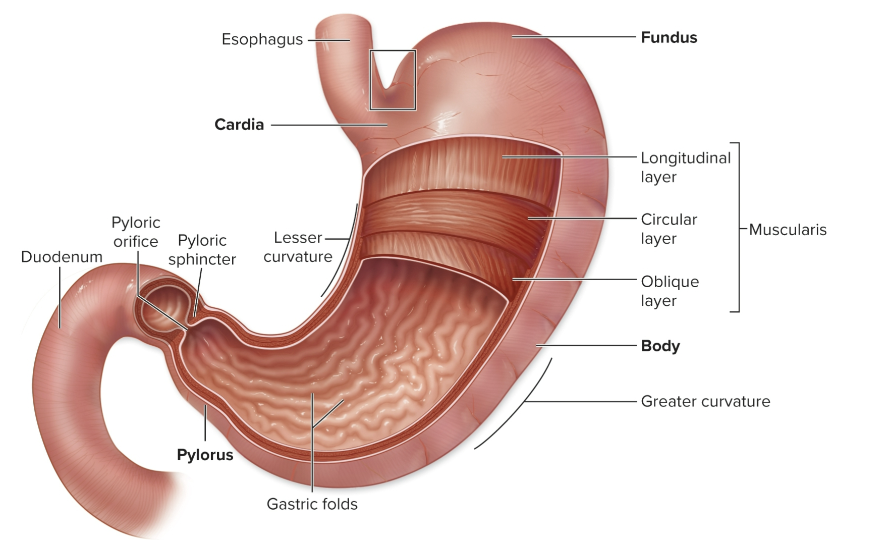
Part | Description / Function |
|---|
Greater curvature | Long, outer curve (convex, lower left side). |
Lesser curvature | Short, inner curve (concave, upper right side). |
1. Cardia | Entry where the esophagus meets the stomach. Has cardiac sphincter to prevent acid reflux. |
2. Fundus | Extra space.Gases and some food collect here. |
3. Body | Largest section; main area for mixing food with gastric juices. |
4. Pylorus | Funnel-shaped end leading to small intestine (duodenum). Controlled by pyloric sphincter to regulate food release. |
what are Rugae?
Rugae (Gastric Folds)
When the stomach is empty, the inner lining forms folds called rugae.
These allow it to expand when full and return to normal size when empty.
Layer | Features / Function |
|---|
Mucosa | ? epithelium . ? glands extend from the pits into deeper mucosa. |
Submucosa | Connective ? and ? |
Muscularis | 3 layers (not 2 like other GI organs): ? These extra muscles help ? |
Serosa | ? |
Layer | Features / Function |
|---|
Mucosa | Simple columnar epithelium with gastric pits (indentations). Gastric glands extend from the pits into deeper mucosa. |
Submucosa | Connective tissue with blood vessels and nerves. |
Muscularis | 3 layers (not 2 like other GI organs): inner oblique, middle circular, outer longitudinal. These extra muscles help with churning food. |
Serosa | Outermost layer (visceral peritoneum). |
Cell Type | Secretion | Function |
|---|
1. Surface mucous cells | ? | Protects stomach lining from ? |
2. Mucous neck cells | ? | Maintains ? |
3. Parietal cells | ? | - Intrinsic factor: ? |
4. Chief cells | ? | ? ? |
5. G-cells | Gastrin hormone | Stimulates ? |
Cell Type | Secretion | Function |
|---|
1. Surface mucous cells | Alkaline mucus | Protects stomach lining from acid & enzymes. |
2. Mucous neck cells | Acidic mucus | Maintains acidic environment; protects tissue. |
3. Parietal cells | Intrinsic factor and Hydrochloric acid (HCl) | - Intrinsic factor: needed to absorb vitamin B12 (for making red blood cells). |
4. Chief cells | Pepsinogen (inactive enzyme) and Gastric lipase | - Pepsinogen → Pepsin (activated by HCl) to digest proteins. |
5. G-cells | Gastrin hormone | Stimulates HCl release and increases stomach motility (movement). |
The stomach performs two key movements:
Gastric Mixing?
Gastric Emptying
What is it called when chyme moves back for more mixing?
The stomach performs two key movements:
Gastric Mixing
Mechanical digestion → mixes food with gastric juice → forms chyme (a thick, soupy mixture).
Gastric Emptying
Small amounts of chyme move into the duodenum through the pyloric sphincter.
The sphincter then closes again → some chyme moves back for more mixing (retropulsion).
Regulation of stomach
Explain the cephalic phase.
Gastric phase
Intestinal phase
Phase | Trigger / Control | What Happens |
|---|---|---|
1. Cephalic Phase (“thinking of food”) | Triggered by smell, sight, or thought of food → brain CNS (hypothalamus → medulla) → vagus nerve (long reflexes) | Increases stomach acid and motility → your stomach "growls." stimulates mucous, cheif and parietal cells |
2. Gastric Phase (“food in stomach”) | Food stretches stomach (baroreceptors) + proteins raise pH (chemoreceptors) → brain & hormone release gastrin → activate cheif cells → sells break down protein. | Increases HCl, motility, and gastric juice secretion. Pyloric sphincter tightens to slow emptying. |
3. Intestinal Phase (“chyme enters small intestine”) | Chyme entering duodenum → triggers intestinal reflex and hormones CCK- detects presence of presence of lipids & secretin- decrease PH and GIP - detects presence of carbohy | short reflex: inhibits stomach activity to allow small intestine time to digest. chemoreceptors—because chyme has entered the ph has changes |
Small Intestine
Divided into three parts and function :
Large Intestine
Absorbs ?
Forms and eliminates ?
Small Intestine
Divided into three parts:
Duodenum (1st): receives chyme (partially digested food) from the stomach and secretions from the liver, gallbladder, and pancreas.
Jejunum (2nd): main site of chemical digestion and nutrient absorption.
Ileum (3rd): continues absorption and ends at the ileocecal valve, which connects to the large intestine.
Large Intestine
Absorbs water, electrolytes, and some vitamins.
Forms and eliminates feces through the anus.
What are the accessory Organs and what do they do?
Accessory Organs
Liver: makes bile, which helps digest fats.
Gallbladder: stores and releases bile.
Pancreas: produces digestive enzymes and an alkaline (basic) fluid to neutralize stomach acid.
The small intestine is designed to ?
Circular folds?
Villi
Each villus has:
Microvilli – ?forming a?
Intestinal glands?
Cells ? name the 4
The small intestine is designed to maximize surface area for absorption.
Circular folds – large internal ridges (“speed bumps”) that slow chyme and increase contact time.
Villi – tiny, fingerlike projections of mucosa that absorb nutrients.
Each villus has:
Blood vessels (for carbs & proteins)
Lacteal (a lymph vessel for fats)
Microvilli – microscopic extensions on epithelial cells forming a brush border.
Contain brush border enzymes that finish digestion.
Intestinal glands – secrete intestinal juice for digestion.
Cells:
Goblet cells: produce mucus.
Enteroendocrine cells: release hormones (CCK, secretin).
Paneth cells: secrete antibacterial substances.
Duodenal glands: make alkaline mucus to neutralize acid from chyme.
Movement in the Small Intestine
Segmentation:?
Peristalsis: ?
Motilin hormone: ?
Gastroileal reflex: ?
Movement in the Small Intestine
Segmentation: back-and-forth mixing motion (helps absorption).
Peristalsis: wave-like contractions that move chyme forward.
Motilin hormone: triggers waves to push chyme toward large intestine.
Gastroileal reflex: when new food enters stomach → ileum contracts → ileocecal valve opens → chyme moves into large intestine.
Movement of bile
From Liver to Major Duodenal PapillHepatopancreatic sphincter
Liver → Right & Left Hepatic Ducts → merge to form Common Hepatic Duct→Joins with Cystic Duct (from gallbladder) → forms Common Bile Duct→Common Bile Duct joins the Main Pancreatic Duct → empties into duodenum at Major Duodenal PapillHepatopancreatic sphincter controls release of bile and pancreatic juice.
What is the largest internal organ and where is it located ?
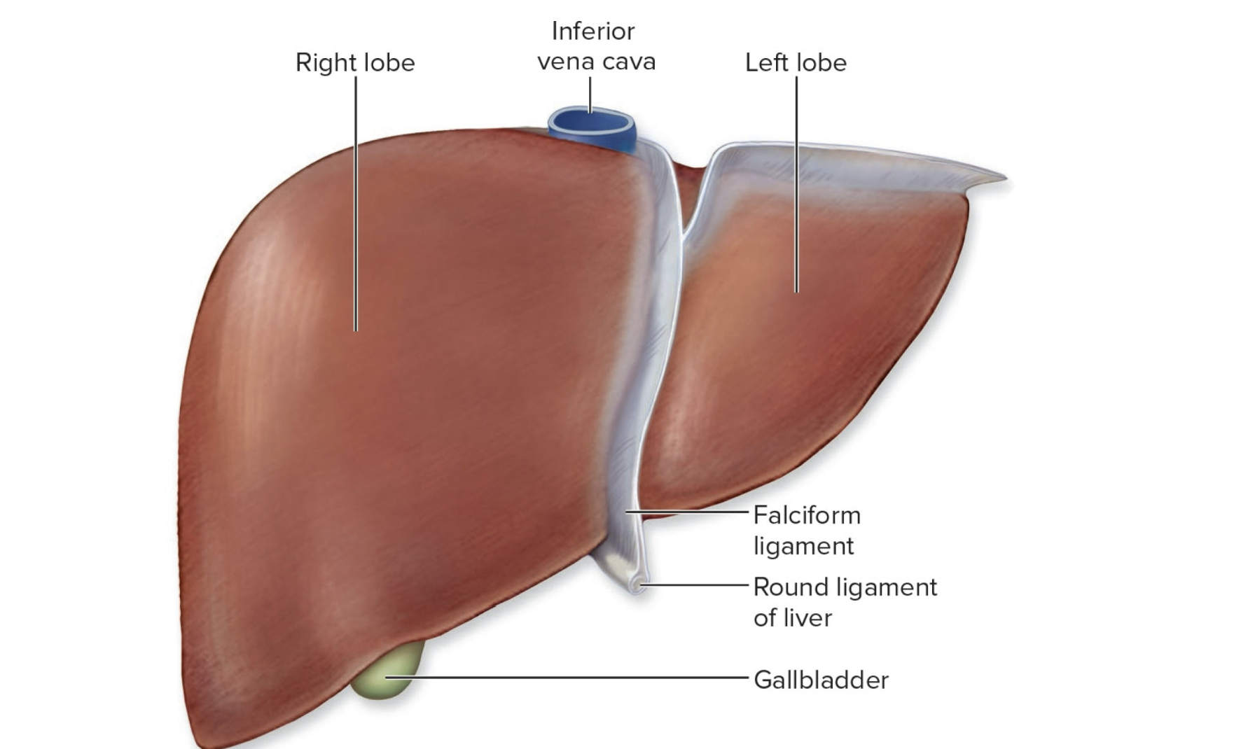
Largest internal organ, located under the diaphragm in the upper right abdomen.
Liver
Main Function:
🔹 Structure:
4 lobes:
Falciform ligament:
Round ligament:
Porta hepatis:
Main Function:
Produces bile for fat digestion.
🔹 Structure:
4 lobes: right, left, caudate, quadrate
Falciform ligament: separates right & left lobes; attaches liver to abdominal wall.
Round ligament: remnant of fetal umbilical vein.
Porta hepatis: entry/exit for blood vessels and ducts.
Blood Flow in the Liver:
Hepatic artery:
Hepatic portal vein:
Hepatic sinusoids:
Hepatic veins:
Kupffer cells (stellate cells):
🔹 Bile
Made by
Contains:
Bile salts
Blood Flow in the Liver:
Hepatic artery: brings oxygen-rich blood.
Hepatic portal vein: brings nutrient-rich blood from GI tract.
Hepatic sinusoids: spaces where blood mixes and nutrients are processed.
Hepatic veins: carry blood to inferior vena cava.
Kupffer cells (stellate cells): macrophages that clean the blood.
🔹 Bile
Made by hepatocytes (liver cells).
Contains: water, bile salts, bile pigments, cholesterol, lecithin, mucin.
Bile salts emulsify (break down) fats into smaller droplets.
Gallbladder
location.
Stores and concentrates :
Connected via :
?controls bile release.
Gallbladder
Small sac under the liver.
Stores and concentrates bile until needed.
Connected via cystic duct to the bile ducts.
Sphincter valve controls bile release.
Pancrease
Located ?, it has two jobs:
🔹 Endocrine (hormone function)
?
🔹 Exocrine (digestive function)
Produces ?
Made up of ?
The ducts add ?
Located behind the stomach, it has two jobs:
🔹 Endocrine (hormone function)
Releases insulin and glucagon into blood to control sugar levels.
🔹 Exocrine (digestive function)
Produces pancreatic juice that helps digestion.
Made up of acinar cells, which release enzymes.
The ducts add bicarbonate, making the juice alkaline to neutralize stomach acid.
Pancreatic Juice Contains:
Component | Function |
|---|---|
? | Digests starch → sugars |
? | Digests fats |
? | Digest proteins (activated in small intestine) |
? | Digest DNA & RNA |
? | Neutralizes acid from stomach |
Pancreatic Juice Contains:
Component | Function |
|---|---|
Pancreatic amylase | Digests starch → sugars |
Pancreatic lipase | Digests fats |
Proteases (inactive) | Digest proteins (activated in small intestine) |
Nucleases | Digest DNA & RNA |
Bicarbonate (HCO₃⁻) | Neutralizes acid from stomach |
Food passage from duodenum to elimination as feces
Chyme enters duodenum from the stomach.
Liver & gallbladder add bile → breaks down fats.
Pancreas adds digestive enzymes → digests carbs, fats, proteins.
Jejunum absorbs most nutrients.
Ileum absorbs remaining nutrients → passes leftovers to large intestine.
Large intestine absorbs water → forms feces → eliminated.
Accessory Digestive Organs and Their Regulation
Cholecystokinin (CCK)
Released when
Main actions:
Makes the
Causes the
Relaxes
Slows down the
Cholecystokinin (CCK)
Released when fatty chyme enters the small intestine.
Main actions:
Makes the gallbladder contract → releases bile (which breaks down fats).
Causes the pancreas to release pancreatic juice (enzymes for digestion).
Relaxes a valve called the hepatopancreatic ampulla (so bile and pancreatic juice can flow into the small intestine).
Slows down the stomach by inhibiting its movement and acid release — this gives the small intestine time to digest.
Accessory Digestive Organs and Their Regulation
Secretin
Released when
Main actions:
Makes the liver and pancreas….
Also inhibits ….
Secretin
Released when acidic chyme enters the small intestine.
Main actions:
Makes the liver and pancreas release an alkaline (bicarbonate-rich) fluid to neutralize acid.
Also inhibits the stomach’s activity (so the acid doesn’t overwhelm the small intestine).
The large intestine is size compared to the small intestine?
It runs from ? to the anus.
Main Functions:
Absorbs
Compacts
Stores
The large intestine is shorter and wider than the small intestine.
It runs from the ileocecal junction (where the small intestine ends) to the anus.
Main Functions:
Absorbs water and electrolytes from the leftover material.
Compacts chyme into feces.
Stores feces until defecation (elimination).
Parts of the Large Intestine
Cecum
Connected to the small intestine via the….
Attached to it is the appendix ……..
Parts of the Large Intestine
Cecum
First section (a pouch in the lower right abdomen).
Connected to the small intestine via the ileocecal valve.
Attached to it is the appendix — a small, thin sac that may store beneficial bacteria and has immune tissue.
Parts of the Large Intestine
Colon – The main part of the …., shaped like …..”
name the 4 parts of the colon
Colon – The main part of the large intestine, shaped like an upside-down “U.”
Ascending colon – goes upward on the right side.
Transverse colon – goes across the abdomen.
Descending colon – goes down the left side.
Sigmoid colon – S-shaped section leading to the rectum.
Parts of the Large Intestine
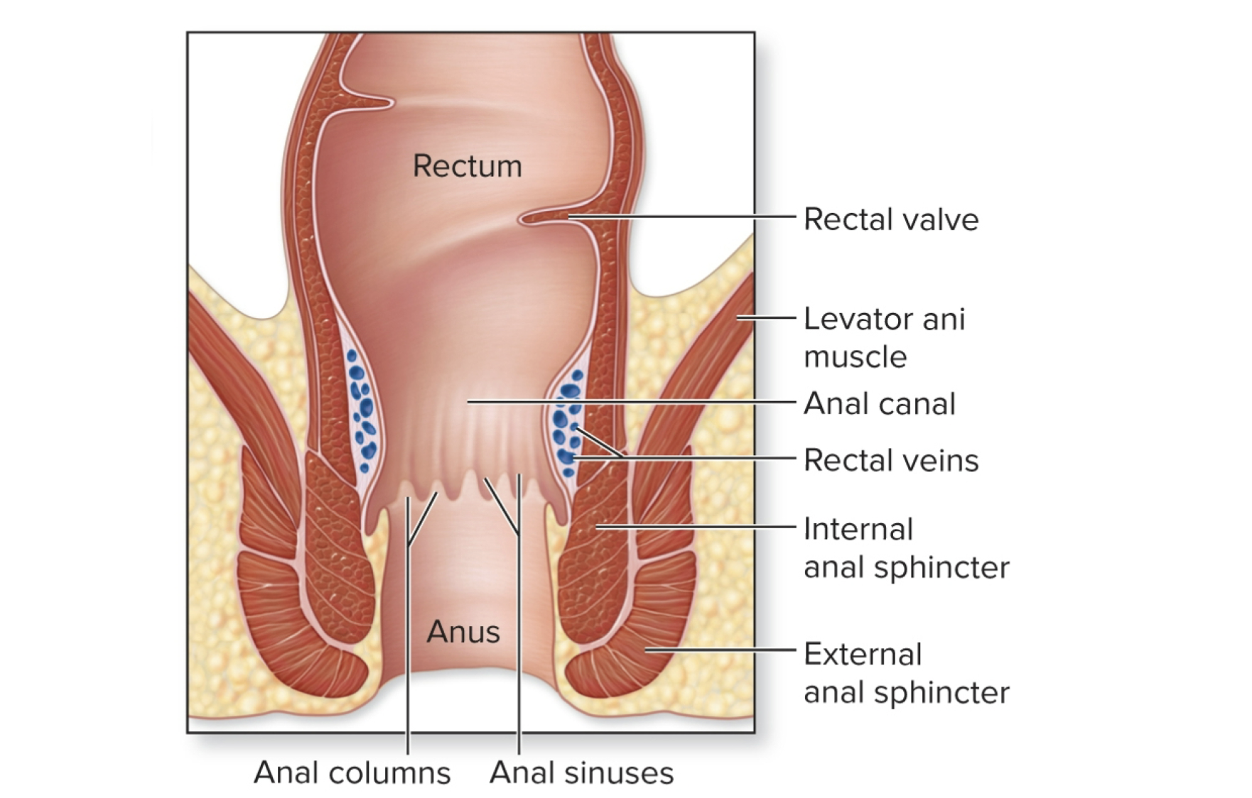
Rectum
Stores…
Has rectal valves that help…
Anal Canal
Has two sphincters?
Both keep the anus ?
Rectum
Stores feces.
Has rectal valves that help separate gas from feces so you don’t accidentally pass stool when releasing gas.
Anal Canal
Final part.
Has two sphincters:
Internal anal sphincter (involuntary, smooth muscle)
External anal sphincter (voluntary, skeletal muscle)
Both keep the anus closed until it’s time to defecate.
Large intestine
Teniae coli?
Haustra: ?
Omental appendices:?
Teniae coli: 3 bands of smooth muscle that “bunch up” the colon into sac-like pouches.
Haustra: the bulges formed by teniae coli.
Omental appendices: small fat-filled pouches hanging off the colon.
Large intestine
Bacterial Action
The large intestine contains helpful bacteria (normal flora) that:?
Bacterial Action
The large intestine contains helpful bacteria (normal flora) that:
Break down remaining carbs, proteins, and fats.
Produce gas (CO₂, H⁺) and vitamins B and K, which are absorbed.
Help form feces (made of water, salts, bacteria, dead cells, and undigested material).