Cell reproduction
1/53
There's no tags or description
Looks like no tags are added yet.
Name | Mastery | Learn | Test | Matching | Spaced |
|---|
No study sessions yet.
54 Terms
cell cycle
the events that take place from one cell division to the next
cells reproduce so that organs can grow larger
cells that are damaged, worn out or diseased must be replaced
some cells have a short lifespan and some have much longer
cells in the stomach lining → 2 days
nerve cells in the brain → lifelong
G1 + G2 phase
growth phases (separated by synthesis)
cell produces new proteins, grows and carries out normal tasks
phase ends when the cell starts duplicating DNA
s phase
synthesis phae
DNA molecules in the cell nucleus form exact duplicates of themselves
m phase
mitotic phase
after division cells may continue the cycle and re-enter G1 → some cells may stop dividing (G0 phase)
mitosis
the process by which a single parent cell divides to produce two identical daughter cells, each containing the same number of chromosomes as the original cell
it is used for growth, repair and replacements of cells in multicellular organisms
mitosis ensures genetic consistency, meaning the DNA in each new cell is identical as the original cell
chromosome structure
if it was possible to see chromosomes in a non-dividing cell (its not as they are in their chromatin form) they would look like the adjacent figure, each chromosome consisting of one chromatid
just before cell division occurs the DNA duplicates, when the DNA condenses into chromosomes during prophase they consist of two chromatids (the original and the copy) joined by a centromere
interphase - not a stage of mitosis
cell goes through G1, S and G2 phases
in the S phase: DNA molecules duplicate themselves → the quantity of DNA is doubled due to DNA replication
some cells will be in the G0 phase which is when cells are not preparing to divide and are performing normal cellular functions
during interphase the centrioles replicate, DNA replication occurs (DNA is still in the form of chromatin) and the nuclear membrane is clearly visible
prophase
2 pairs of centrioles become visible and move to opposite ends (poles) of the cell
microtubules begin to radiate from them
nucleolus disappears and the nuclear membrane begins to break down
chromatin becomes tightly coiled (condenses) and can be seen as chromosomes
each chromosome is comprised of a pair of chromatids due to the prior DNA replication
By the end of prophase:
centrioles have reached opposite poles and microtubules radiate from them to form a spindle
nuclear membrane has disappeared completely
spindles begin to attach to the centromere of the chromosomes
metaphase
spindle fibres move chromosomes towards the centre of the cell
centromere is attached to a spindle fibre
the chromosomes (two chromatids) line up at the equator of the cell (metaphase plate)
the centrioles are at opposite ends and the spindle fibres are attached to the centromere
anaphase
the spindle fibres contract causing the centromeres to break, dividing the sister chromatids
the centrioles ‘pull’ on the spindle fibres
the chromosomes are pulled to opposite poles
each chromosome goes from having 2 sister chromatids to being two separate chromosomes
telophase
two sets of chromosomes form tight groups at each pole of the cell
nuclear membrane forms around each group
nucleolus appears in each new nucleus
spindle fibres disappear
chromosomes gradually uncoil to become chromatin threads again
cytokinesis
involves the division of the cell contents (cytoplasm + organelles)
occurs concurrently with telophase
a furrow develops in the cytoplasm between the two nuclei. The furrow deepens until it cuts the cytoplasm into two parts resulting in two identical daughter cells, each with a full set of chromosomes / DNA
mitosis
mitosis and cytoplasmic division have resulted in the formation of 2 daughter cells
each chromosome was duplicated
each daughter cell has identical number and type of chromosomes as the parent cell
the genetic information is therefore passed from parent cell to daughter cells and without change
cancer
when normal differentiation of cells goes wrong
this results in a tumour → an abnormal mass of tissue from uncontrolled division of cells
how does cancer form
cells failing to follow normal cell division and multiply excessively into a mass of proliferating cells
normal cells die when they lose contact with surrounding matrix
carcinogens cause mutations where DNA is altered changing the expression of certain genes
certain genes produce proteins that are essential for cell division, growth, cellular adhesion and other things
exposing these genes to carcinogens lead to the failure of producing these genes leading to the formation of cancer
malignant tumours
cells are able to spread to other parts of the body (metastasis)
secondary tumour can develop well away from the original tumour
cancerous type of tumour
benign tumours
cells are not able to spread to other parts of the body
they grow and press on surrounding tissues
normally have a capsule surrounding them making them easier to remove
non cancerous type of tumour
causes of cancer
certain environmental factors (carcinogens) can trigger malignant tumours:
UV radiation
X rays
ionising radiation (radium, uranium) → single exposure to high dose may result in leukaemia
viruses (e.g. HPV)
chemical carcinogens (e.g. alcohol, asbestos, soot, tar, organic solvents in glue and paint, tobacco tar)
cancer prevention methods
education: advertising and educational programs to limit exposue to carcinogens (e.g. Slip, Slop, Slap to limit UV exposure)
Legislation: laws to control exposure to carcinogens
smoking being banned in many public places
tobacco advertising is not permitted
cigarettes must be sold in plain packages / images
standards for manufacture and operation of X ray machines
banning products containing asbestos
reducing UV exposure: sunscreen, sunglasses, long sleeved clothing, shade and hats, stay out of direct sunlight between 10am and 3pm
diet: adequate fibre and low fat, not overweight / obese, limit alcohol
protective clothing when handling chemicals
avoid smoking
cervical cancer
caused by HPV → some people who have HPV may have cervical cells change and later become cancerous
pap test: cells collected from cervix smeared on microscope slide and examined. This detects early changes in cervical cells
breast cancer
mammogram: X-ray of breasts → tumours as small as 1cm in diameter can be detected
bowel cancer
bowel cancer: most bowel cancers develop from polyps, it not all polyps become cancerous
Faecal Occult Blood Test (FOBT): at home tests for blood in faeces, mail to lab for analysis → can detect small amounts of blood not visible to the naked eye
if the test is positive, referred for a colonoscopy (visual examination of the intestine)
prostate cancer
Digital rectal examination (DRE): insert finger into anus to feel surface of prostate → swelling, hardening or irregularities of surface may indicate cancer (some irregularities may be beyond reach)
prostate specific antigen (PSA): blood test for presence of protein produced by prostate, if PSA rises it may indicate presence of cancer
Biopsy: several small samples of prostate tissue checked for cancer (used once the other 2 methods have indicated positive)
meiosis interphase
similar to mitosis interphase
chromosomes replicate (in chromatin form) in the s phase
each duplicated chromosome consists of two identical sister chromatids attached to their centromeres
meiosis prophase 1
spindle fibres form
centrioles move to the poles
nuclear envelope dissolves
chromatin condenses into replicated chromosomes (2 sister chromatids)
homologous chromosomes pair up
in prophase 1 ‘crossing over’ occurs:
during crossing over segments of chromosomes break off and reattach to the paired homologous chromosome → this leads to greater genetic diversity
meiosis metaphase 1
the shortest phase
spindle fibres attach to the centromere of each homologous chromosome
pairs of homologous chromosomes line up at the equator of the cell
meiosis anaphase 1
homologous chromosomes separate and move towards the poles
sister chromatids remain attached at their centromeres
there is no separating of chromatids
meiosis telophase 1
chromosomes uncoil into chromatin and spindle fibres break down
nuclear envelopes form around the DNA at each pole creating 2 nuclei
each pole now has one of the 2 homologous chromosomes consisting of 2 sister chromatids
cytokinesis occurs and 2 haploid daughter cells are formed
meiosis prophase 2
the same of prophase in mitosis
nucleolus and nuclear membrane disintegrate
centrioles migrate to opposite poles, which are at right angles to the previous devision
chromatin condenses to form chromosomes and become visible
spindle fibres develop and attach to centromeres
meiosis metaphase 2
same as metaphase in mitosis
chromosomes are arranged at the equator of the cell in a single file line
each chromosome is attached to a spindle fibre at the centromere with the centrioles at opposite ends
meiosis anaphase 2
same as anaphase in mitosis
spindle fibres constrict ‘breaking’ chromosomes to separate sister chromatids. Each chromatid is now considered a chromosomes and are pulled to opposite poles
sister chromatids separate
meiosis telophase 2
chromosomes uncoil into chromatin
nuclear membrane and nucleolus reform around each set of chromosoems
spindle fibres disappear
cytokinesis occurs, resulting in a tetrad of haploid cells
somatic cells vs. gametes
somatic cells | gametes |
|---|---|
normal body cells | sex cells |
contain the normal number of chromosomes - one copy from each parent cell | contain half the normal number of chromosomes |
called the diploid number - 2n | called the haploid number - n |
meiosis
the process by which gametes are produced with half the number of chromosomes (haploid)
during meiosis diploid cells are reduced to haploid cells
diploid (2n) → haploid (n)
gametogenesis
meiosis and the processes that follow result in the formation of ova and sperm, this is collectively called gametogenesis
there are two types of gametogenesis:
spermatogenesis: the formation of sperm in the testes
oogenesis: the formation of ova in the ovary
homologous chromosomes
pairs of chromosomes (maternal and paternal) that are similar in shape and size
each gene is in the same position on homologues
humans have 23 pairs of homologous chromosomes
22 pairs of autosomes, 1 pair of sex chromosomes
sources of variation
crossing over - recombination of chromosomal sections in prophase 1
independent and random assortment of chromosomes into gametes
random fusion of gametes and fertilisation
genetic variation
the advantage of meiotic division and sexual reproduction is that it promotes genetic variation in offspring
the three main sources of genetic variation arising from sexual reproduction are:
crossing over
random assort of chromosome
random fusion of gametes from different parents
crossing over
during meiosis 1, homologous chromosomes (1 from each parent) pair along their length
the chromosomes may cross over at point called chiasma
at each chiasma, the chromosomes break and rejoin, trading some of their genes
crossing over can result in a new combination of alleles along the chromosome, called recombination
therefore, crossing over creates a new combination of genes so that the chromosomes passed on to the offspring are not exactly the same as those inherited from the parents
crossing over - recombination
crossing over is an exchange of segments of chromosome between homologous chromatids during meiosis 1 (prophase 1)
it may occur at one or more places along the chromosome
allele closer together are less likely to be separated
independent (random) assortment
describes how pairs of alleles separate independently from one another during gamete formation → the inheritance of genes / traits is independent to the inheritance of any other gene / trait
this is due to the random orientation of pairs of homologous chromosomes in meiosis 1 → the orientation of each homologous pair is random and is not affected by the orientation of any other
this means an allele on one chromosome has an equal chance of being paired with, or separated from, any allele on another chromosome (the inheritance is independent)
this random, independent assortment takes place for each of the 23 pairs of human chromosomes → any human egg receives one of two possible chromosomes 23 times (possible combinations are 2^23)
random fertilisation
the fusion of two haploid gametes results in the formation of a diploid zygote
this zygote can then divided by mitosis and differentiate to form a developing embryo
as meiosis results in genetically distinct gametes, random fertilisation by egg and sperm will always generate different zygotes
non disjunction (error in replication)
refers to the chromosomes failing to separate correctly, resulting in gametes with one extra or one missing chromosome (aneuploidy)
failure of chromosomes to separate may occur via:
failure of homologues to separate in anaphase 1 (resulting in four affected daughter cells)
failure of sister chromatids to separate in anaphase 2 (resulting in only two daughter cells being affected
aneuploidy - trisomy
trisomy is a condition in which an individual inherits an extra copy of a chromosome - 3 copies instead of the normal 2
one such chromosome defect is down syndrome, or trisomy 21
aneuploidy - monosomy
monosomy is where an individual is missing a chromosome - they have only one copy instead of the normal two
partial monosomy and partial trisomy can also occur
in partial monosomy, part of a chromosome is missing - part of the chromosome has two copies, but part only has one copy
partial trisomy occurs when part of an extra chromosome is attached to one of the other chromosomes
mutation
changes to the DNA code
can happen spontaneously through mutagens (e.g. radiation / chemicals) or errors in replication
if this occurs in gametes, it will be passed onto the next generation
mutation as the source of new variation
many cellular processes exist to repair mutations in DNA because:
harmful mutations can stop a protein, and therefore a cell, from functioning properly
harmful mutations may impair the process of apoptosis, leading to cancer
if a cell cannot repair a mutation it will try to undergo apoptosis
effects of mutation on survival
neutral: does not change the amino acid or changes it to one with a similar shape and charge. Protein essentially unchanged
deleterious: deletes, impairs or enhances the proteins activity in such a way that the organism is adversely affected. Can lead to premature death of the organism
beneficial: changes the proteins activity in such a way that the organism benefits
mutations occur throughout the genome and non coding regions are also affected
variation
while variation between species allows us to tell them apart, variation is a common and important observation within species
this is infraspecific variation
variation in phenotypes can be genetically, or epigenetically, determined
morphological phenotypic variation
shape and structure including internal anatomy
size and shape of noses
biochemical phenotypic variation
chemical structure and composition of organisms including proteins, lipids, carbohydrates, and other molecules
expression of enzymes creating pigments and resulting colour of hair
physiological phenotypic variation
metabolic and other bodily processes
blood group, haemophilia
behavioural phenotypic variation
the ways individuals perceive, think, and react. This includes congnition and behaviour
agression, inquisitiveness, mate selection
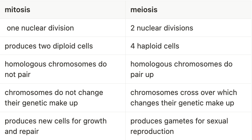
mitosis vs. meiosis