Hole's Essentials of Human Anatomy & Physiology - Chapter 8: Muscular System
 Call Kai
 Learn
Learn Practice Test
 Spaced Repetition
 Match
Match Flashcards
 Knowt Play
1/59

Earn XP
Description and Tags
Comprehensive vocabulary flashcards detailing muscle structure, contraction physiology, cellular respiration, motor responses, and general muscular anatomy.
Name | Mastery | Learn | Test | Matching | Spaced | Call with Kai | Chat |
|---|
No analytics yet
Send a link to your students to track their progress
60 Terms
Fascia
Layers of dense connective tissue that surround and separate each skeletal muscle.
Aponeurosis
A broad sheet of connective tissue that connects muscles to each other or to nearby bones.

Epimysium
The outermost layer of connective tissue that directly surrounds a skeletal muscle.
Perimysium
Connective tissue extending inward from the epimysium that surrounds bundles of skeletal muscle fibers called fascicles.
Endomysium
A connective tissue layer that covers each individual skeletal muscle cell or fiber.
Fascicle
A bundle of skeletal muscle fibers enclosed by the perimysium within a skeletal muscle.
Sarcolemma
The cell membrane of a skeletal muscle fiber.
Sarcoplasm
The cytoplasm of a muscle cell, containing many mitochondria, nuclei, and parallel threadlike myofibrils.

Myofibril
Parallel, threadlike structures in the sarcoplasm active in muscle contraction, composed of thick myosin and thin actin filaments.
Myosin
The protein that makes up the thick filaments in myofibrils, featuring globular heads that bind to actin.

Actin
The primary protein composing thin filaments in myofibrils, arranged in twisted filaments containing binding sites for myosin heads.

Sarcomere
The structural and functional unit of a myofibril, extending from one Z line to the next Z line.
I Band
The light striation band in a sarcomere, composed of thin actin filaments anchored to Z lines.
A Band
The dark striation band in a sarcomere, composed of overlapping thick myosin filaments and thin actin filaments.
H Zone
The central region of the A band that consists strictly of thick myosin filaments.
M Line
A protein structure located in the center of the H zone that holds thick myosin filaments in place.

Sarcoplasmic Reticulum (SR)
A network of membranous channels beneath the sarcolemma that stores calcium ions and serves as the endoplasmic reticulum of a muscle cell.
Transverse Tubules (T Tubules)
Invaginations of the sarcolemma extending inward between cisternae of the sarcoplasmic reticulum, open to the outside of the muscle fiber.

Neuromuscular Junction
A functional synapse between a motor neuron axon and a skeletal muscle fiber.
Motor End Plate
A specialized, tightly folded region of the sarcolemma at the neuromuscular junction containing specific neurotransmitter receptors.
Synaptic Cleft
The narrow extracellular gap separating the motor neuron membrane from the muscle fiber membrane at a synapse.
Acetylcholine (ACh)
The neurotransmitter stored in synaptic vesicles of motor neurons that stimulates skeletal muscle fibers to contract.

Sliding Filament Model
The mechanism of muscle contraction where cross-bridges pull actin filaments past myosin filaments, increasing overlap and shortening the sarcomere.
ATPase
An enzyme located in myosin heads that catalyzes the conversion of ATP to ADP, releasing energy to cock the myosin heads.
Rigor Mortis
A state of partial skeletal muscle contraction and rigidity after death caused by increased calcium permeability and a lack of ATP to break cross-bridge linkages.
Acetylcholinesterase
An enzyme present in the motor end plate that rapidly decomposes acetylcholine to end muscle fiber stimulation.

Creatine Phosphate
A molecule containing high-energy phosphate bonds used to rapidly regenerate ATP from ADP in muscle cells.
Myoglobin
An iron-containing pigment in muscle tissue that temporarily stores oxygen, increasing oxygen availability for aerobic respiration.
Oxygen Debt (EPOC)
The amount of oxygen required by liver cells to convert accumulated lactate back to glucose, plus the amount needed by muscle cells to restore ATP and creatine phosphate concentrations.
Muscle Fatigue
The condition where a muscle loses its ability to contract during strenuous exercise, often stemming from electrolyte imbalances, decreased ATP, or reduced pH from lactic acid.
Muscle Cramp
A sustained, painful, involuntary muscle contraction thought to result from changes in extracellular fluid causing uncontrolled motor neuron stimulation.
Slow-twitch Fibers
Fatigue-resistant muscle fibers with small diameters, abundant mitochondria, and capillary networks specialized for prolonged aerobic endurance activities.
Fast-twitch Fibers
Fatiguable muscle fibers with large diameters, high glycogen stores, and few mitochondria that provide rapid, powerful anaerobic contractions.
Hypertrophy
Enlargement of a muscle resulting from repeated forceful exercise that increases the synthesis of actin and myosin filaments.
Atrophy
A reduction in muscle size and strength caused by physical disuse.
Threshold Stimulus
The minimum strength of electrical or chemical stimulus required to generate a muscle impulse and trigger contraction.

Twitch
A single brief contraction-relaxation cycle of a muscle fiber in response to a single impulse.
Latent Period
The brief delay on a myogram between the time of stimulation and the beginning of muscle contraction.
All-or-None Response
The principle stating that when an isolated muscle fiber is brought to threshold stimulus, it contracts completely and produces maximum force for that twitch.

Summation
The combining of individual twitch forces in a muscle fiber when exposed to rapid, repeating stimuli that prevent complete relaxation.
Partial Tetany
A state of sustained, incomplete muscle contraction resulting from high-frequency stimulation with very brief relaxation periods.
Complete Tetanic Contraction
A continuous, smooth contraction lacking any relaxation phase, achieved experimentally under very high stimulus frequencies.

Motor Unit
A functional subunit consisting of a single motor neuron and all the skeletal muscle fibers it innervates.
Recruitment
An increase in the number of active motor units within a muscle as stimulation intensity increases.
Muscle Tone
A continuous state of partial contraction in resting muscles caused by low-frequency stimulation of a few motor units.
Isotonic Contraction
A muscular contraction where force remains constant while muscle length changes, resulting in movement.
Isometric Contraction
A muscular contraction where internal force is generated without changing the muscle length or causing movement.
Multiunit Smooth Muscle
A type of smooth muscle tissue with separate, unorganized fibers found in blood vessel walls and iris, stimulated individually by nerves or hormones.
Visceral Smooth Muscle
A type of smooth muscle with spindle-shaped cells arranged in sheets, found in hollow organ walls, displaying rhythmicity and peristalsis.
Peristalsis
Rhythmic, wavelike muscle contractions of visceral smooth muscle sheets that propel contents through tubular organs.
Intercalated Discs
Specialized cell membrane junctions in cardiac muscle tissue that join adjacent cells and allow rapid transmission of contraction force and impulses.
Origin
The less movable or fixed attachment point of a skeletal muscle.

Insertion
The movable attachment point of a skeletal muscle pulled toward the origin during contraction.
Agonist (Prime Mover)
The muscle that bears primary responsibility for causing a specific movement.
Synergist
A muscle that assists the prime mover in performing a specific action.
Antagonist
A muscle whose action opposes that of a prime mover.
What are the three types of muscle tissue in the body?
skeletal, smooth, and cardiac muscle
how many skeletal muscles does the body contain
about 600
REMEMBER FTA (FLORENCE TAKES A**)
The three are fascia, tendons, and aponeuroses
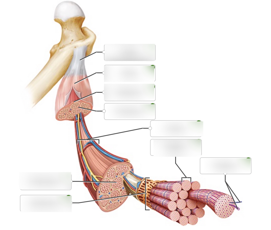
KNOW EACH PART