PART B: Infectious Diseases (copy)
 Call Kai
 Learn
Learn Practice Test
 Spaced Repetition
 Match
Match Flashcards
 Knowt Play
1/23

Earn XP
Description and Tags
PART B of SMALLS STATE EXAM. Bruker oppsettet/question numbers til VM dokumentet & NATH.
Name | Mastery | Learn | Test | Matching | Spaced | Call with Kai | Chat |
|---|
No analytics yet
Send a link to your students to track their progress
24 Terms
1.Infectious & Parasitic skin diseases in dogs and cats 🩸
(Nb: focus on infectious diseases than parasites, see q24 for details on ectoparasites)
Skin problems → parasites, bacterial infections, fungal infections & allergic dermatitis.
Bacterial Skin Diseases (pyoderma)
typically as a secondary issue, so imp. to treat underlying cause.
Skinˋs natural barrier is bypassed due to factors like:
immunosuppression & young age (immature immunity)
breed (skin folds → moisture),
break of skin (scratching due to parasite bite, allergies)
Pyoderma (“pus in skin”) can be on surface, superficial (enters hair follicle) & deep (massive inside follicle → rupture → spills into fat&dermis).
Primary Pathogens: Staphylococcus Pseudointermedius (most common in dogs) incl. Methicillin-resistant strains (MRSP).
2ndary invaders: Staphylococcus aureus (MRSA), Streptococci, micrococcus, Bacillus, corynebacterium, E.Coli, Proteus & Pseudomonas.
Pathogenesis: Bacterial pyoderma occurs when normal resident or transient flora overcolonizes the skin. Common esp. in warm & moist areas, like skin folds (lips, face, neck, axillarym vulvar or tail folds), and interdigital spaces.
Pressure points (hocks and elbows) are also susceptible, due to follicular irriration (tiny skin cavities from which hairs grow → inflamed, sore)
Diagnosis: Physical exam (noting alopecia, pruritus, type of lesion), cytology (swabs, tape impression smears, diff quick), skin scrapings - help rule out parasites, cultivation, histopathology (last option).
Treatment:
Topical: clorhexidine wash, shampoo, gels, sprays - sometimes enough alone or together w/systemic
Systemic: ATB, based on culture (for deep pyoderma)
Viral Skin Diseases
Viral dermatoses - often w/severe systemic signs or specific local lesions.
Canine Distemper: “hardpad disease” → hyperkeratosis (thickening) of nasal planum and footpads, w/pustular lesions on ventral abdomen.
Cutaneous form of distemper = better form of prognosis
Aujeszky’s Disease (Pseudorabies): "mad itch disease” → intense pruritus in dogs, self-multilation esp. on head.
Feline Cowpox: Orthopoxvirus via direct route (bites) → papules & vesicles on head & forelimbs.
Parasitic Skin Diseases (overview)
Mites: inhabit skin, feed on blood, lymph, skin debris or sebaceous secretions.
cause mange → Scabies - typical w/ yellow scabs, wet blood squeezed excoriations, shedding hairs & weight reduction.
Etiology - potential zoonotic, except demodex.
Burrowing Mites:
Sarcoptes: Intense pruritus (pinna-pedal reflex), crust, alopexia. Pruritus - Scabies.
Notoedres (N.cati): “Feline version of scabies”, starts typ. on ear pinnae and face.
Demodex: By commensal mite overgrowth due to immunosuppression (esp. in puppies) → local, general or pododemodicosis (foot) forms. (NOT pruritic!)
Non-Burrowing Mites:
Cheyletiella: "walking dandruff”
(neo)trombicula/Chiggers (edderkoppdyr): larval mites → secrete cytolytic enzymes → intense local dermatitis.
Otodectes - O.cynotis (Otitis externa) → more pruritic in dog, exudate in cat. Local allergic reaction w/inflammation&discharge.
Mites release allergens in feces/other metabolic products → triggers immune system → skin hypersensitvity reaction → itch, scratch, inflammation & exudation. Scabs dries - can be septic complication (w/Lnn enlargement).
Lice → self trauma, hair loss and abrasions on skin. itch/scratch, matted fur, stressed. Sucking lice (feed on blood/secretion) & Biting lice (feeds on skin/debris).
Ticks → hematophagous ectoparasites, local skin irritation. Vectors of disease.
Fleas → Mild irritation or severe FAD. Hematophagous ectop.
histamine-like compounds&enzymes → injected, induce immune hypersensitivity → pruritus, papulocrustous lesions on lower back, tailhead, post.&inner thighs.
Dg: CS, Fleas present. Tx: insecticides - fipronil, ivermectin, selamectin, pyrethroids, imidacloprid.
Fungal infections: Dermatophytes - microsporum, ringworm, trichophyton, Malassezia. (q2)
notes:
Difference of scabies vs. demodex: Scabies = pruritic, demo is not.
Which for of distemper has better prognosis → Cutaneous
Otitis Externa 🩸
(is part of qs.1)
Otitis Externa → inflammation of the external ear canal. Can also involve the pinna (ear flap itself).
Acute or chronic, uni or bilateral
Etiology:
Predisposing factors: Ear conformation (heavy, floppy ears of basset) & excessive moisture.
Primary (changes the environment → allows sec.infections to develop):
1) Atopy (genetic predisp. to develop allergic reactions) > 70% of dogs develop otitis externa → bacterial or yeast infection.
2) Allergies → contact (rare, but can occur shortly after tx. w/topical drug) & food allergy (less common)
3) Immune-mediated (immune system attacks own healthy cells) (ex. pemphigus (group of rare autoimmune skin diseases), discoid or systemic lupus erythematosus, drug reactions,
4) endocrine (hormonal issues - hypothyroidism, may have seborrhea),
5) viral (ex. CDV),
6) Foreign bodies (grass seed) or growths/tumor
7) moisture, overcleaning,
8) Fungal (ex. ringworm, but rare)
9) parasites (Otodectes cynotis) - ear mite, non-burrowing
FH: car, esp. cats
Location: ear canal, outer ear
Transmits by direct contact, very contagious
Patho: Local allergic reaction. Purulent inflammation & discharge of external ear, can perforate the tympanic membrane. → wax which traps bacteria. It sycks lymph&tissue fluid → otitis externa, head shaking - can lead to pinnal hematoma, itching.
CS: Dark, dry, “Coffe-ground” ceruminous exudate in ear canal.
Cats: massive, coffee-ground excretions in ear canal, NO itch/sometimes w/severe pruritus.
Dogs: small excretions w/ear itching.
Dx: detect w/otoscope (see white moving mites), ear-smear w/microscope
Tx: clean ear canal & apply selamectin (spot-on), moxidectin. Amitraz & local w/thiabendazole/acaricide ear drops.
3-4w to cover LC (treat several times as tx does not affect eggs).
10) Otobius megnini - ear tick of dogs & less common in cats → the tick bite itself is the primary cause → creating wound → then secondary is bacterial/fungal infection.
Other - Sarcoptes scabiei, Notoedres cati, demodex canis & cati.
Secondary (The reason that cause disease in abnormal ear):
Bacteria
Dogs: Staphylococcus sp. = Most common, proteus, pseudomonas & other gram-
Cats: staphylococcus spp., pasteurella
fungi (yeast like Malassezia Pachydermatitis) - plentiful dark, sweet-smelling discharge.
General Clinical signs: head shaking, smell, pain on manipulation, exudate, erythema, head tilt

Yellow, oily cerumen → keratinization disorders.
Dg: otoscopy, dermatological examination (cytology of exudate & crust and skin scrape), x-ray
Tx: ensure ear drum is intact before giving drugs, if ruptured → may need oral ATB/antifungal instead.
Clean ears - cerumenolytic agents (cerumarual, dioctyl sodium succinate, carbamine peroxide), irrigate w/cleansing agent (warm water and iso/hypertonic saline).
Topical anti-inflammatory agents: corticosteroids (betamethasone, hydrocortisone)
Topical anti-bacterial agents: gentamycin, neomycin, enrofloxacin)
Topical anti-fungal agents: imidazoles
Antiparasitics: pyrethrin, ivermectin
3.Mycotic diseases of small animals 🩸
Mycoses = Fungi causing infections → affecting skin (superficial) or internal organs (systemic). Zoonotic risk, can be chronic.
1) Dermatomycosis
Fungal infection of skin, hair & nails, by dermatophytes (fungi that use keratin to grow, spore-forming).
Dermatophytes are classified by their source: Anthropophilic (bw.humans), Zoophilic (animals, ex. M.canis, T.verrucosum) & Geophilic (from soil, rarely affecting humans).
Ringworm - Dermatophytosis (Zoonotic)
Tricophyton spp. - T.verrucosum (ru, eq, dog), T.mentagrophytes
Microsporum spp. - M. canis - most common (dog, su), M.gypseum
Transmission: Direct contact, indirect contact with bedding, or via asymptomatic carriers (cats -possible)/spores in the air (1y in environment). More prevalent in the tropics, fall, and winter, worldwide.
Pathogenesis:
Fungi penetrate hair follicles → form hyphae & spores → use enzymes to decompose keratinized infection.
Natural infection: incubation (1-2w, multiplies in S.corneum & hair follicles) → maturation (fungi grow/spores, hair breaks off → small round inflamed patches) → crust forms (day 28-40, pathes → scaly & crusty - parakeratosis, epidermal hyperplasia) → regression (self limiting often in large animals - up to 4mo, crusts → darken, fall off).
Clinical Signs: Round alopecia, scaling, crusts & broken hairs (head, neck, limbs), onychomycosis (nail infection). Not itchy.
Dogs - generalized form, larger patches vs. cats - localized, neck&front of chest.
Diagnosis: Clinical signs, Woodˋs lamp (yellow fluorescence for Microsporum only), Fungal culture (sabouraud agar, min 3w). PCR.
Micrscopy w/KOH (skin scrape/hair pluck w/roots to see hyphae/spores)
Note: vaccine exists, but it is not commonly used.
Treatment: Self-resolving, but treatment speeds recovery and reduces spread. Use topical antifungals (lime sulfur, iodophors, thiabendazole) and systemic antibiotics/antifungals like Griseofulvin (noting nephro/hepatotoxicity).
itraconazole (systemic antifungal) - for ringworm (round alopecia) in cats.
2) Opportunistic Skin Fungi: Malassaziasis
Cause: Malassezia pachydermatis —a normal skin inhabitant that causes disease only during immunosuppression. Causes → overgrowth (increased sebum/moisture in nailbeds, digits, lips, ears).
Clinical Signs: Greasy, smelly (like musty corn chips/old beer🍾), erythema, crusts, pruritus, alopecia, hyperpigmentation, and otitis externa.
Diagnosis: Skin scrapings stained with Diff-Quik (revealing "footprint-shaped/peanut/snowman" yeast ⛄⛄).
Treatment: Topical antifungals (miconazole, ketoconazole) and baths (chlorhexidine, lime sulfur).

3) Systemic Mycoses (internal organs)
Worldwide, all spp. susceptible.
Fungi enter via the lungs, gut, sinuses, or skin, often spreading via the bloodstream, reproduce & metabolize → cause organ failure.
Predisposing Factors: Uncritical antibiotic use, steroids/immunodepressive agents, overcrowding, and environmental/nutrition factors.
Key Systemic Fungi:
Aspergillosis ( Aspergillus fumigatus/flavus ): Opportunistic; causes respiratory signs (dyspnea, coughing, bloody/purulent nasal discharge).
Candidiasis ( Candida albicans ): opportunistic, Secondary mucosal infection after antibiotic use; causes white pseudomembranes, vagina - white, w/creamy discharge, mouth - inflamed mucosa/crust. Skin - folliculitis, moist red lesions, papules/vesicles, white crust
Histoplasmosis ( Histoplasma capsulatum ): Causes pulmonary or generalized disease, respiratory signs, yellow nodules, and lymphangitis.
Coccidioidomycosis: Infects the respiratory tract and other organs.
Cryptococcosis ( Cryptococcus neoformans ): Transmitted via inhaled spores; causes pulmonary or generalized disease.
General Systemic Diagnosis: Microscopy (KOH scrapings from organ - hyphae/spores), Wood's lamp, Sabouraud agar cultivation, serology (ELISA), and cytology.
4.Kennel cough and other respiratory infections of dogs and cats 🩸
A) Kennel Cough
“Canine infectious respiratory disease complex” (CIRDC) - Also known as infectious tracheobronchitis/kennel cough.
Syndrome of diseases - acute, very contagious,
sudden onset + cough + discharge, Multifactorial (environment, stress & Nutrition).
Poly-aetiological & Multifactorial: a number of viral and bacterial organisms cause kennel cough.
Viral: Parainfluenza, Adenovirus-2, Herpesvirus-1, Distemper, Influenza, resp. coronavirus, pneumovirus & Canine influenza virus.
Bacterial: Bordetella bronchiseptica (most common), Mycoplasma spp. (no cell wall) , Streptococcus , Pasteurella, pseudomonas, Klebsiella.
The infection is usually w/more than one of these organisms at the same time - but usually B.bronchiseptica and/or PI virus infection.
Predisposing Factors: High dog density (kennels, shelters), poor hygiene, stress, malnutrition, and immunosuppression, vaccination history. More common in autumn & early spring
Pathogenesis: These agents multiply in epithelial cells of nasal mucosa, trachea, bronchi/bronchioles & peri-bronchial Lnn → massive mucosal damage, later systemic bacterial invasion
Clinical Signs: ranges from asymp. to severe
Persistent Harsh, dry cough (can be stimulated by pinching the trachea), gagging, and eye/nasal discharge (always some from eyes)
Severe cases (puppies/immunocompromised): Fever, anorexia, vomit, pus-nasal discharge/ocular, depression, and productive cough indicating an additional infection like distemper/bronchopneumonia.
Diagnosis: Patient history, clinical signs, thoracic radiography (cloudy), hematology, and CRP measurement (C-reactive protein - senstive biomarkers for systemic inflammation).
Bronchoalveolar lavage (cytology, bacteriological exam) - avoided (invasive).
Treatment:
spontaneous recovery is possible, but the disease may persist for several weeks in young&immunosupp.
Symptomatic: Anti-tussives (hydrocodone, dexametorphan) - avoid if bronchopneumonia is suspected and bronchodilators (aminophyline)
Supportive: Fluids, nutrition, aerosol therapy (O2), and short-term glucocorticoids (avoid if pneumonia is present).
Causal: Antibiotics (eg, Doxycycline for Mycoplasma , Amoxicillin, Azithromycin), although not always required.
Prevention:
Protection against these diseases can be by maternal immunity, natural immunity (dog gets it, recovers → immunity), vaccination (6-12mo lasting).
Vaccines, such as:
DHPPi (core) - Distemper, Hepatitis/adenovirus (CAV-2), Parvo (CPV) & Pi. (Distemper, hepatitis & parvo - every 3y), Pi = every year)
“Duramune Max-5 CVK” - brand name of a 5-in-1 combo shot w/corona too.
BbPi - for B.bronchiseptica & Parainfluenza (Intranasal)
Quarantine, reduce population density, isolate suspected, clean&disinfect.
B) Feline respiratory Disease Complex
Set of respiratory illnesses caused by feline viral rhinotracheitis (FVR), Feline calicivirus (FCV) & Chlamydophila felis.
Both cause General resp. symptoms, classic URI signs - fever, depression, loss of appetite, sneezing, nasal/ocular discharge.
More severe in young - fatal pneumonia
Recovered cats can be carriers - continue to shed virus & infect others long after their own CS are gone
FVR - intermittent, stress induced/corticosteroids, remains latent for life
FCV - continuous, but many completely eliminates virus after weeks/months
Diagnosis: CS, PCR (strain variability in FCV can lead to false negatives), oropharyngeal, conjunctival & nasal swabs. Virus isolation in FCV. NO serology.
Tx: Supportive (nebulization/airway clearance, IV), mucolytics (Bromhexine to break down mucus), ATBs (doxycycline for 2nd bacterial).
Prevention: CORE feline vaccine (In standard FVRCP) - Reduces severity, does not guarantee 100% immunity. + Strict hygiene, 3w quarantine with new cats to house/shelter.
1) Feline Viral Rhinotracheitis (FVR)
By Feline herpesvirus 1 (FHV-1)
Transmit by direct contact w/resp. secretions (seen as severe eye infections in very young). The virus is very fragile outside in environment, survives for a short time (enveloped virus). Airborne - resp. droplets.
CS: Severe conjunctivitis & Acute rhinitis, Ocular discharge, sneezing
Ulcerative dendritic keratitis (Leading cause of feline corneal ulcers = FHV-1)
2) Feline Calicivirus (FCV)
By feline calicivirus - less common, more severe (adult cats). Common to occur w/FHB, chlamydophila/Bordatella.
Transmit also by resp. secretions, directly but also indirectly. Virus (Non-enveloped) - can survive on dry room-temp. surfaces for a month.
CS: Oral ulcers, high fever & chronic stomatitis/gingivitis - often linked.
“Limping kitten syndrome” - transient arthritis → joint pain, lameness.
Virulent systemic FCV (VS-FCV) strain → highly fatal hemorrhagic strain → severe facial/limb edema, cutaneous ulcers, jaundice & multi-organ failure.
5.Chlamydial Infections of Cats 🩸
Chlamydia (Chlamydial conjunctivitis)
Etiology: Chlamydophila felis (Gram-negative, obligate intracellular cocci). Cannot produce ATP; relies on host cell metabolism.
Cycle:
Elementary body: Infectious extracellular form that enters host cells.
Reticulate body: Intracellular form that replicates inside cells and exits via cell lysis.
Feline resp. disease complex (FRDC) - commonly known as feline upper resp. infection (URI) or cat flu → feline herpesvirus 1, feline calicivirus, Chlamydial conjunctivitis, mycoplasma, bordetella bronchiseptica.
when a cat comes into a clinic with red, weeping eyes (conjunctivitis), it is almost always caused by an infectious agent but because the cs of these pathogens overlap → considered primary diff.dx for one another.
Typical of Chlamydial Conjunctivitis - by C.felis
Most common in cats 2-6mo old. Kittens < 8w are protected by maternal Ab. Cats > 5y are less susceptible due to more developed immune system.
Transmission: Ocular/nasal secretions and feces (infected at birth). Recovered animals have weak immunity and remain chronic carriers, recurrent infection is common.
Pathogenesis: Targets mucosal tissue (esp. conjunctiva). IP: 2-5d. Severity depends on elementary body load:
High level: Phagocyte lysis.
Low level: Avirulent form transforming macrophages into epithelioid cells, causing chronic infection. (does not lyse cells).
Clinical Signs: Unilateral (first) or bilateral (later) conjunctivitis.
Ocular discharges - watery to mucoid/mucopurlent
blepharospasms
Edema & redness of conjunctiva, corneal sequestration, sneezing, nasal discharge.
Severe: Massive hyperemia, conjunctival chemosis, keratitis, ulcers, enlarged lymphoid follicles, general malaise, rhinitis, fever over 40C.
If linked with FHV and FCV → pneumonia.
Young kittens: variable pneumonia → death
Other: GIT, abortion, can affect lungs, spleen, kidney, tonsils.
If owner also has ithcy eyes → chlamydia might be the problem.

Diagnosis: Cytology (Giemsa/Diff-Quik showing elementary bodies in epithelial cells 7-14d post-symptoms), Ag detection w/ PCR/ELISA (conjunctival swab), and serology (2 samples at 2-3w interval, check titre - ELISA, IFAT, CFT).
Differential dx; FRDC
Treatment: Conjunctival sac lavage first, topical and oral ATBs - tetracyclines or doxycycline for 2-4+w or enrofloxacin.
Prevention: Attenuated or inactivated vaccines (reduces severity/shedding) and reduced overcrowding.
Compare C.felis to other (need to know which is more severe, virulent, occurrence):
C.felis → targets conjunctiva, severe chemosis (conjunctival edema), starting unilaterally then bilateral. Mild/moderate severity.
FHV-1 → upper resp. tract & ocular epithelium, dendritic corneal ulcers & keratitis. High virulence, severe ocular damage, fatal pneumonia in young kittens. Most common, very widespread.
FCV → oral cavity, upper resp. tract & joints. Oral ulcers, transient shifting-leg lameness. highly variable virulence. Can be mild to highly severe. Virulent systemic can have up to70% mortality in adults (vasculitis, severe edema of head/limbs). Most common.
Mycoplasma → conjunctiva & lower resp. tract, mild uni/bilateral conjunctivitis. low virulence.
B.bronchiseptica
What would the overall dg be of the CS (conjunctivitis) → if cat presents with this (with or without mild upper resp. signs like sneeze or nasal discharge) → approach by: syndromic (overall) dg - patient is diagnosed w/ FRDC / URTI → because these pathogens occur frequently as co-infections and present with similar CS.
presumptive etiologic dx based on ocular presentation - if conjunctivitis & extreme chemosis dominate while no corneal ulcers, oral lesions → presumptive dx is feline chlamydiosis.
Definitive dx: conjunctival swab PCR is Gold standard.
6.Parvoviral and Other GIT Infections of Dogs 🩸
Canine Parvovirus (CPV)
Etiology: Canine Parvovirus type 2 (2a, 2b, 2c). Very contagious.
→ Acute, infectious GI illness in puppies. DNA virus, single stranded - does not change / mutate easily.
Non-enveloped - very resistant in environment (>1y) - disinfected w/peracetic acid.
Severity depends on stressors, strain, immunity, maternal Ab & co-infections. Genetic predisp. in rottweiler & doberman pinnscher.
Transmission: Fecal-oral route, transplacental route & direct/indirect contact w/infected animals & bodily fluids, like vomit..
Pathogenesis: replicates in lymphocytes in the tonsils → viremia → targets rapidly dividing cells (mitotically active cells) - crypt enterocytes of SI, lymphoid tissue, bone marrow
destroys intestinal crypt epithelium, epithelial necrosis, villous atrophy, malabsorption.
Clinical Signs:
In utero or up to 2w of age, + in 6-7w aged puppies: acute myocarditis (sudden death - cardiac failure, acute/progressive). rarely Cerebellar hypoplasia
Puppies/Adults: severe hemorrhagic enteritis (bloody diarrhea, vomit, dehydration - hypovolemic shock); leukopenia, DIC, sec. sepsis.
Diagnosis: History, CS, snap test (fecal Ag - parvo, corona, giardia) or other lab tests, hematology (marked leukopenia).
diff.dx: distemper, corona, salmonella, clostrdium, giardia..
Treatment: Supportive - Aggressive fluid therapy with glucose, restrict movement, gastro diet once vomit stops.
symptomatic: antiemetics, H2 blockers, broad-spectrum antibiotics (metronidazole), NSAIDs, antiparasitics (dewormer as aprt of overall tx plan → adress other issues that lower a puppy`s chances of survival).
Causative: Hyperimmune serum, vaccine, interferons
Prevention: Vaccination (avoid live vaccines in pregnant females; isolate until 2w post-vaccination).
Canine Coronavirus (CCV)
Simple (self-limiting, mild) or mixed infection
By: Canine corona virus (enteric & respiratory) - often complicated by parvo
Transmits by orofecal or contact w/oral secretions
Patho: Infects enterocytes on villi tips → increased young enterocytes (predisp. to parvo).
CS: Simple infection w/mild diarrhea and vomiting (no blood). Or mixed w/parvo, clostridium, salmonella etc. → can be fatal.
Dx: Snap test, hematology (neutropenia); PCR. High LgG = vaccine, High LgM = acute infection.
Tx: ATBs, rehydrate & antiemetics
Prevent by vaccine in high risk groups - local immunity is more imp. (corona does not cause viremia).
Infectious Canine Hepatitis (CAV-1)
Etiology: Canine Adenovirus type 1 (CAV-1).
Type 2 → infectious laryngotracheitis
Transmission: Contact/ingestion of infected urine, feces, or saliva (persists in urine for 6-12mo PI). Reservoir in wild animals (for encephalitis).
Pathogenesis: Replicates in tonsillar crypts/Peyer's patches → viremia → targets liver, kidney, lungs, spleen. Forms immunocomplexes in kidneys, cornea, and uveal tract.
Clinical Signs: Jaundice, mucosal hemorrhages, vomiting, diarrhea (no blood), hepatosplenomegaly, ascites, subcut.edema, DIC (directly correlates to severity), and CNS signs.
Diagnosis: Elevated liver enzymes, hematology - leukopenia, virus isolation, inclusion bodies in liver cells, urinalysis.
Diff dx: leptospirosis (leukocytossi) & intoxications.
Treatment & Prevention: Supportive care & symptomatic, fluids, hepatic diet, antibiotics. CAV-2 vaccine provides cross-protection against CAV-1.
Other infections of GIT (enough to list them):
Bacteria: Leptospirosis, Salmonella, C.perfringens, helicobacter, campylobacter, enterococcus, E.Coli, klebsiella pneumoniae, yersinia enterocolitica, mycobacterium
Virus: Canine distemper virus, rotavirus, astrovirus, calicivirus
7.GIT Infections of Cats 🩸
Feline panleukopenia (FPV), Feline coronavirus (FECV) & FIP.
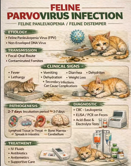
1) Feline Panleukopenia (FPV)
Etiology: Feline Parvovirus. Highly resistant in environment (>1y) - peracetic acid to disinfect.
Transmission: Oro-fecal route (direct/indirect), body fluids or transplacental.
Pathogenesis: Tonsils → replicates in lymphocytes → viremia → enterycytes (SI crypts), lymphoid tissue, bone marrow (targeting mitotically active cells/rapidly dividing cells).
In very young animals - cerebellum & retina
virus can spread transplacentally in pregnant cats → abortion, etc.
Clinical Signs: Depends on time of infection (=which cells are mitotically active)
1st trimester: Fetal death/resorption.
2nd/3rd trimester & neonates: Cerebellar hypoplasia (feline ataxia).
Kittens: Severe bloody diarrhea, vomiting, anemia, leukopenia
terminal cases: septic shock - 2ndary infections, DIC, hypothermia. high death chance 💀
Older: more subclinical - diarrhea, pain, panleukopenia (esp. neutropenia).
Diagnosis: History, CS, snap test (Fecal Ag - ELISA test), oro-pharyngeal swab, hematology - blood count (leukopenia, anemia).
Diff dx: FeLV, FIP, corona, clostridium, toxoplasma..
Treatment:
Supportive: fluids (5% dextrose), blood transfusion, ATBs for 2ndary.
Symptomatic: diazepam, antiemetics
Causal: hyperimmune serum
Prevention: Vaccination (core) - long duration of immunity - 4y. From 6-8w of age till 12w of age, then booster every 3y.
Feline Coronavirus - Feline infectious peritonitis & pleuritis (FIP)
We have two important coronavirus in cats:
Feline enteric coronavirus (Intact 3c gene)
Feline infectious peritonitis virus (Mutated 3c gene)
1) Coronavirus - by feline enteric coronavirus (FECV)
Transmitted oro-nasal, oro-fecal (direct/indirect) & transplacental (from infected mother to kitten)
Patho: Replicates in tonsils (salivary shedding) & SI enterocytes. (Infects & replicates in mature apical epithelial cells of intestinal villi) → brush border shortening & destruction.
CS: asymptomatic or mild, self-limiting enteritis, vomit&diarrhea.
Dg: Serology (ELISA, IFAT), PCR, RIVALTA TEST.
NO TX.
During coronavirus infection → virus may mutate and cause FIP (q13). Mutation occur in the individual host, and infection with FECV does not mean the cat will always develop FIP.
2) FIP - by FIPV (mutation of FECV) - not infectious
Pathogenesis: Mutation allows FIPV to replicate within MACROPHAGES
Strong mucosal or cell mediated immunity (CMI) + low virulence → complete recovery or carriers.
Moderate CMI + high virulence → dry FIP
Low CMI + high virulence → Wet FIP
Is an Ab enhanced disease: High Ab = worse disease
Clinical signs:
Wet: polyserositis (ascites, dyspnea), jaundice, vasculitis, anemia, uveitis
Dry: granulomatous lesions in liver, kidney, CNS (paresis, paralysis), eyes (uveitis).
Dg: history, CS/histology, rivalta test, USG (ascites), possible to detect anti-Abs (difficult), acute phase protein, hyperglobulinemia
Diff dx:
Wet: peritonitis, heart failure, pregnancy, pyothorax, pyometra, abd. neoplasia
Dry: FIV, FeLV, toxoplasma, neoplasia
Tx: No antiviral are approved in europe - but they work: Remdesivir, GS-441524. Can try interferons and prednisolone - not very effective. Euthanasia. (But generally no treatment)!
Prevention: Vaccine - not recommended (humoral immunity is not effective) - Intranasal vaccine maybe (as it is only given at 16w of age (kittens get sick much earlier) + serotype difference - made with serotype 2 strain when it is 1 that is the issue).
Remove seropos. cats - continuous shed on FECV, hygiene, reduce overcrowding and stress.

Other agents causing GIT infection in cat:
Salmonellosis, campylobacter jejuni, enterotoxaemia - Clostridium perfringens, heliobacter, E.Coli
8.Infectious Diseases of Dogs and Cats Affecting the Urinary System🩸
Infections affecting urinary - kidneys → Cystitis, pyelonephritis, prostatitis, borreliosis (Q14), leptospirosis (Q9).
Localized urinary infections:
1) Cystitis
→ Infection & inflammation of the urinary bladder.
Etiology: Primarily bacterial.
Bacteria: E.Coli, staphylococcus, Enterococcus, Streptococcus, Proteus, Klebsiella, Pseudomonas, Psteurella, Mycoplasma.
also secondary to: chronic glucocorticoid use, hyperadrenocorticism, chronic kidney disease, diabetes mellitus, and stress - linked to asympt. urinary tract infections.
Pathogenesis: Typ. ascending infection from the urethra to bladder → resulting in bacterial colonization of bladder epithelium.
Predisp. factors: Abnormal urine flow, decr. immunity, inadequate urine concentration, glucosuria and systemic diseases.
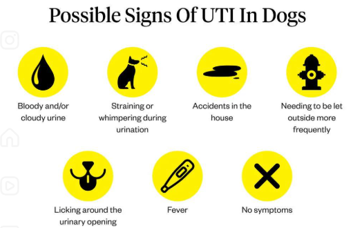
Clinical Signs: Pollakiuria (frequent urination), haematuria (blood in urine), dysuria (painful urination), urinating accidents in the house. The bladder may feel thickened, irregular, and painful on palpation.
Diagnosis:
Urinalysis (cystocentesis/free catch): Increased protein, hemoglobin, pH, WBCs, and RBCs. Reduced specific gravity (SG).
Sediment Microscopy: May reveal struvite crystals.
Other: Ultrasonography and bacterial culture.
Treatment: Antibiotics (Trimethoprim-sulfate, Penicillin, Amoxicillin).
2) Pyelonephritis
→ Bacterial infection of the renal pelvis, often leading to acute illness.
Etiology: E.coli, Staphylococcus, Enterococcus, Streptococcus, Proteus, Klebsiella, Pseudomonas, Mycoplasma
Pathogenesis: usually due to ascending bacteria (similar predisp. factors to cystitis), but hematogenous spread is possible. The
Renoliths/uroliths can contribute by mechanically blocking urine flow.
Clinical Signs: Kidney or flank pain, fever, malaise, vomiting, and PU/PD.
Diagnosis:
Urinalysis: Proteinuria, pyuria, bacteriuria &/ haematuria, and WBC casts in fresh urine sediment.
Bloodwork: Normal, or showing azotemia (renal/postrenal) and hyperglobulinemia. Animal may be in kidney failure.
Treatment: Broad-spectrum antibiotics initially, adjusted based on culture and sensitivity testing. Often requires IV fluids and IV antibiotics (Fluoroquinolones and Beta-lactams are common).
3) Prostatitis (Male Dogs)
→ Inflammation of the prostate gland, commonly due to long-standing, undetected infections.
etiology: E.coli, Staphylococcus, Streptococcus, Mycoplasma spp.
Pathogenesis: Hematogenous spread (acute) or ascending infection from the urethra (chronic).
Clinical Signs: Tenesmus (straining to defecate), stranguria (straining to urinate), pollakiuria, blocked urine flow, haemorrhagic or purulent urethral discharge.
Fever, lethargy, painful gait, and pain upon palpation of the caudal abdomen/prostate.
Diagnosis: Rectal palpation (reveals enlarged, painful prostate), ultrasonography, X-rays, cytology, and bacterial culture.
Treatment: Antibiotics with good tissue penetration (Chloramphenicol, Clindamycin, Erythromycin, Enrofloxacin) and Castration (chemical or surgical).
Borreliosis (Lyme Disease) - Q14.
A zoonotic, tick-borne infection that can lead to severe renal complications.
Aetiology: Borrelia burgdorferi, B. afzelii, B. garinii (Gram-negative, anaerobic, coiled spirochete bacilli).
Pathogenesis: Can lead to renal disease resulting in uraemia, hyperphosphataemia, and severe protein-losing nephropathy (PLN).
Diagnosis: Serology for antibodies (ELISA and IFAT) - enough to diagnose.
Treatment: Antibiotics (Penicillin and Tetracyclines).
Leptospirosis - see. q9
9.Leptospirosis 🩸
(Also as part of q.8 in some doc.)
Leptospirosis → contagious, gram-, motile, spiral bacteria w/flagella.
Common zoonosis. Dog, cattle, su & man. → Kidney&liver failure.
Key Serovars (Dogs) - Of. L.interrogans
Leptospira interrogans serovar Canicola
Leptospira interrogans serovar Icterohaemorrhagiae
Leptospira interrogans serovar Grippotyphosa
L. interrogans serovar Pomona
Distribution: Worldwide. Seasonal outbreaks in temperate areas (peaking during heavy rainfall/flooding) & year-round in tropical climates.
Reservoirs: rodents in icterohaemorrhagiae, livestock in pomona, while canicola = in dogs, grippo in raccoons..
Transmission: Mostly spread by the urine of infected animals.
Direct: Animal-to-animal via bites, venereal spread (semen), vertical transmission (placenta), or eating infected carcasses.
Indirect (Most Common): Environmental exposure via inhalation or ingestion of contaminated water/soil, or entry through cuts in the skin and mucous membranes (eyes, nose, mouth).
Pathogenesis:
Entry - by skin or MM → enters bloodstream (bacteraemia) → spread → affinity for kidney, liver → replicates in nephrons → kidney failure, hepatitis, necrosis.
Produces toxins - hemolysins → endothelium damage & lysis of RBCs → anemia & haemorrhage.
Affinity for pregnant uterus → crosses placenta, abortion.
Severity: Depends on the infectious dose, bacterial virulence, and host immunity (much worse in unvaccinated animals!).
Clinical Signs (Progressive Disease)
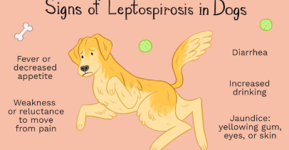
Early/Acute Stage: Fever, lethargy, anorexia, shivering, muscle tenderness (myalgia), and painful inflammation of the eyes (uveitis).
Kidney Damage Phase: Polyuria/polydipsia (PU/PD - increased thirst and urination) → severe dehydration
Later/Severe Stage: Vomiting, diarrhea, and jaundice (icterus) as liver failure progresses.
Oliguria or anuria (decreased/lack of urine) in end-stage renal failure.
Diagnosis
Gold Standard: MAT (Microscopic Agglutination Test) for Serology.
Clinical Nuance: Requires paired sampling (acute and convalescent samples 2-4 weeks apart) to look for a 4-fold rise in antibody titers. A single test might be negative early in the disease (due to intermittent shedding!)
Pathogen Detection (PCR / Culture / Dark Field): PCR of blood (early) or urine (later). Note: Bacteria are shed intermittently in the urine, which can cause false-negative urine PCR/cultures. Dark-field microscopy.
Hematology: Non-regenerative anemia, neutrophilia, and notably thrombocytopenia (low platelets).
Blood Biochemistry: Elevated liver and kidney enzymes (LD, GGT, BUN/Urea, Creatinine, SDMA & ALT/ALP - liver injury)
Urinalysis: Glucosuria (without hyperglycemia), proteinuria, hematuria, pyuria, and bilirubinuria.
Imaging (USG): Ultrasound often shows changes in kidney shape - enlarged, cloud-shaped, irregular margins).
Diff dx: Nephrotoxicosis (toxins), lyme borreliosis, glomerulonephritis, bacterial pyelonephritis, acute pancreatitis, bacterial sepsis.
Treatment:
Isolate animal, measures taken (gloves, gowns, hygiene) to prevent spread.
Causal (ATBs) - penicillin, doxycyline
Supportive: fluids (caution! - must be given carefully due to kidney damage - monitor for oliguria/anuria to prevent fluid overload)
Symptomatic: anti-emetics, low-protein renal diet
Prognosis: depends on early intervention before irreversible renal/hepatic necrosis occur. (fair, guarded).
Prevention:
Vaccination: Initial series of two doses, then every 12 months (or every 6 months in highly endemic areas). The L4 vaccine covers 4 strains (Canicola, Icterohaemorrhagiae, Grippotyphosa, Pomona). Note: There is little to no cross-protection between serovars.
Rodent control, avoid contact w/reservoirs
Inactivation by temperature, UV, disinfection, freezing

10.Infectious diseases of dogs and cats affecting reproduction system 🩸
Diseases affecting reproduction → Canine herpesvirus, Brucellosis & Listeriosis.
1) Canine Herpesvirus Infection (CHV-1)
“Fading Puppy Syndrome” or “Fatal haemorrhagic disease of pups”
Aetiology: Canine Herpesvirus 1 (DNA virus, Alphaherpesvirinae).
Worldwide.
Transmission:
Direct Contact: Aerosol/respiratory droplets, vaginal and nasal secretions, saliva.
Vertical: Transplacental, or puppies are infected while passing through the birth canal or from nasal/ocular secretions from mother..
Pathogenesis: The virus has a high affinity & replicates in mucosa of the upper respiratory tract (URT), genital tract, and CNS. It leads to immunosuppression.
Temperature sensitive - virus replicate optimally at cooler body temp. (under 37C). Becuase neonatal puppies cannot thermoregulate (relies on mother for heat) → their cooler body temp. allows virus to spread systemically → severe vasculitis.
Latency: Following recovery, the virus enters a latent state (Lifelong) in the sensory ganglia (trigeminal/lumbosacral). It can reactivate and shed during periods of stress (eg, pregnancy, drugs, disease).
Clinical Signs:
primary infection is often w/self-limited period of clinical illness, but long-term latency is symptom-free.
Puppies (< 3 weeks old): Fatal haemorrhagic disease
Anorexia, crying/vocalization, growth retardation & systemic hemorrhages (vasculitis), Ataxia. Sudden death within 6-7d.
Adults: Mostly asymptomatic. May show mild respiratory signs (can be a component of CIRDC / Kennel Cough), ocular discharge, and reproductive failure (abortions, stillbirths, mummified fetuses).
Post-Mortem: Haemorrhages in all internal organs.
classic: “Turkey egg kidney” - petechial haemorrhages covering the renal cortex + haemorrhages in liver&lungs
Diagnosis: CSm PCR (using nasal or vaginal swabs in acute infe.), and necropsy.
Serology (ELISA) can detect antibodies (which last 2-5 months). Cannot detect virus during latent phase!
Treatment: Highly debatable due to latency (can be reactivated!) and poor prognosis in pups. Options include placing puppies in an incubator (elevating body temperature to stop viral replication), administering hyperimmune serum, or antivirals (Acyclovir).
Prevention: isolate, stop breeding/castrate the positive female, test others. Vaccinate 1st dose during period from first day of heat until 10d post-mating, 2nd dose 1-2w before birth (to give maternal Ab via colostrum).
2. Canine Brucellosis
A chronic, highly contagious, and zoonotic bacterial disease → Late-term abortion & Reprod. Failure. “BRUCE” - birth material, reproductive failure, Undulating fever - man, C - contagious, E - enlarged Lnn.
Aetiology: Brucella canis (Gram-negative, intracellular bacteria). Worldwide, but most economic dev. countries have eliminated it.
Transmission: Venereal (mating), via ingestion of infected birth material/aborted fetus, infected milk/transplacental(in utero). Can recover but can still shed the bacteria.
Pathogenesis: survive intracellular in phagoyctic host cells/phagocytes (go unnoticed) → high affinity for reprod. tract (forms granulomas).
Clinical Signs:
Females: Late-term abortions (usually 3rd trimester: 45-55 days) often without any other signs, stillbirth, placentitis, metritis, prolonged vaginal discharge usually follows abortion.
Males: Epididymitis, periorchitis, prostatitis
Some get non-specific: lethargy, poor coat, generalized lymphadenopathy/lymphadenitis
Fever IS NOT hallmark finding in dogs as it is in other spp. (man).
Disease can remain subclinical for years in dogs with no reprod. activity or castrated. But should be considered in dogs w/some ocular issues (recu. blepharospasm, uveitis) & musculoskeletal (arthritis & discospondylitis) - due to immune reaction surrounding the Ag-Ab deposition in terminal arteries.
Diagnosis: CS, Slide Agglutination Test (SAT - screening tool), ELISA, AGID. Definitive diagnosis requires PCR (vaginal discharge, aborted material, semen, blood) or blood/tissue culture.
Treatment: difficult to cure because the bacteria hide intracellularly.
Can do long-term ATBs (combo of gentamycin + doxycycline (a tetracycline)) or castrate (to stop transmission).
tx often fails to clear it + due to severe zoonotic risk → Euthanasia can be answer..
3. Listeriosis
rare infection in dogs & cats, more commonly affecting RU & Su. Can be exposed via raw meat diet or farm environment.
By Listeria Monocytogenes (Gram+)
Transmit by eating contaminated stuff (raw meat/ru aborted fetus), vertical & direct contact w/asympt. carriers → hematogenous spread. can cross placenta → abortion.
CS: 3 types → Septicemia, encephalitis (neurological signs), abortion (if infection occurs in the late stages of gestation).
Dx: Bacterial culture, PCR. (ELISA less common - google)
Tx: ATBs (Penicillin). Prevent by not eating ru aborted fetus/raw meat.
Other pathogens to mention:
Parvovirus (primarily enteric/myocardial → but transplacental infection → fetal death, abortion)
Feline leukemia virus (pregnant → cross placenta, embryonic resorption, late term abortion)
Feline immunodef.virus (FIV) (vertical is possible but less common)
Mycoplasma (can overgrow → vaginitis, endometritis, infertility)
Toxoplasma gondii (transplacental)
11.Canine distemper, Rabies, and other diseases affecting nervous system of small animals 🩸
Major Viral Diseases: Canine Distemper, rabies, pseudorabies, botulism, tetanus & other like FIP, FIV, Toxoplasma, neospora, tick encephalitis, infectious canine hepatitis, cryptococcus..
1) Canine Distemper(Canine Morbillivirus)
“hardpad disease” - contagious multi-systemic disease, affecting primarily young, unvax dogs.
Transmission: Direct contact, aerosols, aerogenic / airborne (part of CRDC) - by oronasal secretions, feces, urine, vertical transmission. Viral shedding begins 7d PI, for 3mo.
Pathogenesis: Replicates in macrophages/Lympho/mono within lymphatic tissue → immunosuppression, then spreads to epithelial surfaces of resp., GI & urinary tracts, skin & CNS.
Clinical Forms:
Signs = varies & non-specific. Generally reflects the affected organ systems:
Nervous form: Develops 1–3 weeks PI. Proprioceptive deficits, paresis/paralysis, convulsions ("chewing-gum" jaw fits), ataxia, hyperesthesia, progressive seizures.
Skin: Pustular dermatitis (ventral abdomen), hyperkeratosis of nasal planum and footpads (“hardpad”).
Respiratory: Coughing, dyspnea, oculonasal discharge.
Ocular: Conjunctivitis, corneal ulceration.
GI: Vomiting, diarrhea, enteritis.
Intrauterine: Abortion, stillbirth, enamel hypoplasia (loss of dental enamel) in surviving pups.
GIT & Resp. form has better prognosis, while CNS has worse!
Diagnosis: CS, history (no vaccine = suspect distemper!), PCR, hematology - lymphopenia (decr leukocytes&lymphocytes → because virus destroys them)
biochemistry (some enzymes elevated, not the best way to dg.), cytology/histology - PM (after death, can test ofr intracytoplasmic incl)
DDx: Kennel cough, leptospirosis, infectious canine hepatitis, toxicities.
Treatment: No specific. Symptomatic and supportive (fluids, immune support - supplements, secondary - antibiotics).
Seizures managed with diazepam or phenobarbital (but poor prognosis in CNS form!)
hyperimmune serum at start (can be risky for puppies - allergic reaction/threat by potential patho agents?)
Specific antiglobulin can be attempted.
Prevention: Modified live vaccine (single serotype of virus so vax is very effective) given at 9–12 weeks; annual boosters. Isolation.

2) Aujeszky's/Pseudorabies (Suid Alphaherpesvirus 1)
Fatal neuro disease (“Mad itch”) in dogs/ru. Pigs = natural reservoir - latent/persistent.
Transmission: Direct contact with swine (bites, sniffing, blood contact during hunting) or ingesting raw, contaminated pork.
Pathogenesis: Systemic hemorrhage, ganglioneuritis, brainstem encephalitis, myocardial exudation/necrosis, lymphoid depletion.
CS: Behavior & Neuro signs
Intense localized pruritus (especially head),
anxiety, aggression, self-mutilation, hypersalivation, paralysis.
100% mortality → death after 1-2d after cs begin..
Tx: No treatment; euthanasia recommended.
Diagnosis via clinical signs and history of wild boar/swine contact.
NO Vax for other animals.
3) Botulism & Tetanus

Diagnosis: CS - enough for dg. also in botulism - history, ELISA, culture..
Rabies - Q12 (only mention) - or dont talk about it at all…. ?
Fatal viral infection targeting the nervous system.
Causes hypersalivation, behavioral changes, aggression, convulsions, progressive paralysis, and death in carnivores. Zoonotic.
Other diseases:
FIP - any NS signs due to meningoencephalitis
FIV - behavior changes, dementia, facial twitching, seizure
toxoplasma - ataxia, seizure, circling, paresis, paralysis due to encephalomyelitis
Neospora - progressive lower motor neuron paresis & ataxia starting in hindlimbs → paralysis
Tick encephalitis - apathy, hyperexcitability, ataxia, paresis/paralysis of limbs, abnormal spinal reflexes, nystagmus & paralysis of facial nerve.
infectious canine hepatitis - CNS involved is unusual and is typically the result of vascular injury. Many dogs get convulsions from forebrain damage. Paresis - from brain stemp hemorrhage, ataxia, central blindness.
Cryptococcus - lack of coordination, circling, head rotation, change, paralysis, optic nn damage - blind.
NB - notes:
RNA - single stranded RNA virus in CD, can adapt cross-spp., unstable, relies on direct transmission, via airborne/direct contact.
DNA - double stranded in botulism & tetanus - has good proofreading machine → stable, environmental resistant
distemper part of kennel cough complex - considered one of the core causative pathogens within the CIRDC - alike in initial stages, same transmission (aerosols)…
Tx of tetanus = metronidazole + anti-toxin and diazepam → as metronidazole is used against ANAEROBIC bacteria (as tetanus is!)
causative tx in distemper - interferons? off label (but does not eliminate?)
12.Rabies in Dogs and Cats🩸
Etiology: Lyssavirus (Fam: rhabdoviridae). Zoonotic & 100% fatal once cs appear.
strains: 12+ lyssaviruses, but the “classical rabies virus” = most important. RNA virus.
Worldwide - some exceptions (esp. islands). Almost eradicated in Eur. Some cases recently in Ukraine (due to the war situation) - some spillover. Human deaths every year (asia, africa) - lack of vaccine.
Transmission: saliva from biting animals, car = main reservoir. Virus is found in nervous tissue, salivary gl., saliva & cerebrospinal fluid.
Two main cycles: Urban (stray dogs/cats - 99% of all man rabies death) and Sylvatic (wildlife like foxes, bats & raccoons).
Virus - inactivated in sunlight.
Pathogenesis: rabies only affects NS. IP: varies wildly 2w to mo/years depending on how close bite was to brain/spinal cord.
After bite → enters muscle → replicates → enters peripheral nn (via acetylcholine receptors at neuromuscular junction) → CNS → neurons in brain get infected (cs develop) → via axons to salivary gl.
during 1st course of replication → virus is in limbic system (furious form)
During 2nd course → virus is in brain cortex (dumb form)
Then → virus moves along brain nerves to salivary gl. → excreted.
Clinical Signs (The Two Forms)
Once signs begin = death.
CS often start as non-specific (prodromal phase in both generally?), like restlessness, anorexia/or incr. appetite, vomit, slight fever, pupil dilation, hyperreactivity to stimuli & excessive salivation - last for 2-5d, then can be followed by paralytic or furious form.
Furious form (Limbic)
Prodromal (2-3d) - sudden temperament changes, hyperthermia, mydriasis (dilated pupils) & Loss of corneal reflex.
Excitation stage (last 3d) - snapping at “imaginary objects”, extreme pain sensitivity, hyperreactivity, severe drooling
Paralytic stage - rapid onset of ataxia, convulsions, paralysis → death.
Dumb form / paralytic (brain cortex)
Progressive paralysis, animal remains quiet, lethargic, muscle tremors, paralysis of hind quarters
Typical: paralyzed throat, masseter and tongue → unable to swallow, severe drooling.
Progresses to paralysis of resp. muscles → death.
Diagnosis (Post-Mortem Only)
Testing need brain tissue → dg done PM by opening skull via occipital foramen or retro-orbital approach.
Gold Standard: FAT (Fluorescent Antibody Test) . The tissue is inoculated with a labeled antibody that binds to the rabies antigen. If positive → glows apple green under a microscope. VERY GOOD, QUICK.
Histology: Look for Negri bodies (classic viral inclusion bodies found in the cytoplasm of neurons), multifocal mild encephalomyelitis, and craniospinal ganglionitis.
Other: RT-PCR, ELISA
extra - serology (ex FAVN) - for checking immunity (vax response) - NOT for initial dg of sick animal…
Protocols, Prevention, and Treatment
NO treatment for animals, euthanasia is needed if cs is present. isolate or re-vaccinate.
Vaccination: IM inactivated, dogs & cats older than 3mo can get vaccine. Booster yearly or every 3y. Except in areas where rabies is endemic or where needed by law (sk) - rabies vaccine is considered non-cone.
oral marker vax for wild animals w/live attenuated vax - SAD Bern stain.
qs on state: dates for test and rabies observation: Days 1, 5, 14 (google says 10 day obs. period) idk bro
qs on state: Humans - post-exposure prophylaxis (PEP) - if man is bitten by unknown, suspected, or confirmed rabid animal → hyperimmune serum & vaccine doses at days 0, 3, 7 and 14.

13. Feline leukemia, feline immunodeficiency, infectious peritonitis and pleuritis 🩸
Feline Retroviruses (FeLV & FIV) - RNA viruses:
when entering host cell → uses the enzyme reverse transcriptase to rewrite their viral RNA into DNA. This proviral DNA integrates directly into the host`s genome → allows for long phase latency. Virus is copied every time the cell divided, and can be reactivated.
1. Feline Leukemia Virus (FeLV)
by: gammaretrovirus, retroviridae, affecting usually young kittens (resistance increase w/age). Worldwide. Imp. cause of morbidity&mortality in cats.
Transmission: "Friendly" contact . Shared through saliva (grooming, shared bowls, biting (immunodef.)), nasal secretions, feces, and milk. & blood transf. (rare).
Pathogenesis: replicates in oropharyngeal lymphoid tissue → carried in blood to spleen, Lnn, epithelial cells of intestine, bladder, saliv. gl & bone marrow (targets wbc - b&t-Ly, bm stem cells, epithelial).
Clinical Signs:
common:
immune suppression (predisp. to other infections) - suppressed bm, anemia, lymphopenia, neutropenia
Neoplasia: seen as lymphoma (mediastinal, peripheral, spinal), leukemia, infiltration of bm, hepatosplenomegaly
anemia (non-regenerative)
Less common: other neoplasia, uveitis, reprod. disorders in females, peripheral neuropathies (mydriasis, paresis, paralysis, hyperasthesia). Most develop cs, but some remain healthy for yrs, with proper care & indoor life.
The 3 Outcomes of Infection (important!):
Abortive infection: Good immunity. Virus is stopped; no viraemia. The test is always negative.
Regressive: Transient viraemia followed by latency. Test is initially negative, becomes positive upon reactivation.
Progressive: Poor immunity. Persistent viraemia leading to FeLV-associated disease. The test is always positive (as it is present in blood).
Diagnosis: test for the Antigen (p27 rapid blood test).
also ELISA (But does not detect latent infections)
in FeLV = detect Ag, while in FIV = detect Ab. In FeLV = viremia, usually alot in blood
treatment: supportive, tx of sec.infections, feline interferon, avoid immunosupp. drugs!
Prevention: vaccine (non-core) - test them first, if already infected → can make them sicker!
spay and separate to prevent spread, no need for euthanasia, only when tumors has gone too long, but no need if you just have positive snaptest.
2. Feline Immunodeficiency Virus (FIV)
By Lentivirus (closely related to HIV) - Retroviridae, RNA virus → attacking immune cells → immunosuppression.
→ affects usually older, outdoor, territorial male cats
Transmission: "Unfriendly" contact. Deep bite wounds, fighting and mating from PI cats. Looses its infectioness outside host, susceptible to all disinfectants incl. soap. Vertical (milk) can occur.
Patho&CS: chronically destroys immune cells → T/B cells (T-helper lymphocytes mainly) & macrophages - leads to lifelong infection
Acute primary: 4-6w after inf, transient period of fever, lymphadenopathy & neutropenia
asympt. phase: latent for yrs (infected can go years without cs)
AIDS related complex - “acquired immunodef. syndrome” - reactivation in middle aged/older & immunosupp. cats.
→ Opportunistic infections & chronic illness in resp. tract, GIT, CNS, Renal & neoplasia. Symptoms are typ. secondary to the ruined immune system (chronic gingivostomatitis, lymphadenopathy, rhinitis, weight loss, renal failure (immune med. glomerulonephritis).
Diagnosis: You test for the Antibody (ELISA) - Ab can take 12mo to develop so repeat test every 2mo, kittens can be false pos due to MDA). FIV immediately hide inside host cells → no free ag in blood. Must test for body`s humoral response + hematology (NR anemia, lymphopenia, neutropenia), combo rapid test (test for leukemia w/ag, FIV Ab).
Tx: ATB for sec. infections, corticosteroids to moderate cs.
Vaccine if they do not have cs (even if infected) & vaccinate against everything else as well.
Vaccine at 6-9w of age → after 3-4w → yearly booster. Aim to give last booster no earlier than 16w of age (applies to all vax!)
3. Feline Infectious Peritonitis & Pleuritis (FIP)
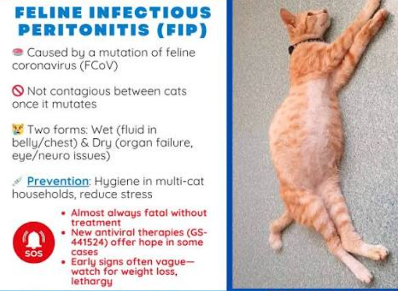
FIP is caused by a mutated form of the Feline Coronavirus (FCoV)
FCoV (non-mutated type) itself only causes mild enteritis. During heavy replication in macrophages/monocytes, it can mutate (only a few of infected cats develop FIP! - uknown why). FIP virus is not infectious. Feces = main source of FCoV, transmit by saliva/pregnancy is rare. + indirect (litter trays, shoe).
Affects: Usually pedigree cats under 1 year old from multi-cat households (high stress, crowded).
Transmission/Patho: FIP is not infectious like FCoV, mutation occur → allows for replication in MF & monocytes. Cats w/FCoV variant - no role in spreading FIP, under natural conditions (as it does not/poorly multiplicate in gut).
Viral load & immune system of cat = factors on whether FIP will develop.
CS: FIP - wet, dry, ocular & Neurological
early signs → undulating fever, lethargy, loss of appetite, slow weight loss, dull coat..
Effusive (Wet) Form: The mutated virus attaches to the endothelium of venules (peritoneum/pleura), triggering cytokines & TNF (tumor necrosis factor) produced → vasculitis and fluid leakage - fluid is collected in abdomen or chest (pot belly/breathing issues).
Signs: we will see a swollen, pear-shaped/pot-bellied abdomen that feels fluid filled (polyserositis, ascites), labored/rapid breathing if fluid is in the chest (pleural effusion), pale/yellow gums (jaundice), anemia
Non-Effusive (Dry) Form: granulomatous lesions (pyogran.) forming in various organs (renomegaly, chronic diarrhea, Lnn enlargement), CNS (paresis, paralysis), eyes (uveitis)..
Diagnosis (combo of findings):
Bloodwork: High total serum protein, hyperglobulinemia, low albumin-globulin ratio & high FcoV Ab titers + CS. (Note: Snap tests only confirm FCoV, not mutated FIP, can suspect it).
The Rivalta Test (wet): by using effusion fluid
Positive for FIP (Exudate): The drop - cloudy shape (like a jellyfish or mist) that sinks.
Negative (Transudate): The drop dissappears
Look at history, cs, USG (ascites)..
No PCR test to distinguish mutated from non-mutated, many snaptest for FcoV but cannot confirm mutated.
Tx: Majority of FIP cases are too late = euthanasia is best option. Non-curable. Poor prognosis. Supportive - not proven to work.
no antiviral approved in Eu, but they do work: NB: FIP tx → currently can use antiviral drug remdesivir (GS-441524) - cure rate of 80%.
Prevent: Reduce contamination - hygiene, small groups of cats, clean litter.
NO vaccine. possible intranasal vaccine (Vanguard) - but not recommended. (questionable efficiency + most get infected before 16w…)
NB! NOTES:
qs: what happens w/cat that develops AIDs → everything basically, all systems can be affected.
vaccines or not? - important before FeLV vax - test that it is not sick already.
Is mutation to FIP a rule - NO it is not. Mutation occur/what makes mutation happen → in Macrophages and white blood cells.
14.Infectious diseases of dogs and cats characterized by natural focality (leptospirosis, lyme borreliosis, tick encephalitis, ehrlichiosis) 🩸
Natural Focality: A pathogen is part of the ecosystem and spreads under favorable conditions. It is geographically and seasonally limited. It consists of an etiological agent, host-donor, vector, recipient, and environment. (Note: A vector is not always present, eg, Leptospirosis).
1) Lyme Borreliosis
Etiology: Borrelia burgdorferi sensu lato (3 types cause disease: B. burgdorferi sensu stricto, B. afzelii, B. garinii).
Vector: Ixodes ricinus tick (need min. 24h attachment to transmit).
Pathogenesis:
Tick expresses Outer Surface Proteins (OSP A adheres to midgut, OSP C binds to salivary glands) → once in the host, borrelia changes antigenic structure (VlsE type) & shape to avoid immune system.
Bacteria binds to proteins, CT (plasminogens, integrins) - can remain dormant for life, causing cs only when animal becomes older & immunosupp.
Prevention: A non-core vaccine (OSP A/C) exists to kill the bacteria while tick is sucking blood, but tick control is the preferred method.
Clinical Signs (Only 10% of infected dogs show signs):
Initial signs (2-5 months post-bite): Fever, inappetence, thrombocytopenia, lameness.
Polyarthritis: Usually starts in the joint closest to the tick bite.
Renal Disease: Acute renal failure (similar mechanism to Lepto).
Meningitis: Can cross the blood-brain barrier (poor prognosis).
Dermatitis: Hard to tell if from bacteria or bite itself.

Diagnosis: Serology is the main method: C6 ELISA rapid test is widely used (detects Ab 3-5 weeks post-infection). Western blot can determine the disease stage.
other: Tick bite, PCR, IFAT (ab usually enough to dg), BSK-II agar, Cross reactions w/lepto, treponema. LgG peaks in 4-6w PI, remains for a year..
Treatment:
ATBs for seropositive dogs w/cs. - Doxycycline (5-10 mg for 4 weeks). Relapse occurs if treated for <30 days.
Crucial Rule: ONLY treat seropositive dogs with clinical signs. Do not treat asymptomatic positive dogs (monitor instead).
Supportive care: NSAIDs, fluids, and treating specific kidney/skin failure.
Other Focal Diseases
Leptospirosis: Leptospira interrogans sensu latu. Affects the liver and kidneys. (in q8&9).
Tick Encephalitis: Dogs can be affected by this → Flavivirus transmitted by ticks OR by dogs drinking unpasteurized infected milk. Can be asymptomatic or cause severe encephalitis/multifocal neurological signs and fever.
Ehrlichiosis (Canine Hemorrhagic Fever): Obligate intracellular bacteria infecting leukocytes. Transmitted by Rhipicephalus sanguineus → immunosuppression, bleeding (nose/mouth), anemia in dogs. Diagnosed (C6 ELISA) and treated (Doxycycline) the same way as Lyme.
Anaplasmosis: Intracellular bacteria transmitted by Ixodes. Zoonotic. Diagnosed via rapid test (differentiate types by clinical signs). Know the two types:
Anaplasma phagocytophilum: Affects phagocytes → immunosuppression.
Anaplasma platys: Affects thrombocytes → hemorrhagic disease (only in ~10% of cases).
Tularemia: Francisella tularensis. Hares carry it; infects hunting dogs → multifocal abscess and fever (only mention name)
Bartonellosis: B. henselae (Cat scratch disease). Transmitted by cat fleas (ctenocephalides felis), does not occur in our territory.. few infected have signs..
NOTES:
values for kidney failure? - Urea, CREA, SDMA
prove lepto? MAT
how to dg and treat borreliosis? ELISA (lgG+LgM Abs.doxy for tx)
Just list the other diseases… (FOCUS On borrelia&Lepto)
15. Preventive, control and eradication measures at infectious diseases of dogs and cats. Epizootological and epidemiological activities in disease reporting and protection of state territory and transport of animals. Registration of companion animals. Pet passports 🩸
Prevention, Control, and Eradication measures:
based on specific immunoprophylaxis (vaccination).
when 50% of animal pop. is immunised = reduces risk of infection by alot.
Good vaccine: Must be safe, affordable, and stimulate a strong, long-lasting immune response.
Rules: Animals must only be vaccinated if they are healthy and not pregnant to avoid adverse reactions or clinical disease, accurate timing of vaccine.
other Measures: Isolation of infected animals & strict hygiene
Epizoo&Epidem activities in disease report/protection: (International Transport & Border Protection)
Protection of country/territory against intro of disease = priority (from exotic or zoonotic, esp. rabies)
Animals must be certified fit for travel & disease-free by a veterinarian in the original country.
Vaccination schedule depend on housing environment & epizoo situation of the exporting & importing countries
import regulations (in Norway) as a standard example.
Traveling from EU or listed Non-EU countries to Norway:
Must have microchip
EU pet passport
tapeworm treatment (echinococcus) in dogs - given 24-120h before arrival (not needed if traveling from ireland/finland/matla/norway). Must be recorded in passport, signed and stamped by vet.
Rabies vaccine - must be followed by a 21d waiting period (unless traveling from rabies-free country)
Must enter via an authorised route, approved transport company
Traveling from Unlisted Non-EU Countries:
Requires all of the above, plus a health certificate if a passport is unavailable.
Stricter Rabies Protocol: Requires a blood titer test at least 30 days after vaccination. The titer must be > 0.5 IU/ml . This is followed by a 3-month waiting period before travel.
If re-entering norway → if microchipped and blood sampled 30d after rabies vax gave positive titration result (over 0.5) before leaving norway (or other eu country) - can re-enter norway without having to meet 3-mo waiting period (must record all in passport)
Disease Reporting & if rules are broken: animal is sent back to departure country, isolated at border or quarantined if infected. Vets are legally obliged to report smuggled or unvax animals to relevant authorities (ex. FSA, OIE, police)
Registration of companion animals & Pet Passports:
Proper identification and documentation are mandatory for companion animal transport.
Microchipping: mandatory in some, chip number must be recorded on vaccination record or pet passport. Must be chipped before rabies vaccine - if not, vaccinate again! (always read chip before rabies vac)
Tattoos: An alternative identification method, although not accepted in certain countries (eg, the UK and Ireland).
Central Registers: Databases where microchip numbers and owner details are officially recorded.
EU Pet Passport: document w/ microchip number (date and location of insertion), owner & animal information, vaccination records (product, batch number, next booster date), deworming history, and general health checks.
Echinococcus deworming needs to be registered in passport
Vaccination Guidelines
Animal | Core Vaccines (Essential) | Non-Core Vaccines (Situational) |
Dogs | Canine Distemper Virus (CDV), Canine Adenovirus 1/2 (CAV), Canine Parvovirus (CPV). Rabies&lepto = also core in SK! | Rabies, Lyme borreliosis, Leptospirosis, Parainfluenza virus, Bordetella bronchiseptica |
Cats | Feline Panleukopenia (Parvovirus), Feline Herpesvirus, Feline Calicivirus | Rabies, Chlamydophila, Feline Leukemia Virus (FeLV), |
NOTES:
WOAH Diseases - rabies & trichinella (can ask about this)
Core: Essential for all. vs. Non-core: lifestyle dependent.
Time frames for giving vaccines:
puppy/kitten: start at 6-8w of age
intervals: giving boosters every 3-4w
final dose - of primary core must be given at or after 16w of age to ensure maternal Ab do not neutralize the vaccine
Rabies: given as a single dose, no earlier than 12w og age (typ 12-16w).
revaccination: first booster for core is given 12mo after final dose at around 16w. After the 1year booster, core vaccines (CDV, CAV, CPV, FPV) & rabies are given every 3y.
Non-core bacterial (like lepto) need yearly boosters
Norway: DHPP (or DHPPi) (8w, 12w, 16w) → første voksenboost 12mo → then every 3rd year, yearly is Pi. Cat is tricat at 8 + 12w, ducat yearly + every 3rd year tricat.
Any adverse reactions to vaccines: normal/mild reactions like lethargy, mild fever, decr. appetite, slight swelling/tender at injection site (resolve within 24-48h). Severe reactions: anaphylaxis (type 1hypersensitivity) - shortly after, symptoms like facial swelling, hives, vomit/diarrhea, collapse → need immediate tx (epinephrine, antihistamines)
feline injection-site sarcoma (FISS) (RARE)
When should we not vaccinate - sick, pregnant, immunosuppr. (undergoing high dose corticosteroid therapy fro ex.)
14. Principles of specific immunoprophylaxis. Vaccination of dogs and cats and actual legislation. Principles of infectious disease prevention in young animals 🩸
Immunoprophylaxis: Enhancing a specific immune response in an animal by vaccine exposure. Vaccines cause mild infection w/no cs, allowing animal`s immune system to respond when exposed to same infectious agent.
Types of immunization:
Active: animal`s own immune system does the work. Can be exposed to a pathogen (natural) or vaccine (artificial) → body makes Ab, builds memory cells.
Passive: animal`s own immune system does NO work, it gets “ready-made” Ab made by a donor ex. mother via colostrum (natural) or hyperimmune serum/immunoglobulins (artificial)
Immunity is diffferent depending on life periods: early post-natal (Ab by colostrum, immature), Adult (developed, but compromised by external or internal factors) & aging (older pets, decline in overall immunity).
Vaccines (“suspensions of MiOs”)
Modified live (multiples in host - almost all vax for small animal med.) or inactivated (does not multiply - triggers immune resp.)
live (rarely used as they can be dangerous, but has longer/stronger immunity)
subunit vaccine (using a piece of the pathogen - the protein that triggers immune response). ex. canine herpesvirus.
Recombinant & vector vaccines (3rd gen) - delivery virus, vector that carries the genetic info for a specific Ag from dangerous patho (ex. rabies). + DNA vaccine.
Factors that may affect efficacy of vaccine:
Polyvalent (many agents) vs. mono (single agent)
Route: SC, IM (lower efficiacy, but no crisis), Intra-nasal, oral - fox rabies
Age - presence of MDA & immune system of young - younger than 6w - vax isnt working that good, should repeat.
Nutrition, pregnancy (do not vaccinate w/modified live → can give disease), only vax healthy. Diarrhea, resp. disease & fever = can influence efficacy. Immunosuppression (using corticosteroids) → must stop 2w, do vaccine → wait another 2w l then start up w/corticosteroids again.
can add adjuvants (heps to boost immune response, but risk of reaction) or non-adjuvant vaccine
Goal: make strong, humoral & cell.mediated immunity, memory cells = husker patho når de møtes igjen! Immunity is present in abscence of humoral Ab.
Duration: 3y for CDV, CAV, Parvo,1y for Pi
Storage of vaccine: In fridge, take them out before use, isolation of vaccinated animals, young, vaccinated are still suscpetible for a few days.
Vaccination Protocols:
Window of Susceptibility: The critical period (typically 8–10 weeks, up to 16 weeks) where Maternally Derived Antibodies (MDA) drop too low to protect against wild infection, but remain high enough where they may neutralize incoming vaccine antigens (but susceptible). We repeat doses spaced 2-4w apart to ensure at least one vaccine dose is given after MDA clears.
Core Protocol (Dogs & Cats):
Primary Series: Start at 6–8 weeks , repeat every 2–4 weeks , with final dose given at 16 weeks (Standard optimal: 8, 12, 16 weeks ).
Booster given at 12 months of age - to ensure full immunity (or 12 mo after 16w dose).
🐶doe pups, where rabies is endemic → get 1 dose at 12w, then in high risk - 2nd dose given 2-4w later.
Adult Revaccination: Core vaccines protect for at least 3 years (revaccinate every 3 years ). Non-core bacterins ( Lepto, Bordetella, Parainfluenza ) require 6–12 month boosters
🐕Canine Vaccines (DHPPi + L, R)
Global Core (WSAVA): Canine Parvovirus ( CPV-2 ), Canine Distemper Virus ( CDV ), Canine Adenovirus ( CAV-1/CAV-2 ).
Non-Core: Rabies, Leptospira, Parainfluenza (Pi), Bordetella bronchiseptica (IN), Canine Coronavirus, Microsporum.
SK: Rabies & Leptospira are treated as CORE due to endemicity/natural focality. DHPPi+L/R is the standard protocol.
🐈Feline Vaccines (RCP)
Global Core (WSAVA): Feline Panleukopenia ( FPV ), Feline Calicivirus ( FCV ), Feline Viral Rhinotracheitis/Herpesvirus ( FHV-1 ). Vaccine = RCP.
Non-Core: FeLV, Chlamydia felis, Dermatophytosis, Rabies.
Not Recommended: FIP (MLV), FIV.
FeLV: MUST test for FeLV antigen before vaccination (vaccinating an infected cat speed up infection!).
Adverse Vaccine Reactions
Immune-Mediated (Hypersensitivity):
Type I (Anaphylaxis): IgE-mediated within minutes to 24 hours. Facial edema, hyperemia, shock & death. Tx: Corticosteroids to acutely treat infla/shock, antihistamines as a preventative before next vax.
Type II: Rare hemolytic anemia.
Type III (Immune Complex deposits in tissue): CAV-1 live vaccines caused "blue eye" (corneal edema), uveitis, nephritis, vasculitis.
Type IV: Delayed local hypersensitivity.
Vaccine-Induced Disease: Mild clinical signs (fever, lethargy). Intranasal vaccines cause mild sneezing/nasal discharge.
Local Reactions & Neoplasia: Injection-site pain/edema/redness, hair discoloration, and Feline Injection Site Sarcoma (FISS) (linked directly to adjuvant-induced inflammation, we do not use these adjuvant vax anymore in cats).
Qs - NOTES:
time after vaccine for developing immunity in dog: Parvo & panleukopenia - 3-5d, adeno - 5-7d, Calici & Herpes - 7-14d
Expect from core vax in terms of efficiency:
CPV, CDV, FPV, CAV = 98%
herpes and calici = 60-70% (can still get symptoms, but good enough)
Antiparasitic tx need to be mentioned in passports? → ecchinococcus
Diff. bw quarantine and isolation in terms of symptoms? → isolation is for animals that are already sick and have cs, while quaratnine is for animal that are not sick (no cs) but may have been exposed to pathogen.
What to do if we have an animal with unknown vaccine status? → start with primo vaccination, do all vaccinal again.
Why is 4w too much between primo and second vax? → immune system is no longer as active so there will be less of an immune response from the subsequent vaccination, so you basically have to restart again.
why is 2w too little between primo and second vax?→ response from the body can produce substances after the first dose that may interfere with next dose (like interferon)
Why is just one primo vaccine not enough? → addiitonal Ag presentation is needed to stimulate the immune memory cells.
If dog has lepto vax, why can it still get infected? → because vaccine only contains certain serovars, not all. Can become infected at the end of the vax before booster (immunity lasts 6mo to 1y).
talk about vaccination only ! - when to vaccinate & window of suscept. ! which are non-core and core, why it differs in some countries like rabies, lepto and leishmania
mention other vaccines? → bordatella, nasal vaccine.
SCHEDULE - TABLE (REPETITION):


17.Prozooses of GIT (giardiasis, cryptosporidiosis, izosporosis, sarcocystosis, toxoplasmosis, neosporosis) 🩸
GIT Protozooses:
Parasite | Zoonotic Potential? | Key Diagnostic Stage & Size | Diagnostic Method | Treatment (1st Choice) |
|---|---|---|---|---|
Giardia | Yes (A & B) | Cyst: 8–12 µm (4 nuclei) | Faust float (40x), SNAP/ELISA | Metronidazole |
Cryptosporidium | Yes (C. parvum) | Oocyst: 4–6 µm | Ziehl-Neelsen (Acid-fast) - fecal smear | Supportive / Nitazoxanide |
Cystoisospora | no, not from spp. of dog/cat. | Small Oocyst: ~25 µm | Faust float | Toltrazuril (Baycox) |
Sarcocystis | no, not from spp. of dog/cat. (but can from eating pork/beef - suihominis ex.) | Sporocyst only | Faust float | None for carnivores |
1) Giardia (giardiasis)
Order: Diplomonadida, fam: hexamitidae, genus: giardia
Species: G.duodenalis/intestinalis (mammals/man) w/several assemblages (A-H):
A&B: mammals&man, zoonotic - 99% of human cases. (g.enterica)
C&D: specific to dogs/canids (g.canis)
F: cats (g.cati)
E (livestock), G (rodent), H (marine mammals)
other: G.agilis (amphibians), G.ardae & psittaci (bird), cricetidarum (hamster), muris, microti..
Target: Duodenum, jejunum (opportunistic/young animals) - located on surface of SI
Food-borne & Water borne disease - oral-fecal transmission, ingestion of contaminated food/water (cysts)
Morphology:
Trophozoite (12–20 µm): 2 nuclei, ventral adhesive disc. Multiplies by binary fission. Colonize intestinal mucosa
Cyst (8–12 µm): 4 nuclei. infective immediately & environmentally resistant
PPP: Dogs = 8 days, Cats = 10 days.
Life cycle: Direct w/binary fission. Cyst eaten by host → trophozoite excyst in SI, then divide by binary fission, later encyst → feces (survive for weeks, 1mo).
Clinical Signs: generally asympto. or w/SI infection signs
Chronic - malabsorption syndrome → steatorrhea, typical foul smell
Acute: diarrhea, vomit, nausea, anorexia
Diagnosis: Faust flotation (look for the "green moon" osmotic artifact; use 40x). Test 3 samples from different days due to intermittent shedding. + snap test, ELISA (expensive) for coproag, nested PCR for assemblage testing.
Treatment: Metronidazole (5–7 days) or Fenbendazole (3 days)
2) Cryptosporidium ( Cryptosporidiosis )
Order: cryptosporida, fam: cryptosporidae, coccidean parsite, apicomplexa (subphyl)
Species: C. parvum (most dangerous for man), C.canis, C.meleagridis & C.muris in dogs. cats - C.felis & C.Muris.
Food-borne & water disease, opportunistic - if healthy then no problem for animal (same as giardia), worldwide. Targets enterocytes, intracell. & extracytoplasmic. SI.
Morphology: Oocysts are tiny ( 4–6 µm ) and contain 3–4 sporozoites. They are immediately infectious when passed.
Life Cycle: All stages = Endogenous/inside host!
Merogony: sporozoites invade enterocytes → asexual prolif. → 3rd gen merozoites
gametogony: gamonts → oocyst, thin (cause autoinfection) and thick walled made.
sporogony: form 4 free sporozoites within oocyst, already infective when passed in feces (the thick walled)
Clinical Signs: Usually asymptomatic.
Hemorrhagic diarrhea in young, immunosuppressed (ex. parvo, FIV, FeLV)
Necrosis & villous atrophy, long-lasting diarrhea (watery, mucoid, bloody), weight loss..
Diagnosis: stained fecal smear w/Kinyon or Ziehl-Neelsen (acid-fast). ELISA - for coproag (expensive), PCR for species, genotypes.
Treatment: No direct anticryptosporidial drug exists. Immunocompetent dogs can be given Nitazoxanide or a combination of Paromomycin and Azithromycin. Tylosin. Supportive for diarrhea.
3) Cystoisospora
order: eimeriida, fam: sarcocystidae, genus cystoisospora. Coccidean parasite, apicomplexa (subphyl).
Targets Enterocytes in SI, C.ohioensis = cecum&colon. Same fecal-oral transmission, but also PH (rare).
Morphology: Oocysts contain 2 sporocysts and 4 sporozoites. Not immediately infective (requires days in the environment).
The "Size"
Small (~25 µm) = Pathogenic. Treat these. Dogs: C. ohionensis complex (c.ohioensis, C.burrowsi & neorivolta), Cats: C. rivolta
Large (~50 µm) = Non-pathogenic. Do not treat unless highly symptomatic. (C.canis & C.felis)
Life Cycle: Monoxenous (1 obligate host), but can use rodents as PH (dormozoites).
Merogony & Gametogony in host: sporozoites → intracell. multiplication in enterocytes (3rd gen is merozoites) → gametogony → unspor. oocyst in feces
Sporogony in environment
Clinical Signs: usually asymptomatic if >6m, similar to giardia (but affects deeper tissues). Diarrhea (usually not bloody), vomit, anorexia, abd.pain, malabsorption. Can be severe in high infective dose → death.
Diagnosis: Faust flotation. You must measure the size to determine if the species is pathogenic.
Treatment: Toltrazuril (Baycox).
4) Sarcocystis ( Sarcocystosis )
Order: eimeriida, genus sarcocystis. FH: car, man, raptors. Several IH like preys.
Very host-specific, often named based on IH+FH → S.bovicanis, S.suicanis etc. S.cruzi (car, cattle), S.tenella (car, sheep). Targets SI (intracell.)
Life Cycle: Obligate heteroxenous (Predator-Prey).
Merogony occurs in the intermediate host (muscle cysts, 3 phases, 2 in muscle, 3rd in circulated lymphocytes).
Gametogony and Sporogony occur in the final host (carnivore). Oocyst w/2 sporocysts and 4 sporozoites each.
PPP: 5–7 days.
Clinical Signs: Usually asymptomatic and non-pathogenic in carnivores.
Diagnosis: The sporulated oocyst wall is incredibly thin and ruptures before or during flotation. You will only find sporocysts in the feces on a Faust floatation.
In IH: serology, dg method..
Treatment: None needed for carnivores
Neospora & Toxoplasma in qs. 18!
18.Systemic protozooses (toxoplasmosis, neosporosis, leishmaniosis, encephalitozoonosis)🩸
Diagnostics across the board: SEROLOGY is the golden standard for almost all of these! Fecal tests are rarely helpful.
Most important: Toxoplasma & Leishmania
1. Toxoplasmosis (Toxoplasma Gondii)
Hosts: FH = Cats (shed oocysts). IH = Mammals/birds (form tissue cysts).
Location: Forms cysts in tissue IH, replicates intracellularly. FH:SI
Transmission: Fecal-contamination → water w/oocysts, eating IH (tissue cysts) & transplacental (tachyzoites)
Life cycle:
IH: Merogony (sporozoits → tachyzoites multiply in organs → slowed by immune response → bradyzoites in cysts
FH: gametogony (bradyzoites → SI → unsporulated oocysts)
Environment: Sporogony → sporulated oocyst (2 sporocysts, 4 sporozoites
FH can be accidental IH (IH is not essential for completion of LC)
Clinical Signs: usually asymptomatic, cs in young or immunosupp. kittens w/coinfection w/FIV, uveitis for kittens
Cats: intestinal (enlarged abdomen, ascites, diarrhea) or systemic (acc. to organ) - resp. distress, CNS signs etc. Transplacental (stillborn/weak kittens)
Dogs: rare to have cs, only in immunosuppressed
Diagnosis:
Serology! IgG = chronic/but also found in clinically healthy (immune, no longer shedding). IgM = increased levels in acute infection.
REMEMBER → dg. relies heavily on serology. If a cat tests positive (has lgG Ab), it is generally considered safe. Cats only shed infective oocysts in their feces once in their entire lifetime (for about 21d during their first infection).
MAT is the gold standard. Ascites w/ abdominocentesis (sometimes free tachyzoites)
Fecal float = Low importance (shedding period is too short + only when infected for first time)!
Treatment: Clindamycin. Note: Does not kill tissue cysts. symptomatic.
2. Leishmaniosis
By Leishmania infantum (zoonotic) - FH:Dogs,cats, wild mammals. In tropical/temperate areas.
Transmission: Vector-borne via Sand flies (Phlebotomus - europe, Lutzomyia). Other: blood transfusion, vertical (mother→baby), venereal (sexual)
Location: FH: MF, monocytes, Langerhans cells → organs (Lnn, spleen, BM). IH:Intestine.
Life cycle: The infective stage is the promastigote (from the fly), which turns into an amastigote inside the dog's macrophages.
Clinical Signs - Canine visceral Leishmaniosis:
Cutaneous: Alopecia (around eyes/blepharitis), ulcerative lesions on nose/paws, onychogryphosis (abnormally long, curved nails), papular dermatitis
progresses to: popliteal lymphadenomegaly, epistaxis (nosebleeds), uveitis, purulent conjunctivitis → cachexia (wasting), chronic renal failure (immune complexes clogs kidneys) - fatal.
Left untreated → death within 2yrs. Inc:1-6mo.
Diagnosis: Serology: ELISA, IFAT, rapid test for initial dg. Serum - sample.
Cytology (MF w/amastigotes by sample of Lnn), PCR, histo
Treatment & Prevention:
Treatment: Allopurinol (often combo w/meglumine antimoniate/miltefosin)
Prevention: Vector control (Deltamethrin collars, Permethrin spot-ons). Vaccine!
3. Neosporosis (Neospora caninum)
hosts: FH: Dogs, IH: dogs, cattle, other mammals/birds
Location: SI in FH, Skeletal mm./CNS in IH.
Transmission: fecal-oral, Ingesting cysts in meat/feces. Dog-to-dog is vertical (transplacental or milk).
LC: indirect
IH: merogony - sporozoites → tachyzoites, multiplies intracell. (infect nucleated cells) → form cysts in neuronal tissue & muscle
FH: gemetogony - bradyzoites/tachy → SI → unsporulated oocyst
Environment: Sporogony → sporulated oocyst (2 sporocyst w/4 sporozoites)
Clinical Signs: dogs older than 6mo - asymptomatic.
Puppies: Neuromuscular signs! Hind limb paresis, muscle atrophy, dysphagia.
enteritis, dermatitis, bronchopneumonia.
Old dogs/<6mo puppies - abortion & encephalitis
Diagnosis:
Serology: IFAT is the gold standard (test 2-3 weeks post-infection) & ELISA
Fecal float = Useless (clinically ill dogs do not shed oocysts).
Treatment: Clindamycin (long course, >8 weeks). Cannot cure tissue cysts. symptomatic.
4. Encephalitozoonosis (Encephalitozoon cuniculi)
Fungal disease - microsporidiosis
Target: Highly deadly in rabbits; dogs depend on immune status.
Transmission: Oro-nasal (inhaling/ingesting spores from infected urine).
Clinical Signs: Parasite destroys lungs, brain, and kidneys. Leads to renal/cardiac failure, and severe neurological signs (seizures, tremors, aggression, blindness).
Diagnosis: Serology (ELISA/IFAT) or PCR of the urine to find spores.
Treatment: Albendazole/Fenbendazole (over 1 month, highly complicated, mostly supportive).
19.Vectorborne blood protozooses (babesiosis, hepatozoonosis, cytauxzoonosis) 🩸
Vector-Borne Protozoal Diseases (Protozooses): caused by blood & tissue dwelling parasites, transmitted by primarily arthropod vectors (ticks).
Comparative Overview Table
Feature | Babesiosis | Hepatozoonosis | Cytauxzoonosis |
|---|---|---|---|
Etiology | Babesia canis (Large) • B. gibsoni (Small) • B. felis (Small) | • Hepatozoon canis (SE Europe) • H. americanum (USA) | • Cytauxzoon spp. (Europe) • C. felis (North America) |
Primary Hosts | Dogs, Cats | Dogs | Cats |
Transmission | Tick bite, blood transfusion, dog bites/wounds, vertical | Ingestion of infected ticks , transplacental | Tick bite |
Vector | Dermacentor reticulatus , Rhipicephalus sanguineus | Rhipicephalus sanguineus (and others) | Ixodes ricinus , Dermacentor spp. |
Localization in Host | RBCs | Macrophages/Monocytes lodged in tissues (Spleen, BM, LN, skeletal muscle) | Phase 1: WBCs / Tissue Macrophages Phase 2: RBCs |
Pathogenesis Focus | Intra-erythrocytic merogony→ RBC lysis | Meront formation in tissues→tissue inflammation | Merogony in vascular WBCs→ parasitic thrombi →multi-organ failure |
1) babesiosis:
→ Intracell. protozoan parasite of RBC, lymphocytes & MF.
Spp. B.canis (L, 2-5um, still hard)) & B.gibsoni (S, 1-2um - small spot in ery, hard to find), B.felis in cats.
Life Cycle & Transmission
Final Host (FH): Dog, Cat.
Intermediate Host (IH): Ticks ( Dermacentor reticulatus - for b.canis , Rhipicephalus sanguineus . in subtropical, for B.gibsoni).
Transmission Modes: Tick vectors, blood transfusion, bite wounds ( B. gibsoni ), vertical transmission.
Development: Gametogony and sporogony occur within the tick (salivary glands). Merogony occurs in the host's red blood cells → hemolysis
Clinical Signs
Non-Specific: High fever (>40C), anorexia, apathy, weakness.
Specific: hemolytic anemia, icterus (jaundice), pigmenturia (hemoglobinuria - brown colored urine), splenomegaly, Lymphadenopathy.
Diagnosis & Treatment
Diagnostics:
Anamnesis (history of tick exposure or blood transfusion).
Capillary blood smear (Giemsa stain) — from ear, tail tip, claw tips = FIRST STEP.
If hstory present, sick dog, but samples/tests are negative, try to treat it w/imidocarb anyways.
PCR (gold st. for spp. ID, By blood & spleen → if. negative microscopy but psence of CS), Serology (IFAT, ELISA)
PCR (gold standard for species identification) and Serology (IFAT, ELISA).
Treatment:
Imidocarb dipropionate (primary agent for dogs). - reduces CS but does not treat infection. Dogs are permanent carriers after infection.
Doxycycline (considered in feline protocols).
Prevention: Year-round tick control (eg, fluralaner, isoxazolines, bravecto), vaccination (where available), immediate tick removal.
2. Hepatozoonosis
Hepatozoon is transmitted via host ingestion of the tick rather than a tick bite.
Etiology: exotic parasite (not so common in europe), Hepatozoon canis (eur, asia) & H.americanum (america, more patho).
Vector: R. sanguineus
Life Cycle & Transmission
Transmission Mode: Ingestion of infected adult ticks or nymphs ( R. sanguineus ), transplacental transmission.
Development: Ingested sporozoites enter monocytes/macrophages → transported to tissues → form parasitic cysts - meronts in bone marrow, lnn, spleen → meronts rupture → merozoites → gamonts infect circulating neu & monocytes (leukocytes)
Tissue Localization:
H. canis: Spleen, bone marrow, lymph nodes.
H. americanum: Skeletal muscle tissue (onion-skin meront cysts).
Clinical Signs: mostly in puppies (<6mo) and immunocompromised dogs. Otherwise, dogs are usually resistant. Usually non-specific, anemia
Blood phase → anemia
Tissue phase → pyogranulomatous inflammation - fever, weight loss, muscle atrophy, mucopurulent ocular discharge, bloody diarrhea, severe hyperasthesia, pain in para-spinal region, cervical, joint or generalised pain, stiffness.
Species Variance:
H. canis: Often subclinical or mild;
H. americanum: Severe, often fatal tissue phase.
Diagnostics:
Microscopy blood smear (Giemsa stain) identifying characteristic bean-shaped gamonts inside neutrophils/WBCs
PCR, serology & possible muscle biopsy (h.americanum meronts)?
Treatment:
Imidocarb + Tetracycline / Doxycycline . - complicated tx, can last up to 8w until all the gamonts are eliminated from peripheral blood
3. Cytauxzoonosis
acute, feline protozoal disease, typ. by vascular occlusion & severe hemolytic state.
Spp: C.Felis (america) & Cytauxzoon spp. (europe)
Transmission Mode: Tick bites ( Ixodes ricinus , Dermacentor spp., Amblyomma americanum ).
Location: Merozoit/gamont - ery (1-2um), schizonts/meronts - liver, lung, spleen, Lnn (50-250um)
Life cycle: Sporozoites invade host WBCs/macrophages → asexual merogony → schizonts, lining vascular endothelium → these schizont-infected WBCs act as “parasitic thrombi” → ischemia & tissue necrosis - main cause of disease & death.
ruptures and release merozoites → infect erythrocytes (RBC phase).
First infect WBC → affect Lnn, spleen, liver, lung & BM by parasitic thrombi → invade, burst RBC.
Clinical Signs
Cytauxzoon spp. (European strains): Often subclinical to moderate course; presents with hemolytic anemia and icterus.
Cytauxzoon felis (American strains): Acute & fatal, cats die within 1w usually
generally: hemolytic anemia, icterus & non-specific signs - cats that survive infection → can become life-long carriers (merozoites in peripheral blood)
Diagnostics:
Cytology - Fine Needle Aspirate (FNA) - large tissue mass, schizonts
Blood smear & microscopy
PCR confirmation (best)
Treatment:
Atovaquone + Azithromycin w/supportive care (IV fluids, antithrombotic therapy like low-dose heparin).
NOTES:
Focus on babesia, talk about hosts, cs, dx, tx.
Briefly mention about hepatozoon and cyaxzoon.
Asked about prevention → tick control!
20. Vector transmitted helminthoses (dirofilariosis, thelasiosis) and spirocercosis 🩸
1. Dirofilariosis
Causative Agents (zoonotic):
Dirofilaria immitis (Heartworm): Right ventricle/heart & pulmonary arteries.
Dirofilaria repens: Subcutaneous tissue (occasionally ocular migration in dogs/humans).
Life Cycle & Vector: Indirect;
vector/IH: mosquito
vector ingest microfilaria → L3 → FH during blood meal
Immitis: In FH: L3 → L4 in subcut → migrate through tissues to right heart & pulmonary artery → adult → microfilariae in blood
mechanical damage & arterial inflammation (vascular endoarteritis) → thromboembolism, right-sided heart failure, pulmonary hypertension, vessel narrowing
Repens: In FH: L3 → move to subcutan. tissue & grows → adults live in nodules under skin & release microfilaria which circulate in blood.
Clinical Signs:
D. repens: Mostly subclinical; painless subcutaneous nodules. (local, mild inflam.)
D. immitis: dyspnea, exercise intolerance, ascites, hepatomegaly. Massive infections → Vena Cava Syndrome (hepatic/renal failure, hemolysis, hemoglobinuria, icterus, circulatory collapse).
Diagnosis:
Heartworm Antigen (Ag) Test (detects adult female antigen).
Modified Knott's Test or Diff-Quik blood stain (detects and differentiates microfilariae).
radiography/angiography (evaluate cardiac enlargement, pulm.vessel changes..)
Treatment & Control:
Melarsomine (adulticide) - IM
Requires strict cage rest to avoid fatal pulmonary thromboembolism (as worms die → risk of blocking). Pre-treat/co-treat with Doxycycline and Macrocyclic Lactones (e.g., Ivermectin).
Larvicide: giving after adults are killed → Selamectin, Moxidectin, or Milbemycin.
Surgical: Physical removal of worms in caval syndrome cases.
Prevention: Insect repellents and macrocyclic lactones (ivermectin, milbemycin, moxidectin)
2. Thelasiosis
Causative Agent:
Thelazia callipaeda & T.californiensis (Eye worm).
Found in eyes, eyelids, tear ducts (conjunctival sacs, nictating membranes, lacrimal ducts of dogs, cats, and humans (zoonotic).
Life Cycle: Indirect;
IH/Vector: Dipteran flies/face flies (Musca autumnalis, M.domestica, Fannia spp.) FH: mammals.
eye worm → embryonated eggs into lacrimal fluid → develops into infective → L1 taken up by fly (mouth) → L3 → To eye of FH during feeding, mature in eye.
Clinical Signs: Excessive watering of eyes, conjunctivitis, itchy, opque cornea w/slow healing sores, worms visible in eye folds
Diagnosis: Seeing worms in eye, L1 in tears (sediment of flushing fluid) - microscopy.
Treatment & Control:
Mechanical: Manual removal of adult worms/surgical (local anesthesia)
Medical: Topical/systemic Macrocyclic Lactones - moxidectin/milbemycin (removing L1). ATB eye prep. to prevent 2nd infections.
Prevention: Vector avoidance via repellent spot-ons during fly season.
3. Spirocercosis
Causative Agent:
Spirocerca lupi (Nematode).
adults: esophageal wall (in fibrous nodules)
immature stages: lesions in stomach/aorta.
Life Cycle: Indirect.
IH: Coprophagous (dung) beetles.
PH: Reptiles, frogs, rodents, poultry.
Transmission: FH ingests beetle or raw meat of PH w/L3
Migration: L3 released in stomach → wall of thoracic part of aorta (L4) → form nodules in submucosa of esophagus (adult) → shed eggs into esophageal lumen → feces
Clinical Signs: can be asymptomatic.
Dysphagia (painful swallowing), vomit, weight loss, regurgitation
Aortic lesions → can cause severe damage, aneurysm formation → can rupture → fatal.
chronic tissue irritation in esophagus → can lead to esophageal sarcoma/carcinogenesis
Diagnosis: Endoscopy (esophageal/gastric nodules), Radiography/CT, Fecal flotation (sporadic shedding)
Treatment & Control:
Surgical excision of large nodules;
Doramectin or Ivermectin for early/small lesions.
prevent monthly tx w/topical moxidectin/imidacloprid
Other eye parasites (not mentioned in qs, but)
Onchocercosis (onchocerca lupi) - nematode, in europe, vector - diphteran but can be more spp. like simullean flies, biting midges like culicoides. Microfilaria in vector → L3 → into host → adult, ocular signs like conjunctivitis, tears, photophobia, edema, ocular nodules → dg by finding worm → tx similar to thelazia, remove surgically and then antiparasitic tx like ivermectin.
4. Onchocercosis (Onchocerca lupi)
General: Nematode infecting dogs and humans; reported in southern and central Europe.
Life Cycle & Vector: Transmitted by Dipteran vectors (simuliid blackflies or Culicoides biting midges). Females are viviparous, releasing microfilariae in ocular tissues/skin, which are ingested by vectors and develop into infective L3.
Clinical Signs: Periocular/ocular nodules, conjunctivitis, photophobia, lacrimation, retrobulbar swelling.
Diagnosed by identifying adults in excised periocular tissues or microfilariae in skin biopsies/tears.
Treatment: Surgical removal of ocular nodules followed by antiparasitic agents (Melarsomine, Ivermectin, Selamectin, or Milbemycin)
NOTES:
LC - only need to know vector, which forms are in animal
no qs. about spirocerca, but d.repens, thelasia (signs and dx. surgical and non-surgical tx. Heartworm - where in body, issue it cause, dg - esp. radiolgoy for caval syndrome - non-surgical and surgical tx.
21.Cestodes (Dipylidium, Mesocestoides, Taenia and Echinococcus) of dogs and cats 🩸
Cestodes:
transmission: IH infected by eggs, FH by eating IH w/metacestode
Location: FH - SI
Tx: Praziquantel (or pyrantel secondary) - Ectoparasite control (spot-ons, collars) - against fleas.
Diagnosis: generally flotation
orders:
Cyclophyllidea - incl. dipylidium, mesocestoides, taenia and echinococcus
Pseudophyllidea - incl. diphyllobothrium & spirometra (2IH)
1) Dipylidium
etio: Dipylidium caninum (dogs, cats, humans),
D. sexocoronatum (cats).
IH: Fleas (ctenocephalides) & chewing lice (Trichodectes canis) - larval stages!
Life Cycle: IH (flea larva) eats egg w/oncosphere → cysticercoid → adult flea or lice is eaten by FH → cysticercoid attaches to SI → mature → gravid proglottids move out of anus.
Clinical Signs: Mostly asymptomatic. Perianal irritation leads to "scooting/sleighing", slight infl. changes in SI → loss of appetite, vomit/diarrhea
Diagnosis: we see “moving rice” on anus or “cucumber-seed-like” proglottids in feces, fecal flotation showing egg cocoons.
2) Mesocestoides
Species: Mesocestoides lineatus (zoonotic), M. literatus, M. corti.
FH: Carnivores
1st IH: arthropods (oribatid mites/ant/beetles) - cysticercoid
2nd IH: Small vertebrates (birds/rodents) - tetrathyridium stage
PH: humans (tetrahyridia migrate to internal organs and reproduce asexually)
LC: 1st IH eat egg → cysticercoid → 2nd IH → tetrahyridium → lungs & liver → FH eats → attach to SI, mature.
Clinical signs: asymptomatic to diarrhea, mucoid feces, vomit/ascites/resp. signs
Diagnosis: Flotation (gravid proglottids w/parauterine organ w/lots of eggs)
3) Taenia
Key Species & Hosts:
Species | Larvocyst Stage | Final Host (FH) | Intermediate Host (IH) |
T. hydatigena | Cysticercus tenuicollis | Dog, cat, fox | Ruminants, pigs |
T. pisiformis | Cysticercus pisiformis | Dog, cat | Rabbits/Lagomorphs |
T. multiceps | Coenurus cerebralis | Dog | Ruminants (brain) |
T. taeniaeformis | Cysticercus fasciolaris | Cat, dog, lynx, fox | Rodents |
T. serialis | Coenurus serialis | Dog, canids | Lagomorphs |
CS - FH: Usually asymptomatic or mild mucosal inflammation. (diarrhea,abd.pain). Rectangular proglottids in feces.
4) Echinococcus
Both: FH - usually asymptomatic, or diarrhea, abd.pain.
Dg: Flotation (taenia spp. eggs), coproAg & CoproDNA detection, serology in IH. Usually accidental finding in intestine during necropsy
→ Echinococcus granulosus (hydatid cyst)
FH = car/dog | IH = Ru, su,eq,man
Larval Stage: Single-cavity Hydatid cyst (enlarges slowly, endogenous budding, fluid-filled cyst).
Pathogenesis in Humans (Paratenic Host): Slow-growing cysts in liver, lungs, brain, or bones. Cyst rupture leads to severe anaphylactic shock or secondary cyst dissemination.
Diagnosis & Tx: Serology, imaging, mucosal flecks during necropsy (extremely small tapeworms), albendazole, surgical removal (avoid puncture to prevent anaphylaxis).
→ Echinococcus multilocularis (Alveococcus)
FH (liver, lung/brain) = car/dog/cat | IH = Rodents,man
Larval Stage: Multi-cavity alveolar cyst (behaves invasive, metastatic, tumor like structure in liver, exogenous budding of daughter cysts)
can lead to metastatic lesions into lungs&brain.
Order Pseudophyllidea (Diphyllobothrium & Spirometra)
need two intermediate hosts (1st IH: Copepods/water crustaceans; 2nd IH: Fish, frogs, or reptiles).
Diphyllobothrium latum: Fish-eating mammals and humans (FH) → competes for vit.B12 → leads to macrocytic anemia. Dg by sedimentation method (operculated eggs)
Spirometra erinacei: Carnivores, pigs, humans (FH) → cause painful fluctuating infl. reactions in tissues (sparganosis in non-FH)
NOTES:
dont need much detail - for dipylidium → wanted way of infection - FH gets infected by ingesting the adult flea w/cysticercoid during grooming/licking.
How does flea get infected and where? → gravid proglottids in feces or crawl out of anus → environment (bedding, soil) → egg packets released containing eggs → adult fleas cannot eat eggs due to their piercing-sucking mouthparts. So THE FLEA LARVA (chewing mouthparts) eats the eggs! → egg hatches inside flea larva into oncosphere → hemocoel → cysticercoid, as the flea matures through pupa to adult.
Name of spp. of echinococcus, CS of FH and how it is mainly found - very small adults in GIT at necropsy
Dg for taenidaee - eggs of taenia & echinococcus are morpho identical - “taenia-type eggs” - cannot be differ visually under microscope.
22. Nematodoses of the small and large intestine of dogs and cats (strongyloidosis, toxocarosis, toxascariosis, ancylostomatidoses: Ancylostoma, Uncinaria, trichurosis)🩸
All of these nematodes:
Location: GIT
SI: Strongyloides, Toxocara, ancylostoma, Uncinaria
LI (cecum & Colon): Trichuris vulpis
infective stage: L3 in environment
Puppies & kittens = most susceptible. Get severe cs like anemia, pot-belly, diarrhea, failure to thrive.
Larval migration:
Most of them (except toxascaris&uncinaria) - Extra-intestinal tissue migration within host before returning to gut → Hepato-tracheal or pulmonary migration (toxocara, ancylostoma & Strongyloides)
Hypobiosis
Zoonotic potential: toxocara (VLM&OLM&Neural larva migrans), Ancylostoma (LMC), S.stercoralis (autoinfection, severe systemic in man).
Tx: benzimidazoles, broad-spectrum anthelmintics - fenbendazole/flubendazole w/spot on (3d cat, 5d dog) - intestinal stages. Human - ivermectin.
selamectin/moxidectin spot on for ancylostoma too.
1. Strongyloides
Etio: Strongyloides stercoralis(order Rhabditida). FH:Man, dog/cat/primates
Transmission: through skin, peroral & lactogenic (dog) - L3
LC (tracheal migration): L3 → skin/or peroral enters lymphatic vessels/capillaries→ right heart → lungs → coughed up and swallowed → SI (only females).
Autoinfection: L1 hatch in intestine → can molt into L3 directly in host to cause autoinfection → hyperinfection syndrome (fatal in immunocompromised hosts, e.g., FIV/HIV).
Clinical Signs:
GIT: Diarrhea, abdominal pain, anorexia.
Respiratory: Dyspnea, cough, wheezing (during lung migration).
Dermatological: Pruritus, erythema, larva migrans cutanea (LMC in human).
eosinophilia
Diagnosis:
Best is to combine Koga agar plate culture (to isolate foot prints of L1/L3) and ELISA.
Serology, PCR. (usually not found in flotation).
2. Toxocara & Toxascaris (Ascaridiae order)
Feature | Toxocara canis | Toxocara cati | Toxascaris leonina |
FH | Dogs / Canids | Cats / Felids | Dogs, cats, wild felids |
Transmission | Ingestion of egg (L3), Transplacental, transmammary/lactogenic, PH. | Ingestion of egg (L3), Transmammary/lactogenic, PH | Ingestion of egg (L3), PH. (NO somatic/tissue migration!) |
Tracheal Migration (EHT) | Only in dogs <3 months old | Occurs in cats of all ages | None (development restricted to gut) |
Egg Morphology | Brown, pitted/rough shell | Brown, pitted/rough shell | Light grey, smooth shell |
Toxocara canis:
Transmission:
Somatic Migration: in dogs >3mo: larvae migrate to tissue/organs → dormant (hypobiotic)
Transplacental (Day 42 of gestation): dormant larvae activates in last 3rd of pegnancy → migrate to fetal liver → after birth, larvae migrate through puppy lungs (tracheal migration) → intestine.
Transmammary: Reaches puppies via milk during the first 3w
Clinical Signs: light-moderate (can be asymptomatic), but in severe - Pot-belly appearance in puppies, diarrhea/vomiting, intestinal obstruction or perforation (peritonitis), pneumonia, and nervous signs (spasms caused by ascaridine neurotoxin).
Puppy Deworming Schedule:
First tx at 2 weeks of age (to kill prenatally acquired worms before they mature and lay eggs).
Repeat every 2 weeks until 12 weeks of age. Monthly is recommended until 6mo. Then every quarter to 1y of age. Then only treat after positive coprology.
Treat the nursing female 2 weeks post-partum.
Toxocara cati:
Transmission:
NO transplacental
It is by Tracheal (EHT) - even in older cats, somatic, lactogenic migration + PH.
Similar CS to Canis, pot-belly or no signs.
Kittens: first tx age 2-3w, at weaning and 14d post-weaning. Then montly are recommended until 6mo. In case of low risk - tx of kittens can be reduced to one at 8 and 12w. Nursing female should also be tx. Adults - copro exam, every 3mo are rec.
Toxascaris Leonina: Most common ascarid of cats. WITHOUT migration in body. Infection only by eating egg in environment or by PH (mouse) → SI, usually asympto.
3. Ancylostomatidoses (Hookworms)
typical strongyle-eggs, thin shell, blastomere
→ Ancylostoma caninum - canids
Transmission: Ingestion of L3, skin, PH, and transmammary (lactogenic) in puppies.
Life Cycle: L3 → blood → lungs -L4 (pulmonary migration) → cough, swallow, into SI, molt to L5, suck blood, adult, make eggs.
Clinical Signs: Pale MM, severe anemia, bloody/dark diarrhea, loss of weight/appetite, respiratory signs during larval lung transit.
→ Uncinaria stenocephalides - dog, rarely cat
Transmission: mainly eating L3 (99%) or skin. No transmammary or prenatal transmission.
Lx: Direct, NO migration. Molts within intestinal tract!
CS: anemia, mucoid diarrhea, pedal dermatitis
Difference: strongyloides = big primordium of L1 (morpho different).
4. Trichurosis (Trichuris vulpis - Whipworm)
Location: Cecum and colon.
Life Cycle: Direct. embryonated eggs w/L1 → eaten → through mucosal gl. of cecum/colon → all 4 molts inside glands → adults into lumen.
Diagnostic Stage: Lemon-shaped (football-shaped) eggs with bipolar plugs via fecal flotation.
Clinical Signs: catarrhal or hemorrhagic diarrhea (colitis), weight loss, anemia, emaciation, abdominal pain
tx: fenbendazole, febantel.. only on adult parasite. So need to repeat tx to remove larval stages too. repeat 3x at monthly intervals - check at 3mo as there is long PP.
NOTE: only wanted to know about ancylostoma apparently..
23.Nematodoses of the respiratory system of dogs and cats (Crenosoma, Angiostrongylus, Aelustrongylus, Filaroides, Capillaria) 🩸
Overview & Classification
Respiratory nematodes of dogs and cats belong primarily to the Order Strongylida (Superfamily Metastrongyloidea) and Order Enoplida (Capillaria).
1) Crenosoma vulpis (Fox lungworm)
FH: Fox, dog, wolf, raccoon. IH = Snails/slugs. PH = Frogs.
Location(FH): Bronchi & bronchioles (True lungworm).
Transmission: Ingestion of infected IH or their slime (commonly puppies/kittens licking/chewing on them) or ingestion of PH
Life Cycle (Indirect): FH eats L3 in snail/PH → EPH migration → (intestine-liver-lung (L1 in bronchioles) -back to intestine). IH: eat L1 → L3
Clinical Signs: Chronic moist cough, pneumonia. Sneeze/discharge/resp. distress.
Diagnosis:
Larvoscopy / Baermann, Radiography (X-ray): Shows interstitial/bronchial pattern, pneumonia, and potential right-sided heart enlargement.
Treatment: Moxidectin or Milbemycin oxime (e.g., spot-on Advocate/Imidacloprid+Moxidectin). Antibiotics - prevent 2nd bacterial from dying adult worms in lung tissue.
2) Angiostrongylidae
Angiostrongylus vasorum (Dog/fox heartworm in right heart & pulmonary artery) - Not a “True” lungworm anatomically, but a cardio-resp. metastrongyloid.
Aelurostrongylus abstrusus (cat lungworm in bronchiole & Alveolar ducts) - True lungworm
IH = Snails/slugs. PH = Frogs.
LC: L1 infect IH → L3 → eaten by FH → Abdominal Lnn → blood → right heart and pulmonary artery (bronchiole in lungworm)
Clinical Signs:
can be asymptomatic or symptomatic w/signs of resp. & cardiovasc. disorders. Coagulopathy & sometimes nervous disorders. severe forms - fatal.
Lungs: Eggs accumulate, surrounded by solid, yellowish, granulomatous nodules (up to 1cm).
Heart: signs of resp. & circulatory disease. Granuloma formation, DIC.
Eggs and larvae in pulmonary arterioles/capillaries → blocks → congestive heart failure.
signs generally of cough, sneezing, nasal discharge, subcutan. hematomas in severe..
Diagnosis: radiography (enlarged right heart), baermann (irregular excretion), serology (ELISA)
Treatment: Fenbendazole, ivermectin, milbemycin, moxidectin. + ATB.
3) Filaroides (Oslerus) osleri
Host: FH = Dog / Canids.
Intermediate Host: NONE — This is the ONLY Metastrongyloidea with a DIRECT life cycle.
Location in FH: granulomatous nodules on the mucosal surface of the distal trachea, tracheal bifurcation, and bronchi.
Transmission: fecal-oral or oral-oral (saliva) - when mother licks puppy
Life Cycle (Direct): L1 in saliva or feces is ingested → gut wall → blood → lungs/trachea → matures into adults within tracheal nodules. Autoinfection can occur.
Cause fibrous nodules in trachea, dead worms → tissue lesions. Immunodepr. → hyperinfection w/severe bronchopneumonia, can die.
Clinical Signs: Persistent cough,respiratory distress (esp. in puppies). But can be asymptomatic usually.
Diagnosis:
Bronchoscopy: Visual identification of characteristic nodules at the tracheal bifurcation
pharyngeal swab (to detect L1 in saliva)
Treatment: Fenbendazole or Ivermectin.
4)Capillaria Species ("Hairworms")
Extremely thin, thread-like nematodes ("hairworms").
Characteristic asymmetrical, barrel-shaped eggs with bipolar plugs (resemble Trichuris egg)
Key Capillaria Species
Species | Final Host (FH) | Target Organ / Location | Life Cycle & Hosts | Clinical Signs & Diagnostics |
Capillaria aerophila | Dog, Cat | Lungs (trachea, bronchi) | Direct (can use Earthworms as PH) | Cough, wheezing, tracheobronchitis. resp. distress. Dx: Eggs in feces or sputum. |
Capillaria plica | Dog, Fox | Urinary Bladder | Indirect (Earthworm) | Cystitis, dysuria, hematuria. Dx: Eggs in urine sediment. |
Capillaria felis | Cat | Urinary Bladder | Indirect (Earthworm) | Cystitis, pollakiuria, hematuria. Dx: Eggs in urine sediment. |
Capillaria hepatica | Rodents, Carnivores, Humans (Zoonotic) | Liver | Direct | Hepatitis, hepatomegaly, anemia. Dx: Liver biopsy / histopathology. |
→ Direct LC but can use earthworm as PH. Infective stage is L1 inside eggs. FH eat infective egg/earthworm → larva hatch → intestinal wall → portal vein system → lungs → mature, adult, lay eggs → cough and swallowed → feces
→ Diagnosis: FLOTATION generally.
Treatment for Respiratory/Urinary Capillaria: Ivermectin, Moxidectin, or Milbemycin oxime (Fenbendazole has limited efficacy for some species).
NOTES:
only wants angio, aeluro, and capillaria species, name FH, IH, CS, TX, dx. wnated to hear x-ray for dx. angio. Names ofr all capillaria (one in urine, one in liver)
Also asked for ancylostoma
24.Ectoparasitoses (scabies, fleas, lice) and other ectoparasites of dogs and cats 🩸
1) Mites (Mange / Scabies)
Burrowing (tunnel into epidermis) & non-burrowing (live on skin surface/follicles/ear canals). All (except Demodex) = high zoonotic potential + are contagious.
Chronic contagious animal dermatitis, typical by skin changes - yellow scabs, wet blood squeezed excoriations, hair shedding, weight reduction
Transmission: contagous (except for demodex) - spread by direct contact bw. animals/environments like fences, equipment → because eggs are very resistant to temp. & humidity.
Etiology - of scabies (6-genera of mites)
Borrowing: Sarcoptes, Notoedres, Demodex
Non-borrowing: Cheyletiella, Otodectes, Trombicula
Patho: mites release allergens in feces/other metabolic products → skin hypersensitivity as it triggers immune system, giving signs like itching, inflammation & exudation. Scabs dries, can be septic complications - by Lnn enlargement.
A. Burrowing Mites
1. Sarcoptes scabiei var. canis (Sarcoptic Mange / Canine Scabies)
Host: Dogs (epidermal tunnels in ear margins, elbows, ventral abdomen, hocks).
Cause pseudoscabies in man (transient pruritic papules)
LC: mites live in skin → forms tunnels in epidermis, mainly on ear tips, elbows, ventral abdomen, hooks. Small foci is formed. Mite feed on cutaneous debris & exudate. After female lay eggs, dies → larvae molt → nymph, LC repeat. (2-3w)
Clinical Signs:
Classical form: intense pruritus & Pinna-pedal scratching reflex → gives urticaria lesions, w/sec. bacterial infection, inflammation w/erosions. crusts/hardening of skin.
Localized: individual red papular crusting lesions around pinna margin, or in elbow.
Juvenile form: Mild pruritus OR very severe systemic form, hardening of skin, crusts, anorexia, fever
Diagnosis: Deep skin scraping (until capillary bleeding) w/microscopy
anamnesis, sometimes can use ELISA (to find Ab with LgG/LgE), skin biopsy
Treatment:
Topical Spot-on: Selamectin or Moxidectin (applied 3 times at 2-week intervals).
Baths: Amitraz 0.05% (dogs only; toxic to cats).
Environment: Permethrin spray (to get rid off eggs)
Pruritus Control: Short-term Prednisolone (7–10 days max).
2. Notoedres cati (Feline Scabies)
Host: Cats (start on ear pinnae/face, eyelids, nose). Smaller morphologically & shorter lifespan than sarcoptes, but similar.
CS: lesions on ear tips, spread to head/neck. Pruritus, alopecia, w/thick yellow/grey crusts, papules. conjunctivitis or keratitis.
Treatment: Selamectin or Moxidectin; Lime sulfur baths; environmental treatment with permethrin.
3. Demodex spp. (Demodicoosis / Red Mange)
Etiology: Demodex canis, D. injai, D. cornei (Dogs); D. cati, D. gatoi (Cats - rare, ~1%).
in Normal commensal fauna (of skin) living in hair follicles and sebaceous glands. Transmitted from mother to puppy during nursing. Disease occurs due to immunodeficiency / T-cell dysfunction allowing massive mite proliferation.
LC: larva → 2 nymphal stages→ adult. Takes 2–3 weeks. entire cycle on host.
Clinical Forms in Dogs: typical hairloss, scaling, hyperkeratosis, non-prurition (pruritus = 2ndary infection)
Localized: Common in young animals (<2y). Erythema, alopecia, mild scaling on head, periocular area ("demodectic spectacles"), or legs. ~95% resolve spontaneously.
Generalized: Covers large body areas. Can w/ 2nd bacterial infection - pyoderma, lethargy, fever, lymphadenopathy..
Pododemodicosis: inflammation of feet/paws.
Diagnosis: Deep skin scraping - every 2-3w + continue tx until getting negative scrapings spaced 1mo apart (squeezing skin to force mites out of follicles - KOH to dissolve keratin), hair plucking (trichogram), or skin biopsy.
Treatment:
Localized: Monitor (often self-limiting); topical benzyl peroxide shampoo (symptomatic)
Generalized: treat underlying issue, Amitraz baths (dogs only), or oral Macrocyclic Lactones (Ivermectin, Moxidectin, Milbemycin oxime). Antibiotics for secondary pyoderma, Fluralaner - highly effective
B. Non-Burrowing Mites
1. Cheyletiella spp. ("Walking Dandruff")
Species: C. yasguri (dog), C. blakei (cat). Zoonotic!
Morpho/location: Large mites with hook-like palps; live on skin keratin surface (whole body)
Transmission: Overpopulation & bad hygiene - contagious.
LC: 3w, entire on host, lay eggs in coccoons on hairs
Clinical Signs: Excessive dorsal scaling/dandruff, pruritus, crusted papules, alopecia, chronic eczema
Diagnosis: Acetate tape impression test or superficial skin scraping. + flea combing, see them in fur
Tx: lime sulphur dips (cat) / pyrethroids (dog) for 6-8w. Fipronil, ivermectin, amitraz.
2. Otodectes cynotis (Ear Mite) - qs. 1&2
3. Neotrombicula autumnalis (Chiggers / Harvest Mites)
Only the larval stage is parasitic (nymphs and adults are free-living in soil).
Feed: Secretes cytolytic saliva into skin for extra-intestinal digestion→ feeds on fluid digested tissue for 3-4d
Clinical Signs: Seasonal (late summer/autumn). Bright orange/red tiny clusters on face, paws, or abdomen; intense localized pruritus. (dermatitis).
Dg: anamnesis, CS
Treatment: Fipronil/selamectin or Permethrin sprays (repellent/insecticidal effect).
2) Fleas & Flea Allergy Dermatitis (FAD)
Etiology: Ctenocephalides felis (cat flea - most common on both dogs & cats), Ctenocephalides canis, Pulex irritans.
Laterally flattened, wingless, jumping, non-species-specific blood-suckers.
Vector for Dipylidium caninum (tapeworm), Bartonella henselae, Rickettsia, and Yersinia pestis.
LC: lay eggs in bedding of host, hatched adults look for host immediately.
CS: After infection - fleas suck blood, animals that are not allergic tolerates the flea ok and have minimal cs. 2 possible outcomes:
1) Flea infestation - see fleas or feces on animals with hot spots on surface&mild/severe scratching and pruritus. In high infection, dogs can self-traumatize skin.
2) Flea allergy dermatitis (FAD) - develops when dog is infected several times (typ. in 3-5yrs old) - saliva of flea has histamin → cause local irritation. Pruritus, erythema, papules, anemia. Non-allergenic = no signs.
miliary dermatitis seen in cats
Acute: pruritus, erythema & papules. Chronic: crusts, purulent dermatitis, folliculitis, 2ndary seborrhea, alopecia
Diagnosis: Direct visualization of fleas or flea dirt (feces turns red when placed on a wet paper towel); intradermal allergy testing.
Treatment
Fipronil, Imidacloprid, Selamectin.
Environmental Control: Vacuuming, synthetic pyrethroids (Permethrin) + Insect Growth Regulators (Methoprene, Pyriproxyfen) to kill larvae/eggs.
3. Insects: Lice (Pediculosis)
Lice = permanent, highly specific-specific (humans cant get lice from pets). Risk increase in winter/overcrowded conditions.
generally: feeding around eyes, perianal area, ear tips… variable pruritus
Type | Species & Hosts | Feeding Habit & Characteristics | Clinical Signs |
Sucking Lice (Order Anoplura) | Linognathus setosus (Dog) | Slow-moving; pierces skin to suck blood. | Anemia, debilitation, papules, erythema. |
Biting / Chewing Lice (Order Mallophaga) | Trichodectes canis (Dog) Felicola subrostratus (Cat) | Fast-moving; feeds on epidermal debris/scales. | more irritation, restless behavior, scruffy coat, pruritus. |
Vector Role: Trichodectes canis is an intermediate host for Dipylidium caninum.
Dg: Microscopy (adult lice/nits - eggs cemented to hair shaft), cs
Tx: Permethrin (dogs only), Fipronil, Imidacloprid, or Selamectin (insecticidal shampoo, dips, spot-on).
4. Flies & Myiasis
Definition: Infestation of live tissues by dipteran fly larvae (maggots) (Calliphora, Sarcophaga, Lucilia).
Predisp.factors: Wet fur, fecal/urinary stained areas, open wounds, poor hygiene
generally are Facultative: Flies lay eggs on animals or not - in pre-existing necrotic wounds or soiled coat.
Obligatory (Wohlfahrtia magnifica): Female flies lay larvae directly onto uninjured mucous membranes or skin; maggots secrete proteolytic enzymes that digest healthy tissue, forming deep necrotic ulcers.
Treatment: Manual removal of maggots/clean and surgical debridement of necrotic tissue, insecticidal washing and spray w/pyrethrin (for dogs). Systemic larvicidal drugs - nitenpyram in cats, ivermectin for dogs.
When myiasis is forming ulcer - need ATBs.
NB: anti-parasitics that are toxic to cats: amitraz, pyrethroids, permethrin, crysanthemum extract
NOTES: ONLY ABOUT MITES & FLIES.
mites: spp., location, dx, and tx. no LC! in depth of demodex, waiting time for next repro cycle and problem in young animals
fleas: spp., location, dx, tx, no lc/cs, FAD, d.caninum diff.dx.