notes
 Call Kai
 Learn
Learn Practice Test
 Spaced Repetition
 Match
Match Flashcards
 Knowt Play
1/54

There's no tags or description
Looks like no tags are added yet.
Name | Mastery | Learn | Test | Matching | Spaced | Call with Kai | Chat |
|---|
No analytics yet
Send a link to your students to track their progress
55 Terms
Cardiovascular System Function
Transporting blood throughout the body
Delivering oxygen and nutrients
Removal of carbon dioxide and other waste products
Goal: provide adequate perfusion to all body tissues
Perfusion
The delivery of blood per time per gram of tissue; typically expressed in mL/min/g. Adequate perfusion involves delivering sufficient blood to maintain the health of all body cells.
Function of the Heart
Muscular pump that circulates blood through vessels
Arteries
Carry blood away from heart; aorta
Veins
Carry blood to heart; pulmonary veins
Capillaries
Exchange nutrients and gases with tissues
Right side of the Heart
Receives deoxygenated blood from the body
Pumps it to the lungs
Blue shading
Left side of the Heart
Receives deoxygenated blood from the body
Pumps it to the body
Red shading
Atrium
Chamber or cavity to which are connected to other chambers or passageways
Ventricle
Cavity within an organ such as the heart or the brain
Pulmonary Circuit
Carries blood from heart → lungs → heart
Systemic Circuit
Carries blood from heart → body → heart
Right Ventricle
Part of the pulmonary circuit that pumps blood to the lungs
Left Ventricle
Part of the systemic circuit that pumps blood to the body
Great Vessels
Large arteries and veins that directly connect the specific chambers of the heart
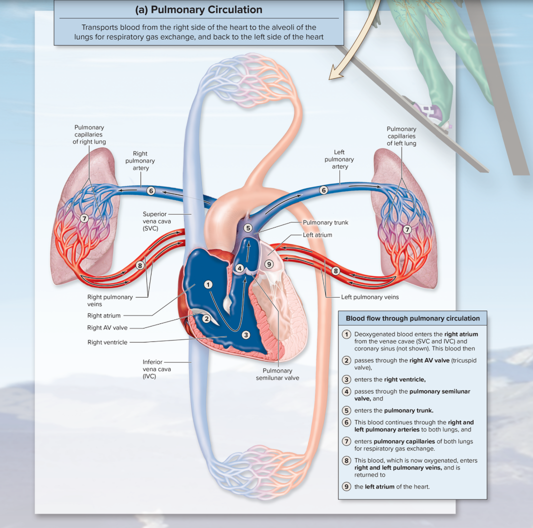
Blood Flow through Pulmonary Circulation
Converts deoxygenated blood from the right side of the heart through blood vessels to the lungs for the pickup of oxygen and the release of carbon dioxide, and then back through blood vessels to the left side of the heart.
Blood Flow through Systemic Circulation
Moves oxygenated blood from the left side of the heart. The blood vessels deliver oxygenated blood to systemic cells, such as those of the liver, skin, muscle, and brain, for the exchange of nutrients, respiratory gases, and wastes, before returning the blood to vessels that enter the right side of the heart.

Location and Position of the Heart
The heart is located posterior to the sternum, left of the body midline, between the lungs in the mediastinum
The posterosuperior surface of the heart is called the base
The inferior, conical end of the heart is called the apex
*Think of the heart's position like an "upside down" pyramid with the apex below the base.
Pericardium
Covering of the heart consisting of fibrous and serous layers
Fibrous pericardium
Composed of dense connective tissue that encloses the heart but isn’t attached
Attached superiorly to base of arterial trunk (pulmonary trunk and aorta) or grest vessels
Also attached inferiorly to the diaphragm
Parietal layer of the serous pericardium
Composed of simple squamous epithelium and an underlying delicate layer of areolar connective tissue
Layer adheres to inner surface of fibrous pericardium
Visceral layer of serous pericardium
Epicardium of the heart
Composed of simple squamous epithelium
Underlying delicate layer of areolar connective tisseue
Forms the external surface of the heart
List the structural components of the pericardium.
The pericardium that encloses the heart includes the pericardial sac, which has an outer fibrous pericardium and an inner parietal layer of serious pericardium, and a visceral layer of serous pericardium (epicardium) that forms the outer layer of the heart wall.

Pericardial Cavity
A potential space between the layers of the serous pericardium that contains serous fluid, produced by the serous membranes, which lubricates the surfaces to reduce friction
Serous Fluid
the space between the parietal layer and visceral layer of the pericardium
Pericarditis
Inflammation of the pericardium, typically caused by viral, bacterial, or fungal infections.
Leaky, excess fluid leaving blood and accumulating within the pericardial cavity
Heart is unable to pump blood
Can cause cardiac tamponade, heart failure and death
Compare the superficial features of the anterior and posterior aspects of the heart
The right side of the heart is more visible from the anterior view, and the left side of the heart is more visible from the posterior view.
Three layers of the heart wall and the tissue components of each
Epicardium: the outermost layer that is also called the visceral layer of serous pericardium
Myocardium: the middle and thickest layer. Contraction of the cardiac muscle in the myocardium generates the force necessary to pump blood
Endocardium: covers the internal surface of the heart and the external surfaces of the heart valves. It is continuous with the inner lining of blood vessels.

Atrioventricular valves
The right AV valve covers the right atrioventricular opening, and it has three cusps; the left AV valve covers the left atrioventricular opening, but it only has two cusps
When open, the cusps of the valve extend into the ventricles, allowing blood to move from an atrium into the opening of a ventricle. This causes the AV valves to close
The papillary muscles secure the thin chordae tendineae that attach to the lower surface of each AV valve cusp, preventing blood flow back into the atrium.
Semilunar Valves
The pulmonary semilunar valve is located between the right ventricle and the pulmonary trunk, and the aortic semilunar valve is located between the left ventricle and the aorta.
Cusps: Each valve has three pocket-like flaps, or cusps, that look like half-moons.
Opening: Pressure from heart contractions pushes the valves open to let blood rush out into the arteries.
Closing: When the heart relaxes, backward blood pressure catches in the pockets and snaps the valves shut.
Heart sound: The snapping shut of these two valves creates the second part ("dub") of the classic "lub-dub" heartbeat sound
Tricuspid Valve
Right atrioventricular valve
Mitral (Bicuspid) Valve
Left atrioventricular valve
General structure of cardiac muscle
Cardiac muscle cells are small, have one or two centrally located nuclei, and are branched.
Aortic Valve
Valve between the left ventricle and aorta
Describe the four chambers of the heart and its function
Right Atrium (Upper Right Chamber): Receives oxygen-poor blood from the body and pumps it down into the right ventricle
Right Ventricle (Lower Right Chamber): Receives oxygen-poor blood from the right atrium and pumps it out to the lungs to pick up oxygen
Left Atrium (Upper Left Chamber): Receives oxygen-rich blood coming back from the lungs and pumps it down to the left ventricle
Left Ventricle (Lower Left Chamber): Receives oxygen-rich blood from the left atrium and pumps it forcefully out to the rest of the body
Hypertrophic Cardiomyopathy
Also known as hypertrophic heart or heart enlargement
Inward growth (hypertrophy) of heart walls, especially in one or both ventricles
Narrows the openings (outlets) for the blood to pass through
Decreases cardiac blood pumped by ventricles per
Describe the location and function of the fibrous skeleton
The fibrous skeleton provides an attachment site for heart valves and cardiac muscle, and prevents action potentials from spreading between the atria and ventricles except through the AV node
Coronary Circulation
Circulation of blood to and from the heart wall
Coronary Arteries
Coronary arteries supply oxygenated blood to the heart wall and include the left and right coronary arteries that branch off the aorta
Describe the specific areas of the heart supplied b their major branches
three major coronary arteries off the right side of the heart (RMP): the right coronary artery splits into the right marginal artery and posterior inter ventricular artery
three major coronary arteries off the left side of the heart (LAC): the left coronary artery, the anterior inter ventricular artery, and circumflex artery
Explain the significance of coronary arteries as functional end arteries
Some arteries may share connections, called arterial anastomoses
Other arteries terminate in capillary beds only, and are called end arteries
The left and right coronary arteries are considered functional end arteries because, although there coronary arteries have anastomoses, if one of the arteries becomes blocked these anastomoses are too tiny to shunt sufficient blood from one artery to the other.
As a result, the part of the heart wall that was supplied by one coronary artery branch will die due to lack of blood flow to the tissue.
Blood Flow through the coronary arteries
Blood flow to the heart wall is not a steady stream; it is impeded and then flows, as the heart rhythmically contracts and relaxes
Heart Sounds
S₁ ("lub") - AV valves close (tricuspid & mitral valve)
S₂ ("dub") - semilunar valves close (pulmonary semilunar & aortic semilunar valves)
Coronary Veins and Specific Heart Drainage
Venous return is through the cardiac veins (great, middle, and small) into the coronary sinus, which collects venous blood and drains deoxygenated blood from the heart wall directly into the right atrium of the heart
Describe the general structure of cardiac muscle
Involuntary, striated muscle found in the heart wall
Are short, thick, and branched (often Y-shaped)
One or two nuclei per cell
Structures of Cardiac Muscle: Intercalated Discs
Desmosomes act as mechanical junctions to prevent cardiac muscle cells from pulling apart.
Gap Junctions provide a low-resistance pathway for the flow of ions between cardiac cells
Allow an action potential to move continuously along the sarcolemma of cardiac muscle cells, resulting in synchronous contraction of that chamber.
How does Cardiac Muscle meets its energy needs
Aerobic cellular respiration for supplying ATP
Creatine kinase transfers Pi from creatine phosphate to ADP, yielding ATP and Creatine
It is also versatile in being able to use different types of fuel molecules, including fatty acids, glucose, lactic acid, amino acids, and ketone bodies.
Myoglobin
Molecule that binds oxygen within muscle
Conduction System
Stimulation of the heart involves the initiation of an action potential at the SA node and its transmission through the conduction system.
Structure of the Conduction System
SA node: located in posterior wall of right atrium, adjacent entrance of the superior vena cava
AV node: located n the floor of the right atrium between right AV valve and the opening for coronary sinus
AV bundle: extends from AV node into and through the interventricular septum (left and right bundle branches)
Subendocardial Branches (Purkinje Fibers): extend from left to right bundle branches beginning at the apex heart
Components of the Heart’s Conduction System
Sinoatrial (SA) node
Atrioventricular (AV) node
AV bundle
Purkinje fibers
Composed of specialized cardiac cells that initiate and conduct action potentials resulting in a heartbeat.
Parasympathetic Innervation of the Heart
Parasympathetic innervation comes from the cardioinhibitory center to decrease the heart rate
Sympathetic Innervation of the Heart
Sympathetic innervation comes from the cardioacceleratory center to increase the heart rate and increase force of contraction
Describe the pumps and channels associated with cardiac nodal cells.
The pumps and channels associated with neurons are also in the plasma membrane of nodal cells
A type of channel unique to nodal cells is the slow voltage-gated Na+ channel that allows the nodal cells to spontaneously depolarize