Anatomy material
 Call Kai
 Learn
Learn Practice Test
 Spaced Repetition
 Match
Match Flashcards
 Knowt Play
1/18

There's no tags or description
Looks like no tags are added yet.
Name | Mastery | Learn | Test | Matching | Spaced | Call with Kai | Chat |
|---|
No analytics yet
Send a link to your students to track their progress
19 Terms
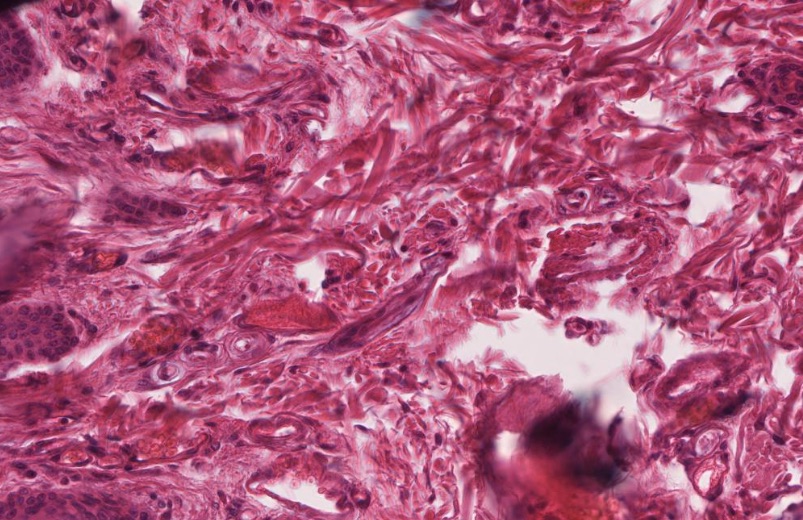
loose connective tissue
Collogen and elastic fibers. binds body parts together and move freely\y. supports epithelial tissue.

dense regular tissue
Lots of collagen fibers. Great for resistance to tension. only a little stretch. Tendons and ligaments.

Dense irregular tissue
LOTs of collagen fibers. Tension in many directions. Covers kidneys, bone, muscle, and cartilage
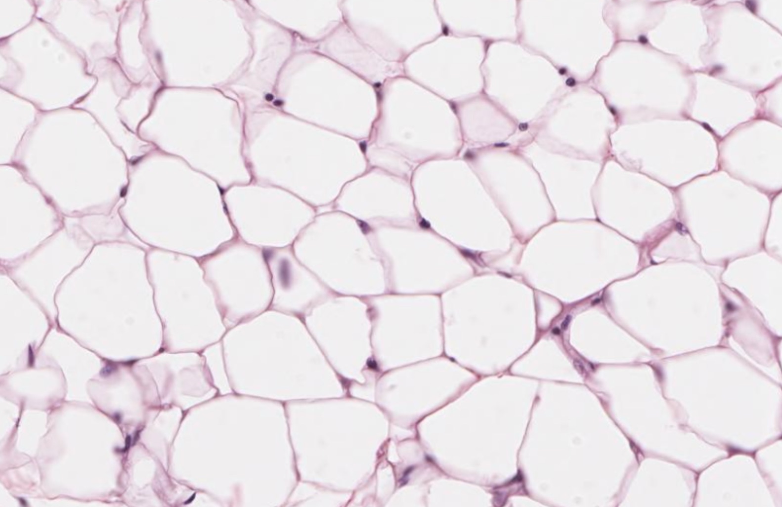
Adipose tissue
composed of Adipocytes. Used for FAT STORAGE. it is the hypodermis (layer under the skin)
Epidermis
epidermis is composed mainly of stratified squamous epithelium. dry, waxy surface

Dermis
The dermis is broken down into two different sublayers. papillary layer is composed of loose connective tissue. reticular layer is made up of dense irregular connective tissue. anchor the epidermis and dermis, holding glands and hair follicles in place!

Hypodermis
resides beneath the skin and works to connect the skin to the underlying tissues. composed primarily of adipose tissue and loose connective tissue. Anchor the skin whereas the adipose tissue in this layer is for insulation, trapping body heat.

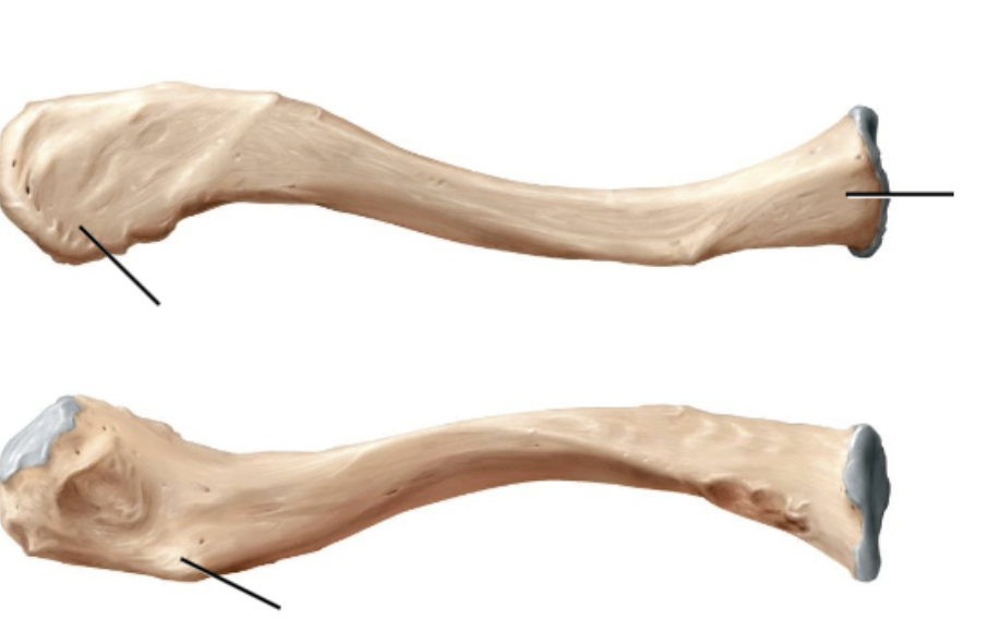
Clavicle: Term A, B, C. Word bank: Sternal end, Acromial end, Conoid tubercle.
Left to right
A: Acromial end
B: Sternal end
C: Conoid tubercle


Humerus
Capitulum
Coronoid fossa
Greater tubercle
Head
Intertubercular groove
Lateral epicondyle
Lesser tubercle
Medial epicondyle
Olecranon fossa
Trochlea


Radius (A, C, G, H)
Head
Radial styloid process
radial tuberosity
ulnar notch

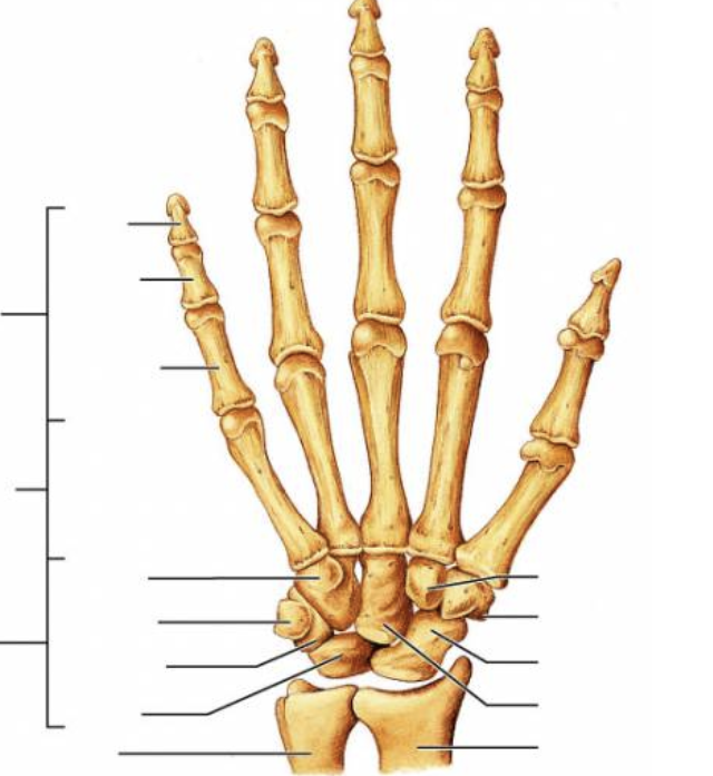
metacarpal/phalanx
distal
middle
proximal


Scapula
Acromion
Coracoid process
Glenoid fossa / cavity
Infraspinous fossa
Spine
Subscapular fossa
Supraspinous fossa

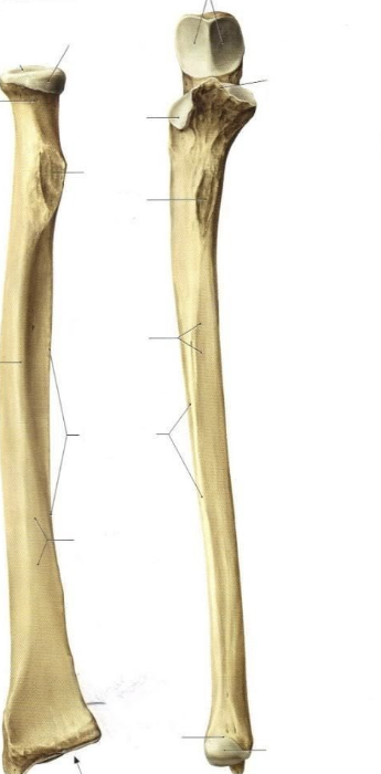
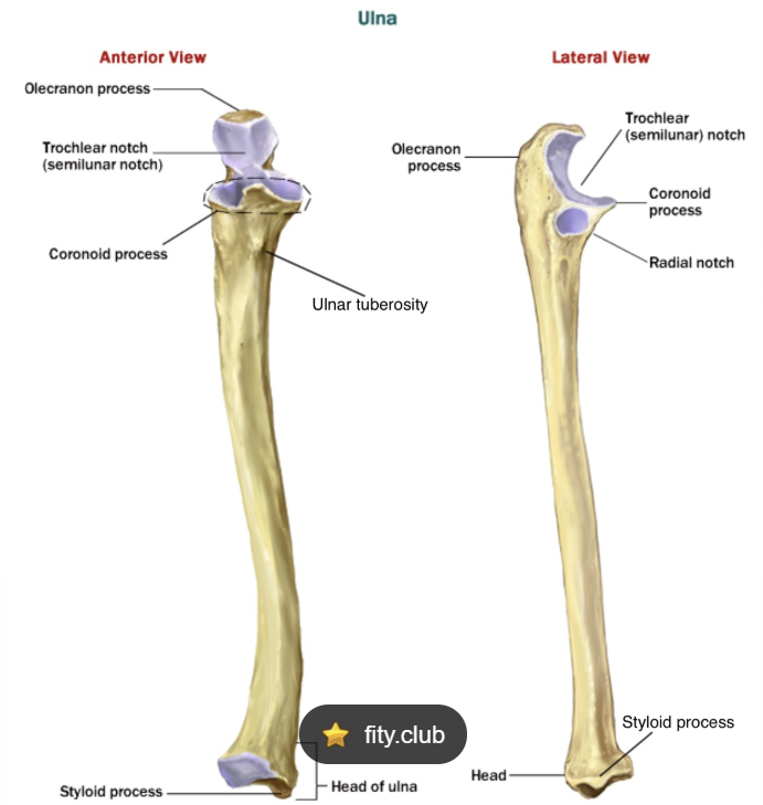
Ulna
Coronoid process
Olecranon process
Radial notch
Trochlear notch
Ulnar styloid process

Humeroradial joint
Capitulum of humerus (LM)
Head of radius (LM)
Synovial (SC)
Hinge (ST)

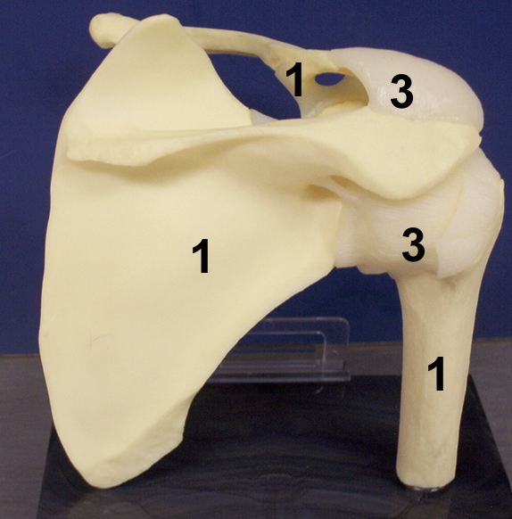
Glenohumeral joint / shoulder joint
Glenoid fossa of scapula (LM)
Head of humerus (LM)
Synovial (SC)
Ball-and-socket (ST)
Glenoid labrum (S)
Joint capsule (S)
Tendon of the long head of the biceps brachii
Humero-ulnar joint
Trochlea of humerus (LM)
Trochlear notch of ulna (LM)
Synovial (SC)
Hinge (ST
Hyaline cartilage
the chondrocytes (cartilage cell
s) appear spherical within their associated lacuna (space), and the collagen fibers, while abundant, are relatively undetectable. Chondroblasts are visible along the periphery of the tissue (appearing narrowed in their lacunae), just deep to the perichondrium (the layer of connective tissue surrounding the cartilage). ends of bones (articular cartilage), connecting the ribs to the sternum (costal cartilage) and in the trachea (respiratory cartilage).

Elastic cartilage
contains collagen fibers, but it also contains more elastic fibers, which makes the ground substance stain darker. Be sure you can distinguish between both chondrocytes and chondroblasts in this tissue as well. There are two main places we find elastic cartilage associated with the skeleton: the external ear and the epiglottis.

Fibrocartilage
ibrocartilage consists of roughly parallel rows of chondrocytes alternating with thick collagen fibers, as such is is designed to withstand both pressure and stretch. You will find fibrocartilage in places such as the menisci of the knee and the discs between vertebrae (intervertebral discs).
