Bio Ch 8
1/191
There's no tags or description
Looks like no tags are added yet.
Name | Mastery | Learn | Test | Matching | Spaced | Call with Kai |
|---|
No analytics yet
Send a link to your students to track their progress
192 Terms
Synaptic transmission
The continuation of a signal at the end of a neuron by release of neurotransmitters into the synaptic cleft to cause hyperpolarization or depolarization of the next neuron
Soma
Cell body of the neuron
Axon
Carries the action potential from the cell body to the synaptic terminal
Bipolar Neuron
has one dendrite
multipolar neuron
has many dendrites
dendrites
receive chemical signals from the presynaptic neuron
synaptic knobs
end of axons that form connections with target cells via a synaptic cleft
synaptic cleft
space between synaptic knobs and dendrites that chemical messengers travel across
Nerve
large bundle of many different axons from many different neurons
Kinesin
protein that drives movement of vesicles and organelles along microtubules in the anterograde direction (soma to terminal knobs) down the axon
Resting membrane potential
-70 mV in most neurons
Two membrane proteins that maintain resting potential
Na+/K+ ATPase
Potassium leak channels
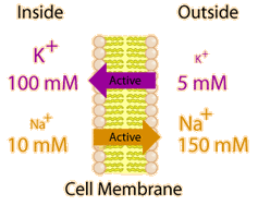
Na+/K+ ATPase
pumps 3 Na+ out and 2 K+ in with every cycle using 1 ATP — primary active transport
Potassium Leak Channels
Allow the potassium pumped in by the Na+/K+ ATPase to flow back out of the cell
Gradient of common ions in a cell
Lots more Na+ outside
Lots more K+ inside
Lots more Ca2+ outside
Lots more Cl- outside
Polarization
The state of a neuron being negative on the inside and positive on the outside
Depolarization
Departure from the resting membrane potential to a more positive potential
Repolarization
Returning to resting potential after a period of depolarization
Threshold potential
The membrane potential a neuron must depolarize to before opening voltage gated sodium channels and depolarizing completely generating an action potential — usually around -50 mV
Reached by opening of ligand gated Na+ channels by binding of neurotransmitters in the axon hillock
Maximum Depolarization Membrane Potential
Usually around +35 mV
Voltage gated Na+ channels inactivate here
Voltage gated K+ channels open here allowing for repolarization
Hyperpolarization
When voltage gated K+ channels open at the peak membrane potential around +35 mV, they stay open for a while depolarizing the cell below resting potential to about -90 mV
After this overshoot of the resting potential, the K+ leak channels and Na+/K+ ATPase restore the resting -70 mV
Schwann Cells
Make myelin membranes that coat stretches of the axon of peripheral nervous system neurons — called oligodendrocytes in the central nervous system
Myelin
Several layers of specialized membrane that wrap around an axon in spaced lengths not allowing any ions to enter the axon portions they cover — myelination increases conduction velocity
Nodes of Ranvier
gaps in the myelin sheath that have high concentrations of ion channels — APs jump from node to node accounting for the faster conduction of myelinated neurons
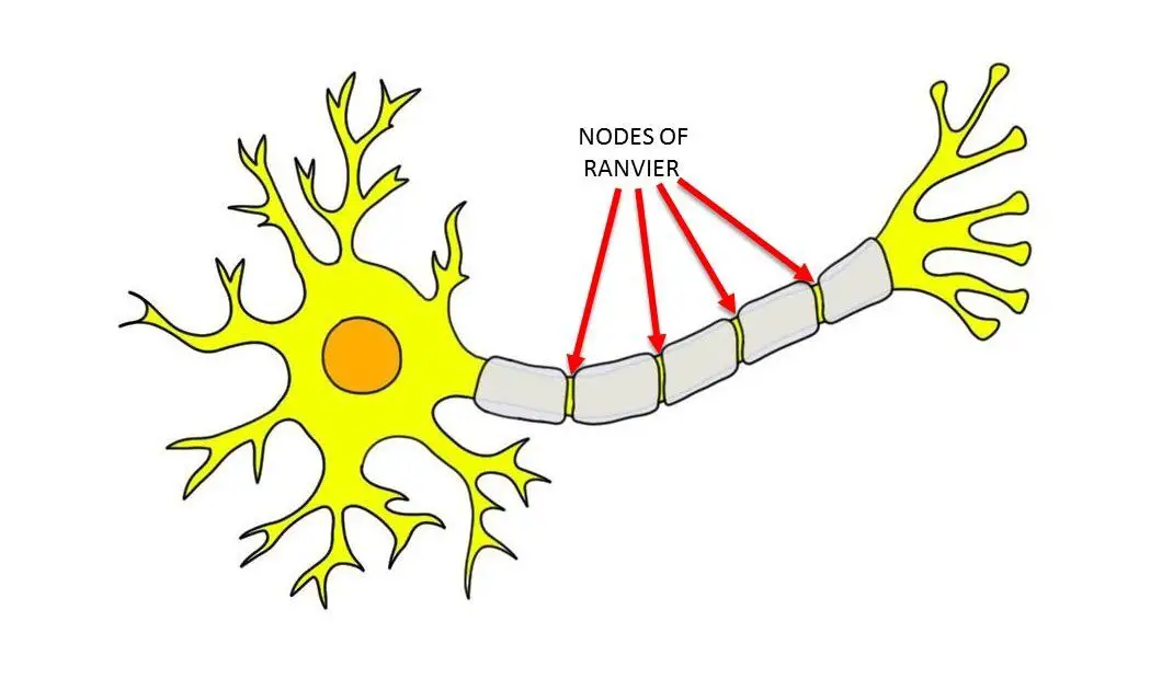
Saltatory conduction
The rapid jumping of action potentials from node to node down a myelinated axon
Glial Cells
Specialized non-neuronal cells that provide structural and metabolic support to neurons such as Schwann Cells
Types of Glial Cells
Cell Type | Location | Primary Functions |
Schwann Cells | PNS | Form myelin |
Oligodendrocytes | CNS | Form myelin |
Astrocytes | CNS | Guide neuronal development, Regulate neurotransmitter levels |
Microglia | CNS | Fight CNS infections Remove dead cells and debris |
Ependymal Cells | CNS | Produce and circulate cerebrospinal fluid (CSF) |
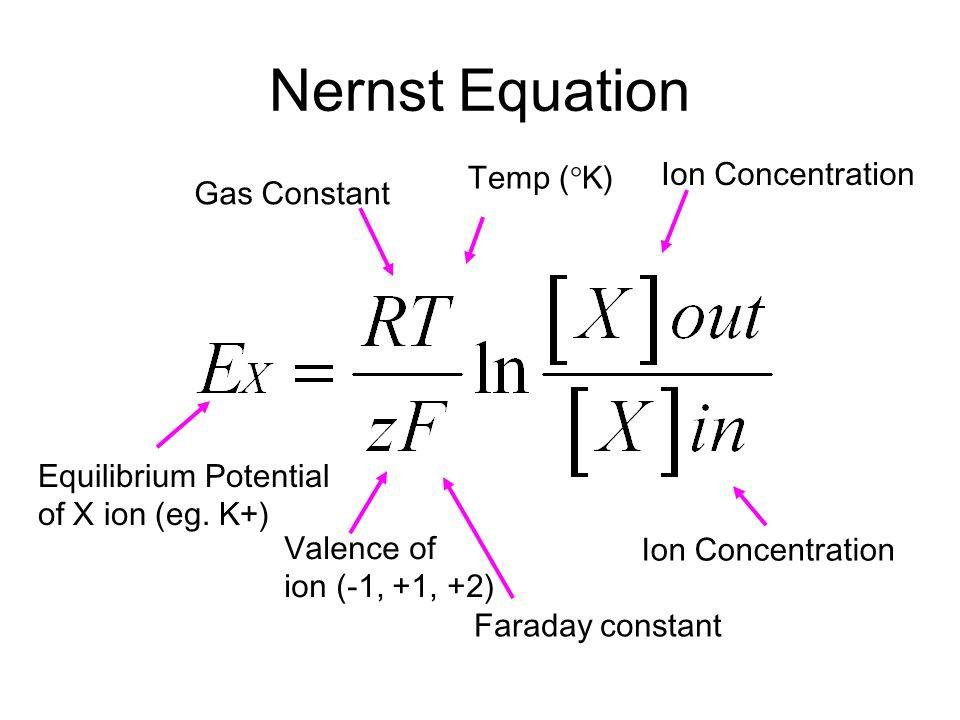
Equilibrium Potential
Membrane potential at which an ion gradient (and thus driving force) no longer exists. Since the ion gradient is different for each ion, each ion has its own equilibrium potential
Na+ = +50 mV
K+ = -90 mV
Nernst Equation
See image
Absolute Refractory Period
Time during which a neuron will not fire another AP regardless of stimulus strength or depolarization magnitude
Happens when voltage-gated sodium channels are in the inactivated state — they cannot open again until they reach the close state
Relative Refractory Period
Time during which a neuron can be induced to fire an action potential but the depolarization required is greater than normal because the neuron is still hyperpolarized after coming down from the last potential and has not yet returned to resting potential
Synapse
Space between an axon terminus and the next neuron’s dendrites, soma, or axon
Can also be the space between the axon terminus and a target organ
Two main types of synapses
Electrical Synapses
Chemical Synapses
Electrical Synapse
Where the cytoplasm of two cells is joined by a gap junction allowing an action potential to seamlessly flow from one cell to the other
Common in smooth and cardiac muscle
Not common in the nervous system
Chemical Synapse
Where an electrical signal (action potential) is converted into a chemical signal (neurotransmitter) which can cross the synapse
Signal transmission steps across a chemical synapse
Dendrites bind neurotransmitter and open ligand gated channels
Ligand gated Sodium channels on axon hillock are bound causing depolarization to threshold
Voltage gated sodium channels open and generate action potential
Action potential propagates by continually opening voltage gated sodium channels as it travels down the axon
Voltage gated potassium channels open at peak depolarization creating a repolarizing wave following the wave of depolarization
Once depolarization reaches the synaptic terminal, it opens voltage gated Calcium channels
Calcium influx causes release of neurotransmitter into synapse
Repeat step 1
Neurotransmitter is reabsorbed through transporters (reuptake)
Neuromuscular Junction
Synapse between neurons and skeletal muscle where acetylcholine is released
Acetylcholinesterase (AChE)
Degrades acetylcholine in the synaptic cleft
Excitatory Neurotransmitter/Receptor
Depolarizes postsynaptic neuron
The neurotransmitter itself doesn’t differentiate between excitatory and inhibitory effects, the receptor it binds to determines which happens
Some neurotransmitters can bind to some excitatory receptors and some inhibitory receptors
Inhibitory Neurotransmitter/Receptor
Hyperpolarizes postsynaptic neuron
The neurotransmitter itself doesn’t differentiate between excitatory and inhibitory effects, the receptor it binds to determines which happens
Some neurotransmitters can bind to some excitatory receptors and some inhibitory receptors
“All-or-nothing” nature of Action Potentials
when fired, action potentials always have the same speed and magnitude of depolarization for a given neuron
Spatial Summation
Every synapse has several presynaptic neurons converging on it
The postsynaptic neuron’s ability to reach threshold potential and fire is dependent upon the summation of effects of all of the presynaptic neurons at any given moment
Some presynaptic neurons may fire inhibitory potentials at the same time others fire excitatory potentials
Excitatory Postsynaptic Potentials (EPSPs)
cause postsynaptic depolarization
Inhibitory Postsynaptic Potentials (IPSPs)
cause postsynaptic hyperpolarization
Temporal Summation
One presynaptic neuron fires a ton of excitatory or inhibitory potentials that all pile up at once creating enough of an effect to single-handedly excite or inhibit the postsynaptic neuron
PNS
Peripheral Nervous system
Carries out sensory and motor functions
CNS
Central Nervous System
Carries out integrative functions, taking sensory signals from the PNS and converting them into motor signals to be sent by the PNS
Effectors (2 types)
The tissues/organs acted upon by motor neurons
Muscles
Glands
Efferent
Out of CNS, motor
Afferent
Into CNS, sensory
Reflex
Direct motor response to sensory input without conscious thought
Usually doesn’t involve the brain and only involves the spinal cord
Usually monosynaptic with the synapse in the spinal cord
Disynaptic Reflex
A reflex where the sensory neuron stimulates two other neurons
Seen in reciprocal inhibition where the sensory neuron stimulates a motor neuron to cause contraction and an inhibitory interneuron to cause relaxation
Reciprocal Inhibition
Sensory neurons allowing for a movement by stimulating a motor neuron to contract one muscle and an interneuron to relax another
Seen with leg extension where quadriceps contract and hamstring relaxes
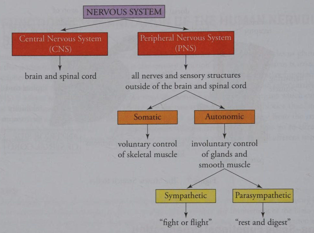
Organization of the Nervous System
See Image
Sympathetic effects often occur as a result of epinephrine release from the adrenal medulla
The enteric system also exists :)
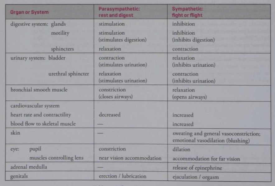
Effects of Autonomic Nervous System on various organs
See image
Tract / Column
White matter (axons) in the CNS
Nucleus
Gray matter deep in the brain
Cerebral Cortex
Gray matter on the surface of the brain
Horn
Gray matter in the spinal cord
Ganglion
gray matter in the PNS
Cerebrospinal Fluid (CSF)
Clear liquid that the CNS (brain and spinal cord) floats in for shock absorption and nutrient / waste exchange
Reticular Activating System (RAS)
responsible for arousal and wakefulness
Mostly contained within the midbrain
Hypothalamus
Controls emotions and autonomic functions
Links the nervous and endocrine systems through its control of the pituitary
Basal Nuclei / Basal Ganglia
Involved in procedural learning and habit forming
Works with the cerebellum to coordinate movement initiated by the frontal lobe
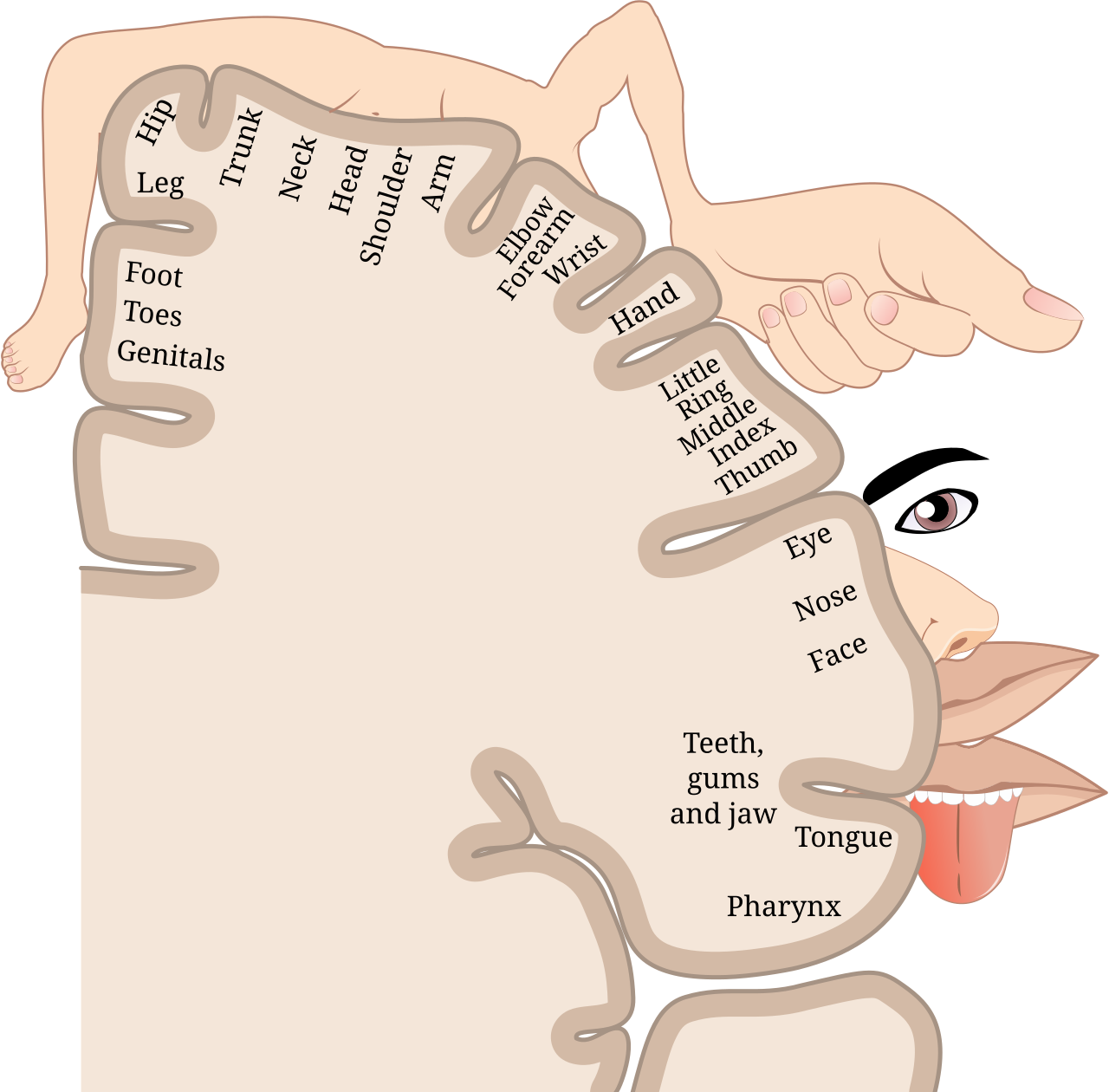
Sensory Homunculus
Depicts which part of the parietal lobe interprets sensation of each part of the body
pairs of cranial nerves
12
pairs of spinal nerves
31
vagus nerve
Parasympathetic cranial nerve
Lowers heart rate
increases GI activity
Somatic Motor Neuron Facts
What do they innervate?
What neurotransmitter do they all use?
Where are their cell bodies located
They innervate skeletal muscle using acetylcholine
Their cell bodies are all in either the brain stem or the ventral root of the spinal cord
ventral vs dorsal roots of the spinal cord
Dorsal = sensory = info into CNS
Ventral = motor = info out of CNS
Somatic Sensory Neuron Facts
Where does the dendrite end?
Where is the cell body?
Where is the first synapse
The dendrite extends from the sensory receptor at some place on the body, to the dorsal root ganglion where the sensory neuron cell body is
The sensory neuron soma is still outside of the meninges, the axon extends into the meninges
The first synapse is either within the spinal cord or within the brain
Meninges
Protective sheath around brain and spinal cord
Autonomic nervous system neurotransmitters
All preganglionic autonomic neurons use acetylcholine
All parasympathetic postganglionic neurons use acetylcholine
Most sympathetic postganglionic neurons use norepinephrine
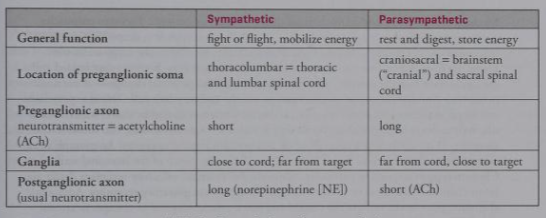
Preganglionic and Postganglionic neuron locations in the efferent autonomic system
Sympathetic starts in thoracic or lumbar vertebrae — thoracolumbar system
Parasympathetic starts in cervical or sacral vertebrae — craniosacral
Sympathetic has a very short preganglionic axon that ends just outside the spinal cord and the postganglionic axon travels most the way to the effector (target organ or tissue)
Parasympathetic has a very long preganglionic axon that travels most the way to the effector with a short postganglionic axon that travels a small distance to innervate the effector

Short Reflex
When an autonomic afferent neuron synapses with an autonomic efferent neuron in the PNS to effect a non-conscious, quicker response than traveling all the way to the CNS
Inner portion of the adrenal gland
adrenal medulla
outer portion of the adrenal gland
adrenal cortex
Main hormones secreted by the adrenal cortex
Cortisol — a glucocorticoid
Aldosterone — a mineralocorticoid
Sex hormones
Hormone secreted by the adrenal medulla
Epinephrine — acts on sympathetic postganglionic neurons to enhance their activity resulting in increased heart rate and the fight-or-flight response
Exteroceptors
Sensory receptors that detect stimuli from the outside world
Interoceptors
Sensory receptors that respond to internal stimuli (within the body)
Mechanoreceptors
Respond to mechanical disturbances
Pacinian Corpuscles
Onion-shaped pressure sensors located deep in the skin
When the onion-like layers of the corpuscular membranes are compressed by pressure on the skin, a graded depolarization results in a nearby neuron that could start an action potential
Auditory Hair Cell
Found in the cochlea, detects vibrations generated by sound waves
Vestibular Hair Cell
Located within semicircular canals of the inner ear
Detect acceleration and position relative to gravity
Intestinal stretch receptor
Type of mechanoreceptor on the intestinal wall that feels the stretch when the intestine is full and stops the signal of hunger and induces satiety
Chemoreceptors
Respond to chemicals
Olfactory receptors
Type of chemoreceptor
Detect chemicals in air to allow us to smell things
Gustatory Receptors
Type of chemoreceptor — taste buds
Bind to molecules in food that cause us to taste
Aortic and Carotid Chemoreceptors
Chemoreceptors in the aortic and carotid arteries detect
pH
pCO2
pO2
Nociceptors
Pain receptors — may be somatic or autonomic
Consist of a free nerve ending that detects chemical signs of tissue damage
Give unclear sensation of dull, aching pain
Can create the illusion of pain on the skin when crossed with other somatic afferent neurons — referred pain
Referred Pain
Pain that is felt in one part of the body despite the tissue damage occurring somewhere else.
If the injury occurs at point A but the pain is felt at point B:
The nociceptor at point A crosses with a sensory neuron that originates at point B, thus causing the body to think that the pain signal from the nociceptor at point A came from the origin point of the somatic sensory afferent at point B
Thermoreceptors (3 categories)
Stimulated by changes in temperature — can be both autonomic and somatic
cold-sensitive
warm-sensitive
thermal nociceptors — detect painfully hot stimuli
Electromagnetic Receptors
Respond to electromagnetic waves — only consist of rods and cones of eye in humans
Four Properties of a Stimulus communicated to the CNS
Modality — type of stimulus — based on which receptor is firing (labeled line)
Location — origin of stimulus — based on which receptive field the sensory neuron comes from
Intensity — strength of stimulus — encoded by the frequency of action potentials
Duration — not always communicated — can be communicated by tonic response receptors that fire APs for the duration of the stimulus
Stimulus Adaptation
Decrease in AP frequency when stimulus intensity remains constant — allows us to get used to consistent stimuli
Does not apply to nociceptors (pain), they never adapt
Proprioceptors
Provide an “awareness of self”
Form the kinesthetic sense
Muscle Spindle
A type of mechanoreceptor and proprioceptor
Detects muscle stretch
Golgi Tendon Organs
Proprioceptors that monitor tension in the muscles to make sure it doesn’t get too great and tear the muscle
Joint Capsule Receptors
Detect pressure, tension, and movement in joints