Lungs and respiration
 Call Kai
 Learn
Learn Practice Test
 Spaced Repetition
 Match
Match Flashcards
 Knowt Play
1/45

There's no tags or description
Looks like no tags are added yet.
Name | Mastery | Learn | Test | Matching | Spaced | Call with Kai | Chat |
|---|
No analytics yet
Send a link to your students to track their progress
46 Terms

Define these structures top to bottom
Trachea/Primary Bronchi/ Secondary Bronchi/Tertiary Bronchi/ Bronchioles/Terminal Bronchioles/Alveoli
Where is level of the sternal angle?
At the level of bifurcation of trachea
What is the Primary, Secondary, Tertiary Bronchi?
-first branch from trachea to lung
-second braches, goes to each lobe of the lung
-More divisions
What is the of terminal Bronchioles?
The smallest branches of the bronchi have no cartilage
What are the functions of alveoli?
-called air sac
-exchange of Co2 and O2
What are the two main pleural coverings
Visceral pleura and parietal pleura
What is the function and location of the visceral pleura?
membrane that tightly adheres to the outer surface of the lungs
What are the functions and location of parietal pleura?
lines the pulmonary cavities, adhering to the thoracic wall, the mediastinum, and the diaphram
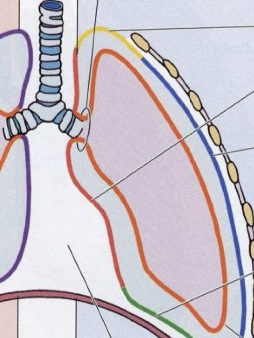
Label the parts of the parietal pleura and its location
Cervical pleura (superior, above collar bone)/Mediastinal pleura(medial)/costal pleura(lateral, reef of the ribs)/diaphragmatic pleura(inferior, above diaphragm)
What happened to the lung during exhalation?
It is collapsed, and the visceral pleura shrinks, and the pleural cavity is shown, which contains pleural fluid
What is the space in the middle part in between the two lungs, what does it contains?
Mediastinum, heart
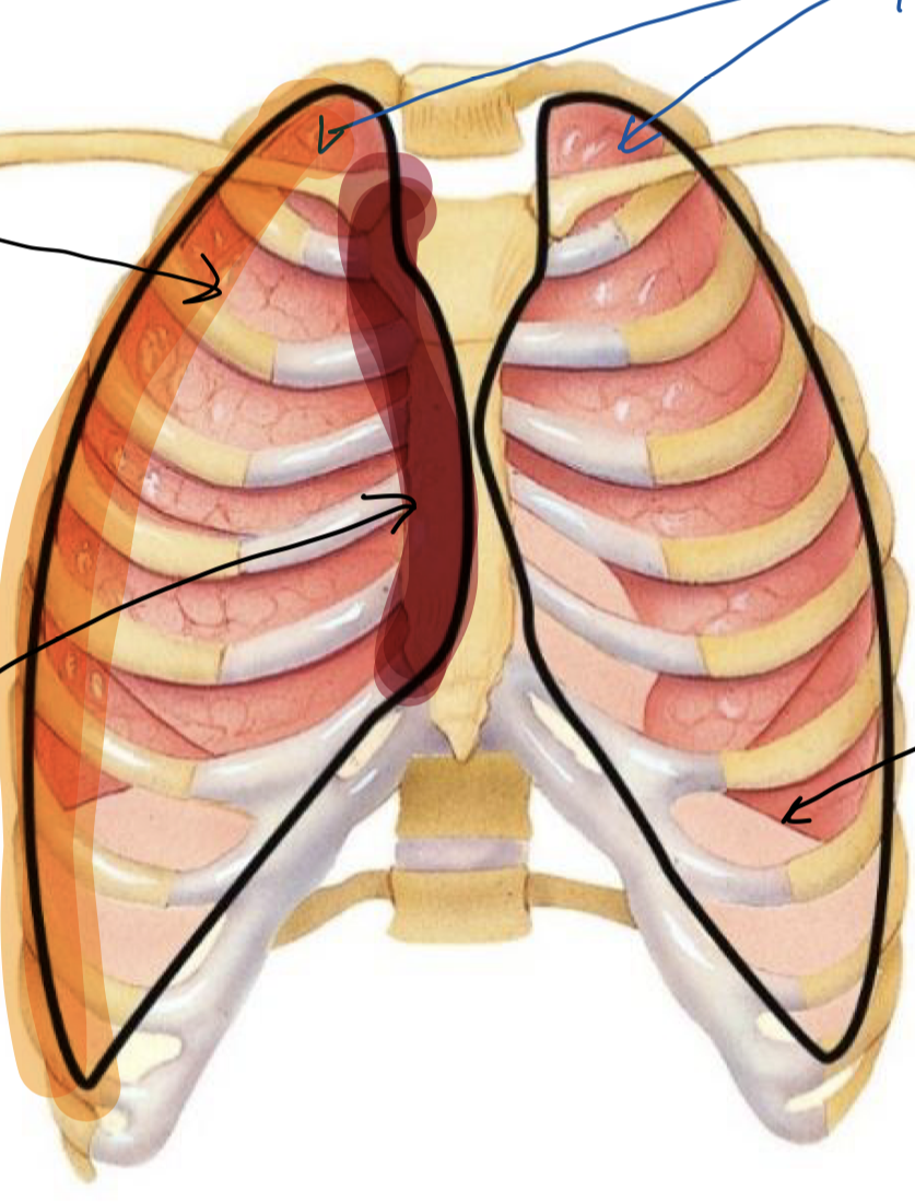
Label these structures (top clockwise)
Apex/Base/Mediastinal Surface/Costal surface
Where does the apex start and end?
it starts above first rib and clavicle and ends at 7th cervical to 1st thoracic
Where does the pleura extend to?
ribs 9-10
What is the location of the base?
ribs 7-8
What is the location of the coastal surface?
smooth outer area that presses directly against the ribs and the chest wall
What is the location of the mediastinal surface?
Located on the medial side of each lung, facing the middle mediastinum

label these part s of right and left lung features, right lung to left lung top to bottom
Right lung: Superior lobe/horizontal fissure/middle lobe/oblique fissure/inferior lobe
Left lung: Superior lobe/ cardia notch/inferior lobe/lingula
What is the function and location of cardia notch?
-Indentation of the anterior border of the superior lobe of the left lung
-provide space of the heart
What is the function and location of the lingula?
-tongue-shaped part of the left upper lobe.
-respiratory unit exchanging oxygen and carbon dioxide
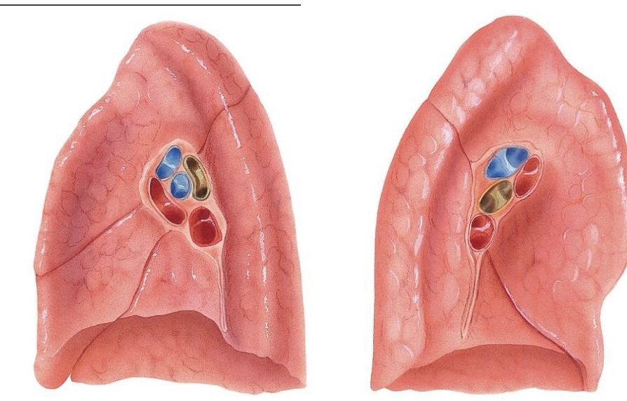
Label the structures of the right lung (outermost, blue, yellow, red)
Hilum/Pulmonary arteries/Bronchus/Pulmonary veins

Label the structures of the left lung (outermost, blue, yellow, red)
Hilum/Pulmonary artery/Bronchus/Pulmonary veins
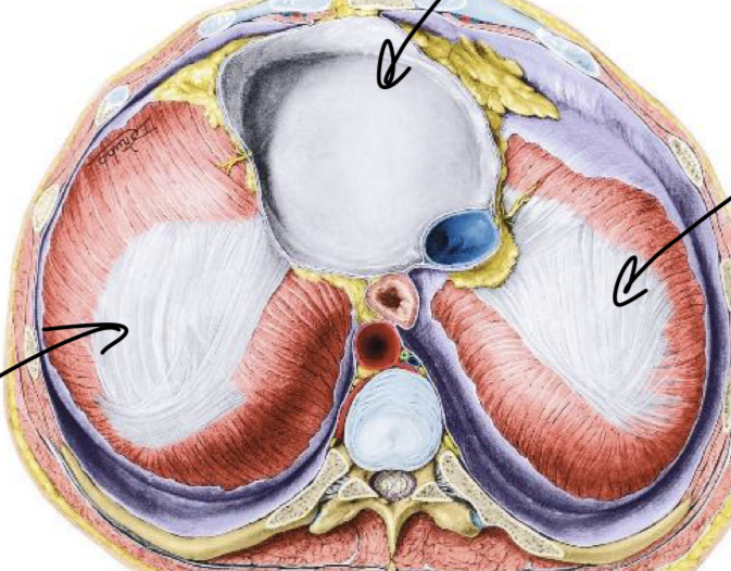
Label these structures
Pericardial sac and central tendon
What is the function and location of central tendons?
-a thin, strong, aponeurotic sheet in the center of the diaphragm
- muscle fibers of the diaphragm converge and attach
What is the function and location of pericardial sac
-middle mediastinum of the thoracic cavity
-lower base fuses directly with the central tendon of the diaphragm
-give downward contraction of the diaphragm during inhalation gently shifts the heart vertically.

label three structures on the left side and the blue green red ring on the diaphragm
Median arcuate ligament/medial arcuate ligament/lateral arcuate ligament
Caval/Esophogeal/Aortic
What is the function and the location of median arcuate?
helps form a protective arch over the arota
What is the location of the medial arcuate ligament?
-Anchors diaphragm to the spine, helps provide protective arch over the psoas major muscle
What is the psoas major muscle?
long, thick muscle that connects your lower back to your upper leg
What is the function and the location of the lateral arcuate ligament?
-Anchors the diaphragm to the posterior abdominal wall
-Helps form a protective arch over the quadratus Lumborum muscle
What is the function of the caval opening?
-in the central tendon
-transmits the IVC, branches of the right phrenic nerve, and lymphatic vessles
What is the function of esophogeal?
-in the muscle of the diaphragm
-transmit esophagus
What is the function of arotic hiatus?
-posterior to the diaphragm
-transmits descending aorta, azygos vein, and thoracic duct

Label the intercostal from out to in
External intercostal/Internal intercostal/inner intercostals
What is the purpose of the external intercostal?
-External intercostals pull the ribs up and outward
-Increases the volume of the thoracic cavity
What is the purpose of the internal intercostal?
Internal intercostals pull the ribs down and inward
-This decreases thoracic volume
-most important during forced expiration
What is the purpose of the innermost intercostal?
-Similar action to the internal intercostals
-helps stabilize the spaces between the ribs
What is the mechanics of ribcage expansion 1 and its activity?
-Anteroposterior dimension
-The expansion if the ribcage comes from the anterior and posterior
-lifting of the sternum and pull of the external intercostal muscles
What is the mechanics of ribcage expansion 2 and its activity?
- Medial-lateral dimension
-Mainly involves the middle/lower ribs.
-the ribs move upward and outward during inhalation.
-This increases the side-to-side (transverse) diameter of the thorax
What is the mechanics of ribcage expansion 3 and it’s activity?
-vertical dimension
-Mainly associated with the lower ribs
-Mainly produced by movement of the diaphragm
-The ribs move outward laterally during inhalation.
-This increases the transverse diameter, particularly in the lower thorax
What muscles does assisting inhalation include?
Sternocleidomastoid, scalenes, Pectoralis minor
What muscles does assisting exhalation include?
Internal intercostals, external obliques, rectus abdominus, internal obliques
What muscles does quiet breathing/regular breathing include?
external intercostals, diaphragm
What is the purpose of the diaphragm and the external intercostals?
Diaphragm-contracts downard and expands the thoracic cavity
external intercostals- elevate the ribs and expands ribcage
What is the purpose of
Internal intercostals, external obliques, rectus abdominus, internal obliques?
Internal intercostals-depress ribs
External obliques- compress abdominal cavity
Rectus abdominus- compress the abdominal cavity diaphragm upward
Internal obliques- compress abdominal cavity
What is the purpose of Sternocleidomastoid, scalenes, Pectoralis minor?
Sternocleidomastoid- elevates the sternum
scalenes-elevate upper ribs
pectoralis minor- elevate upper ribs