Chest Xrays
1/60
There's no tags or description
Looks like no tags are added yet.
Name | Mastery | Learn | Test | Matching | Spaced |
|---|
No study sessions yet.
61 Terms
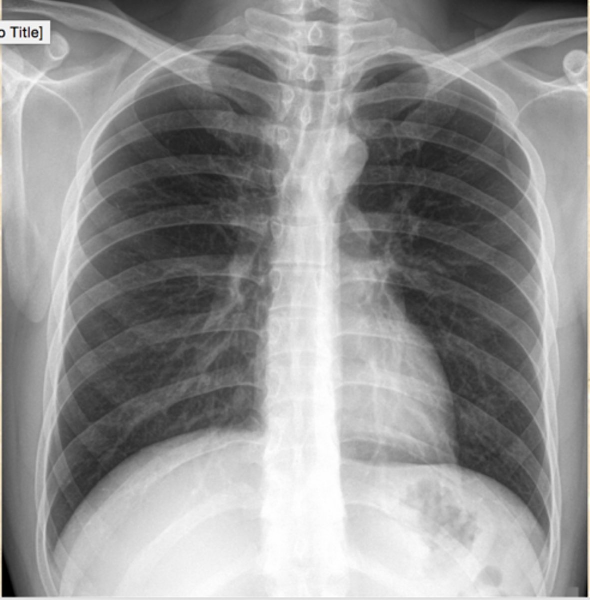
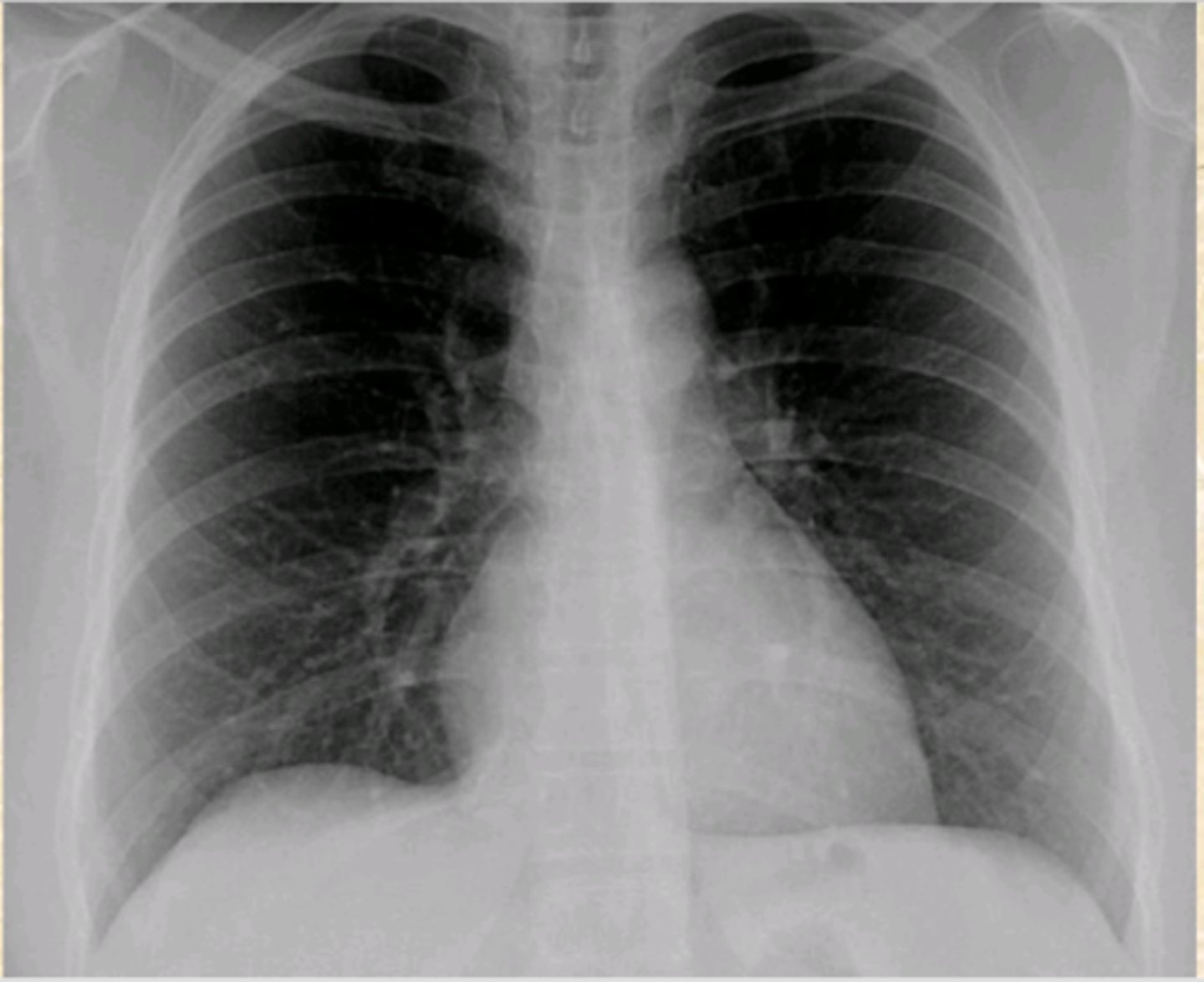
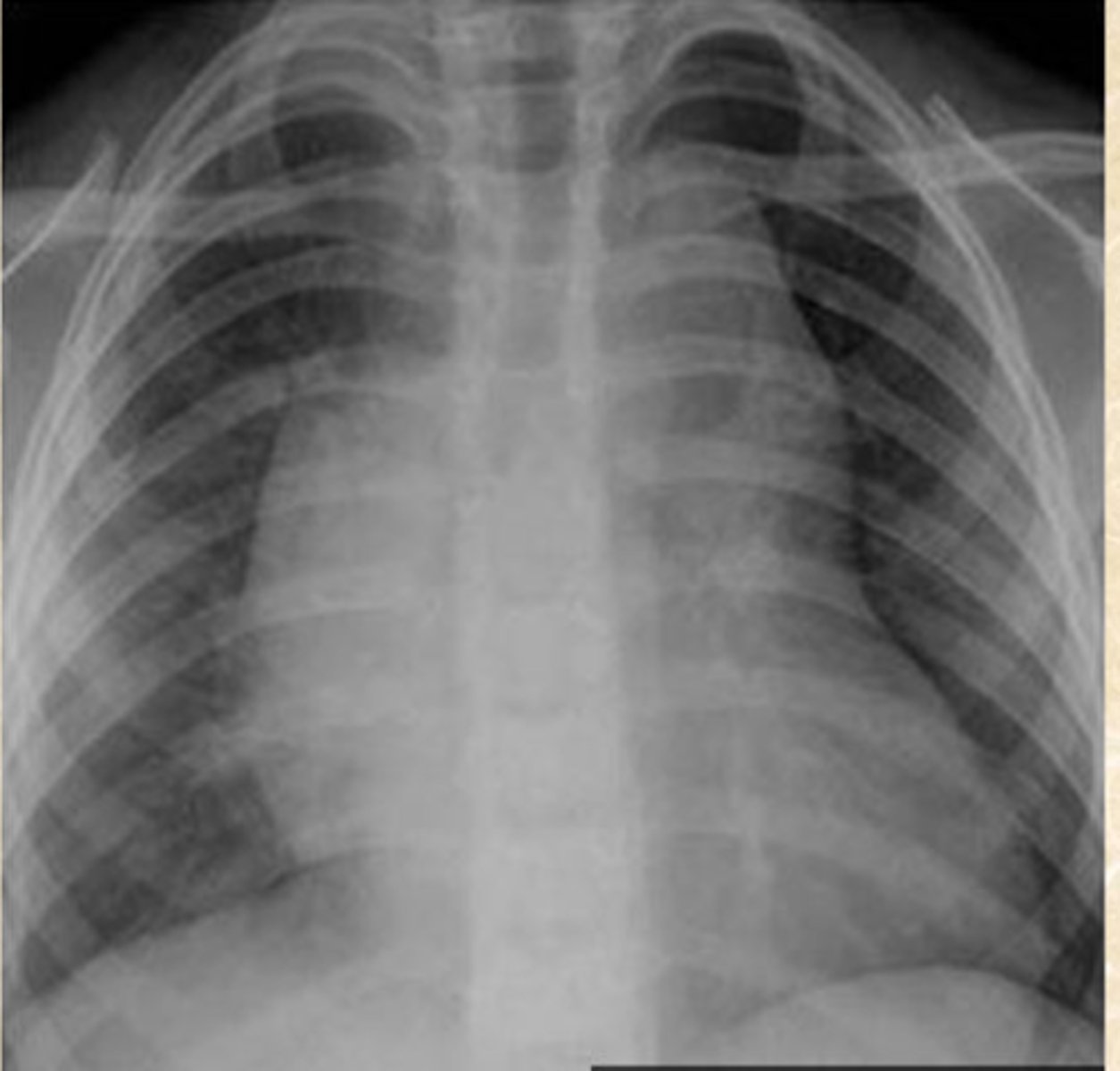
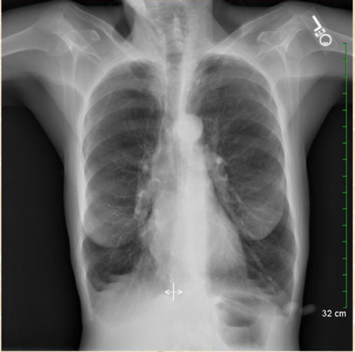
Normal CXR
Diagnosis?
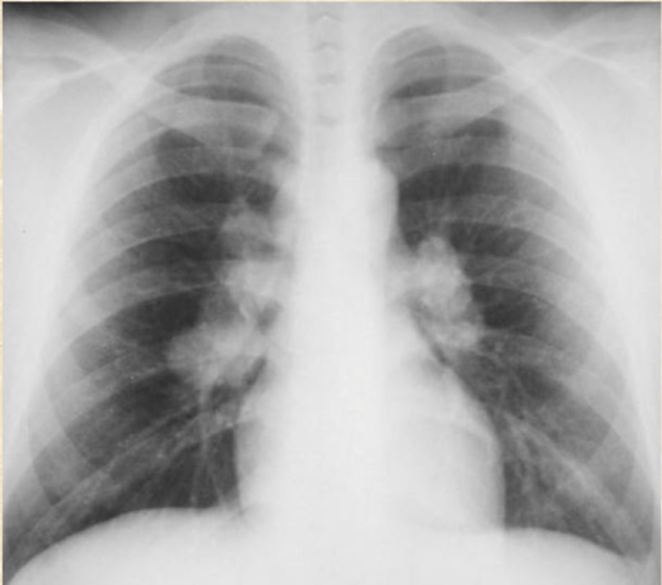
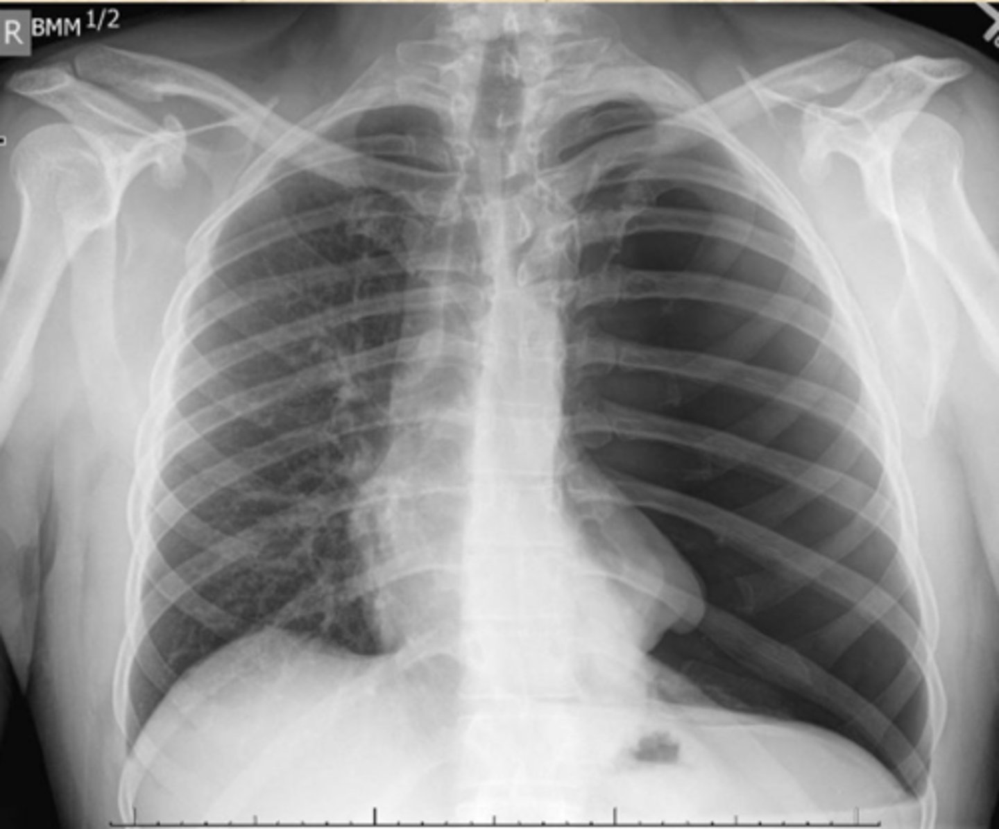
Normal CXR
Diagnosis?
Air bronchogram
tubular outline of an airway made visible by filling of the surrounding alveoli by fluid or inflammatory exudates
Patchy, cloudy lobar consolidation
X-ray findings of pneumococcal pneumonia
Diffuse, fine, and reticular with progression to airspaces
X-ray findings of viral pneumonia
Perihilar, reticular infiltrates
X-ray findings of Pneumocystis jirovechi pneumonia
Pneumocystis jirovechi
Opportunistic lung infection in HIV patients
Pleura, Bronchi
What structures of lungs are typical not visible on radiographs?
PA and left lateral
Components of two-view chest X-ray
adenopathy
What is the most common reason the retrosternal space is obscured?
pleural effusions
What typically causes blunting of the costophrenic angles
half
The heart should take up less than ___ of the thoracic cavity
aortic arch
What is the first convexity on the left side of mediastinum?
pulmonary artery
What is the second convexity on the right side of mediastinum?
Left Hilum
Which hilum is more superior?
Silhouette sign
loss of normal borders between thoracic structures
Right border of the heart
What structure is usually referenced when talking about silhouette sign
It is under-penetrated
What is wrong with this CXR?
It is over-penetrated
What is wrong with this CXR?
PA
What view: Heart is anterior structure and therefore truer to actual size
10
TQ: If _____ Posterior ribs are visible; it is an excellent inspiration
poor inspiration
What is wrong with this CXR?
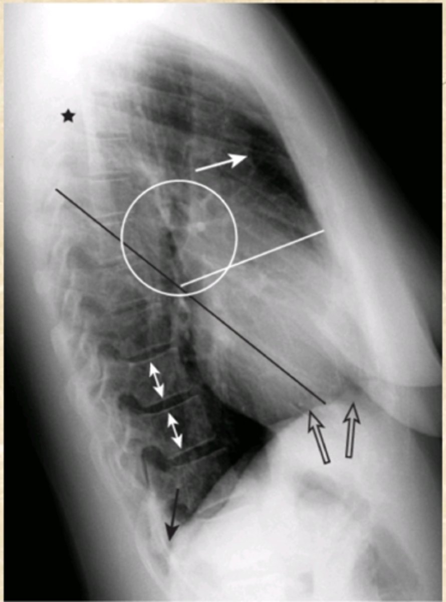
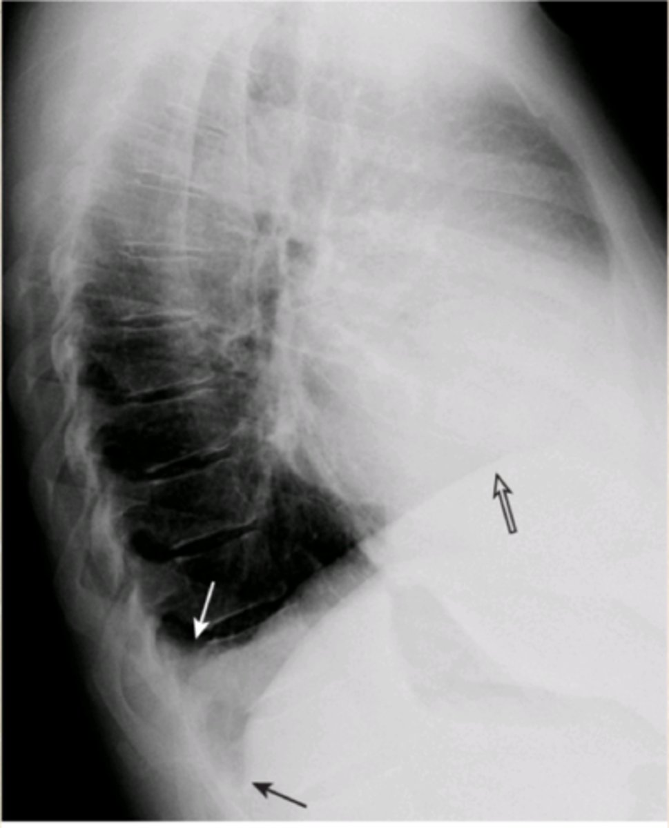
Lateral
What is the best view to look at the thoracic spine?
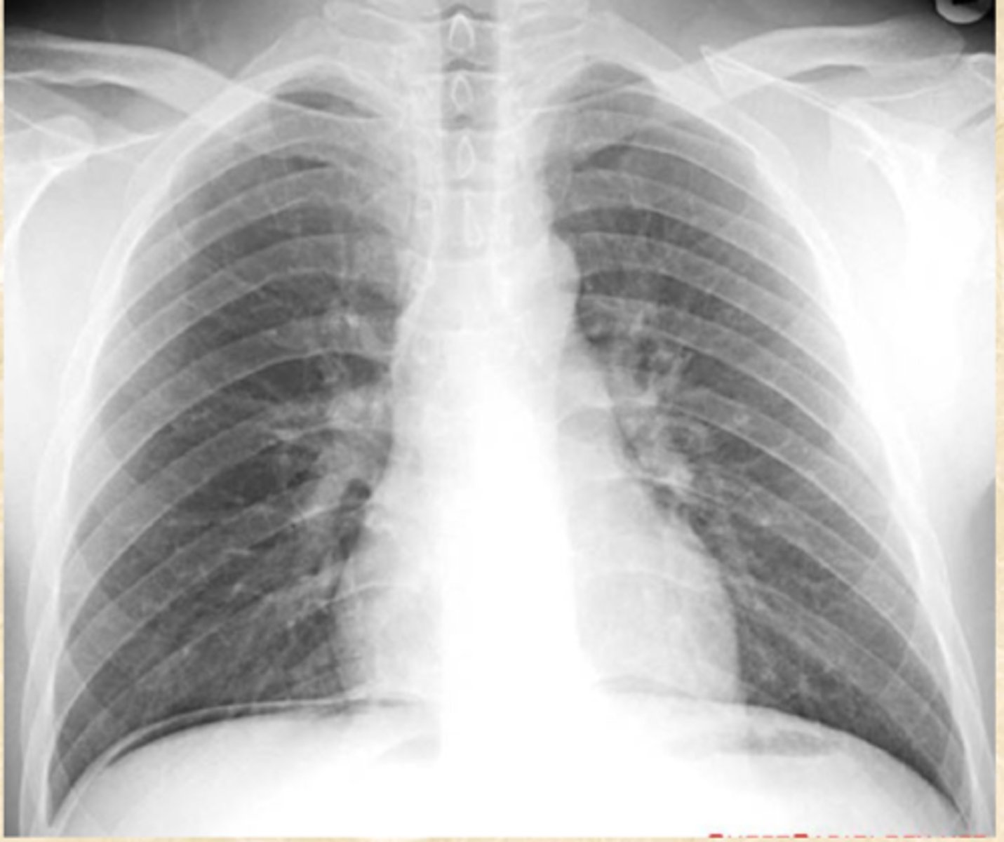
Normal CXR
Lateral View
Diagnosis?
View?
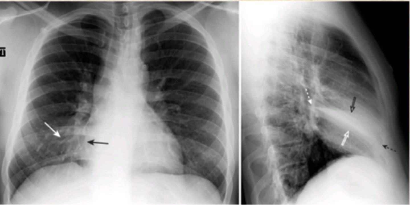
Hilar Mass
Lateral View
Diagnosis?
View?

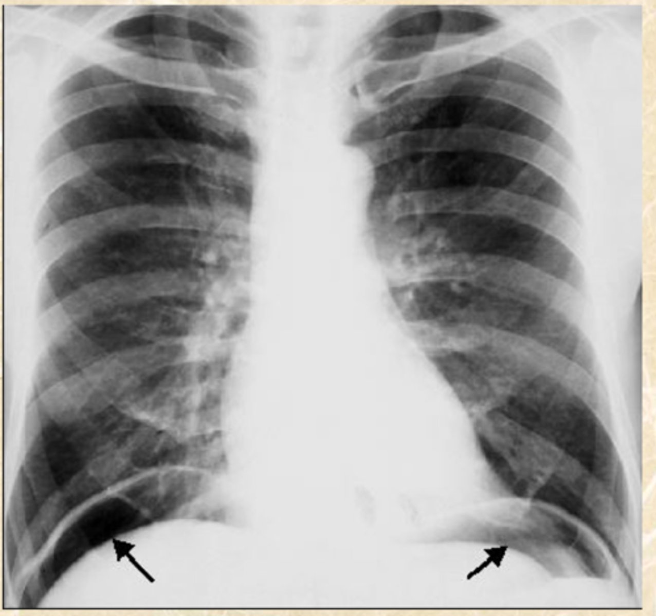
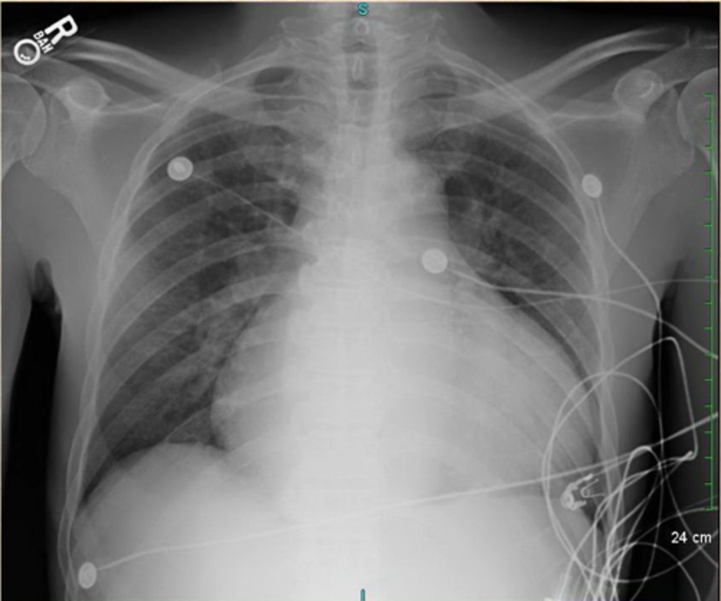
There is blunting of the Costophrenic angle, cause by a Pleural Effusion
Diagnosis?
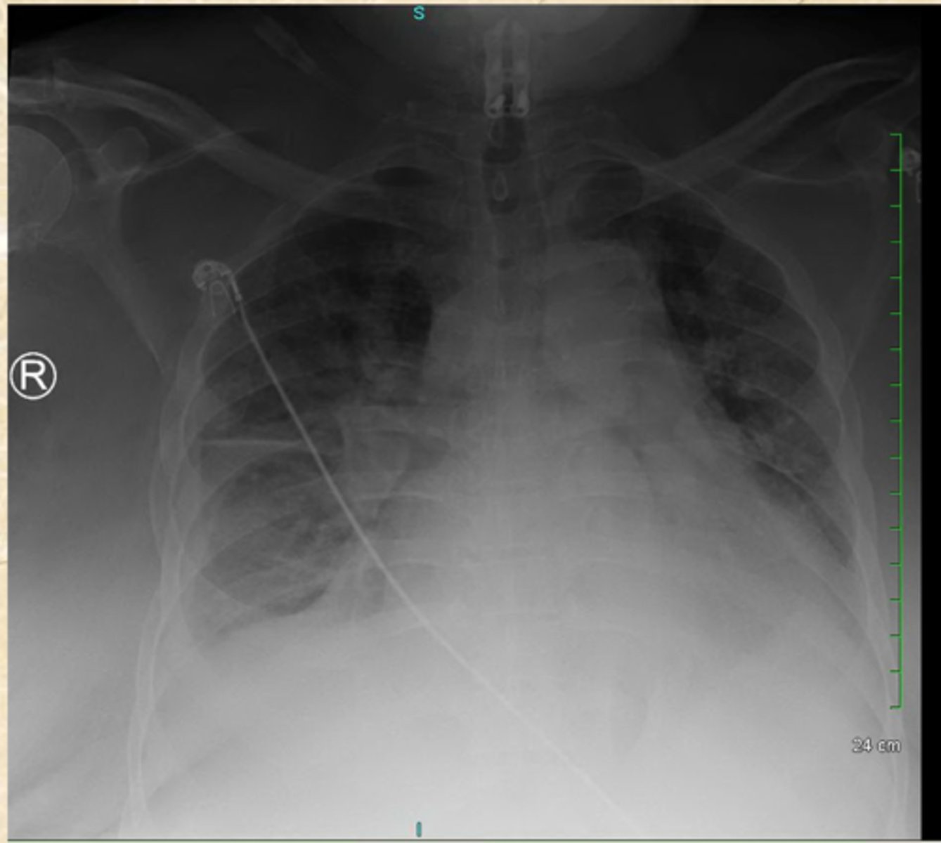
A pleural effusion
What does this image show?
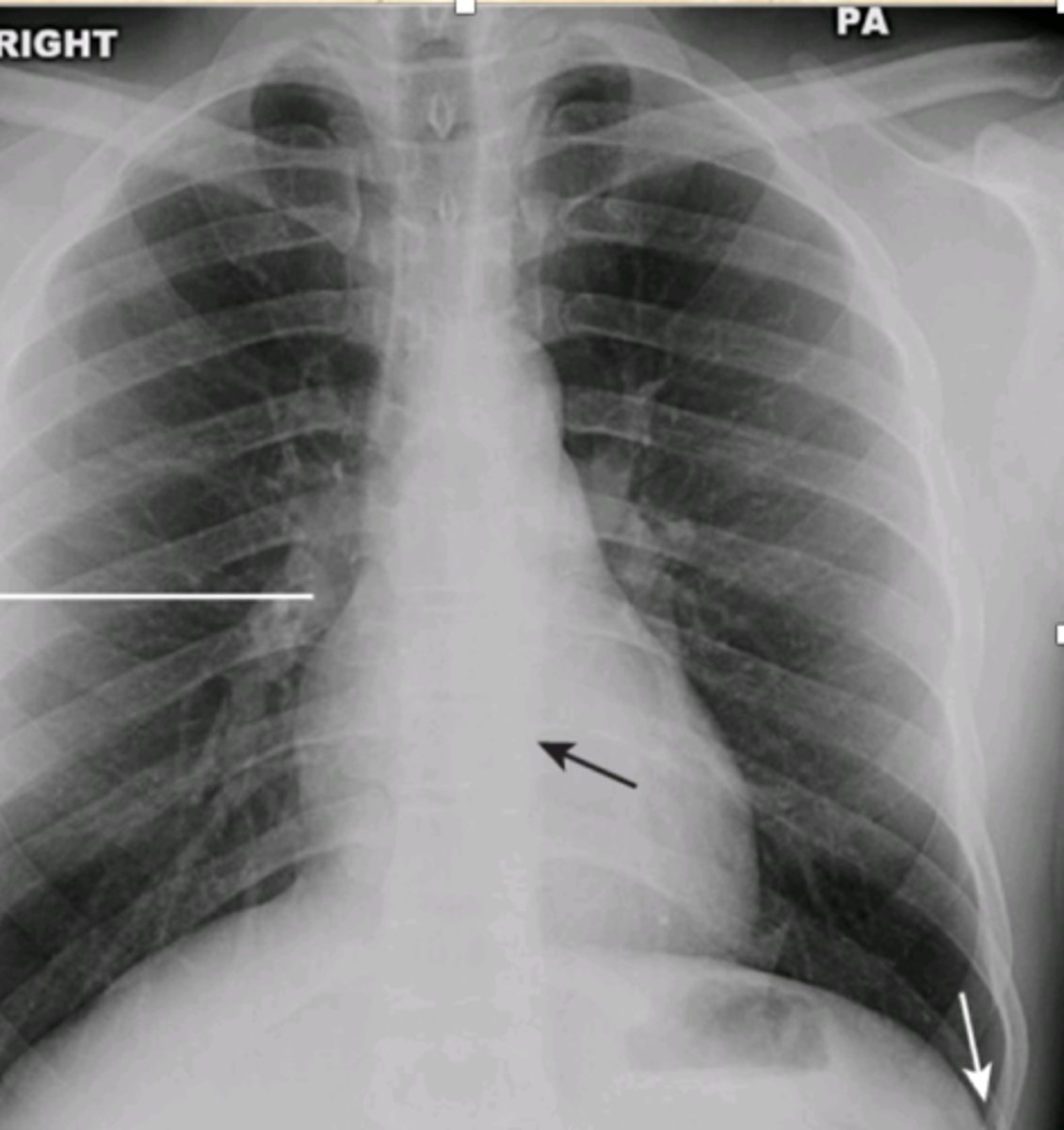
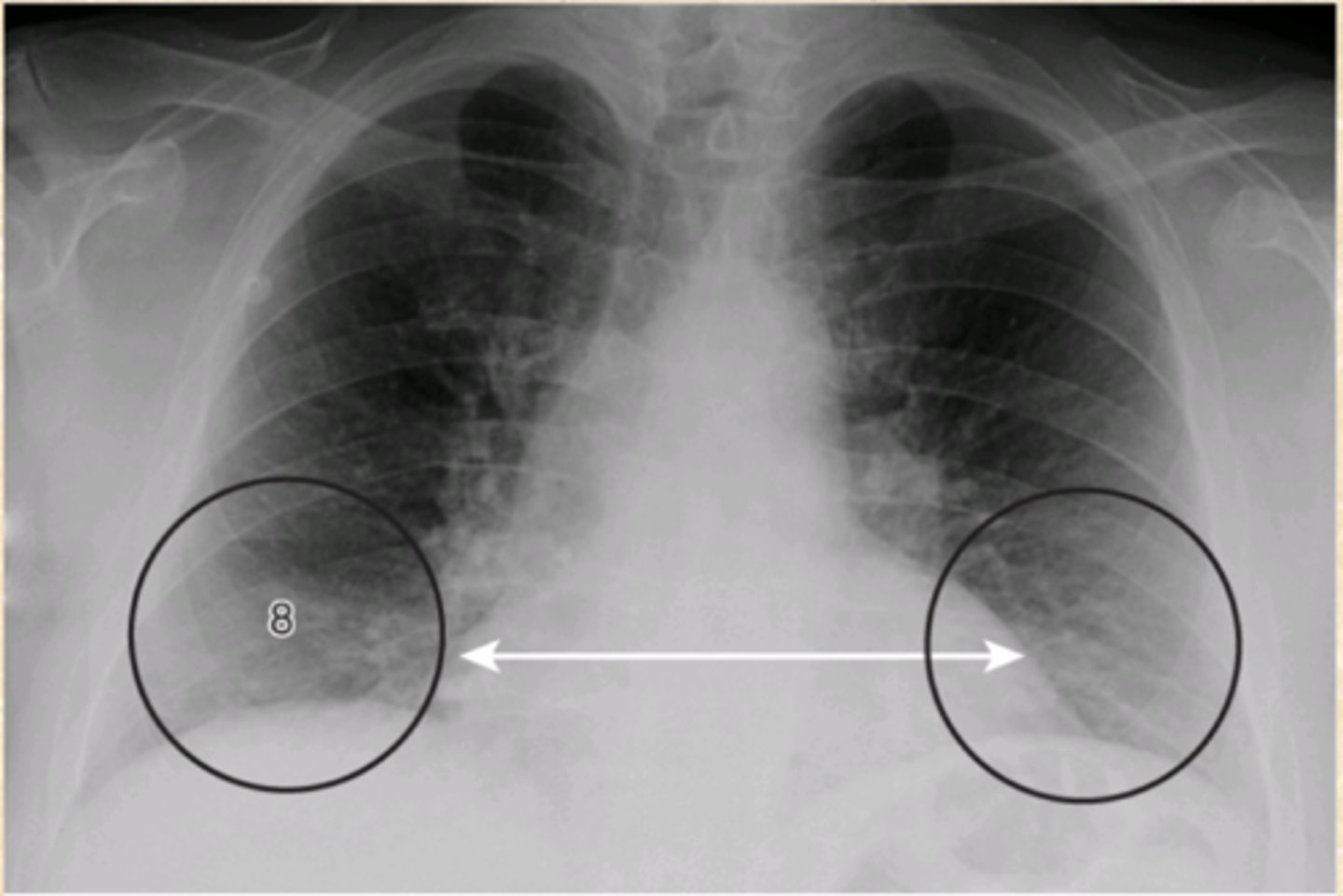
Wide Mediastinum
What does this CXR show?
L - Lymphadenopathy
P - Pneumomediastinum
A - Aortic Dissection
M - Masses
What are the main causes of Wide Mediastinum
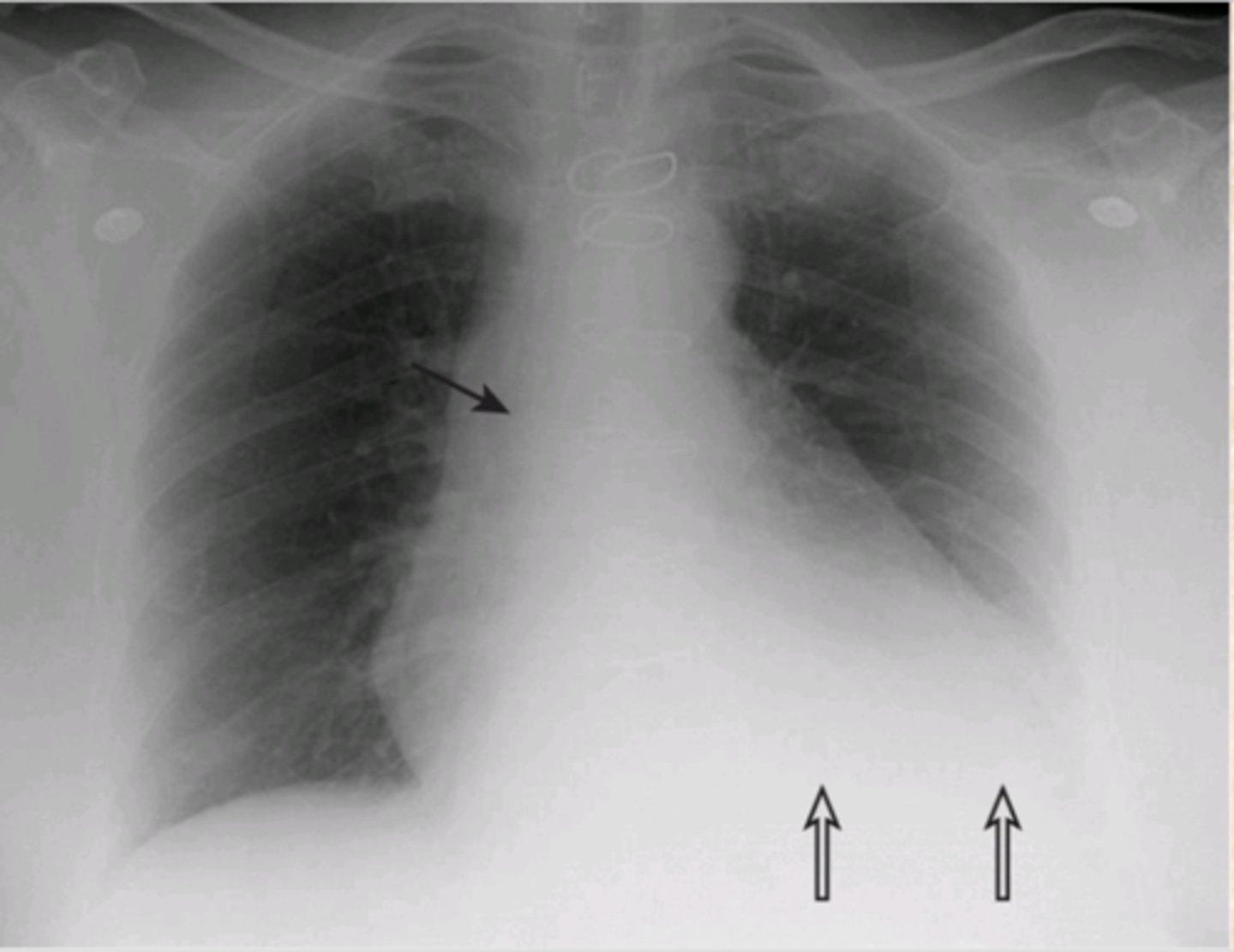
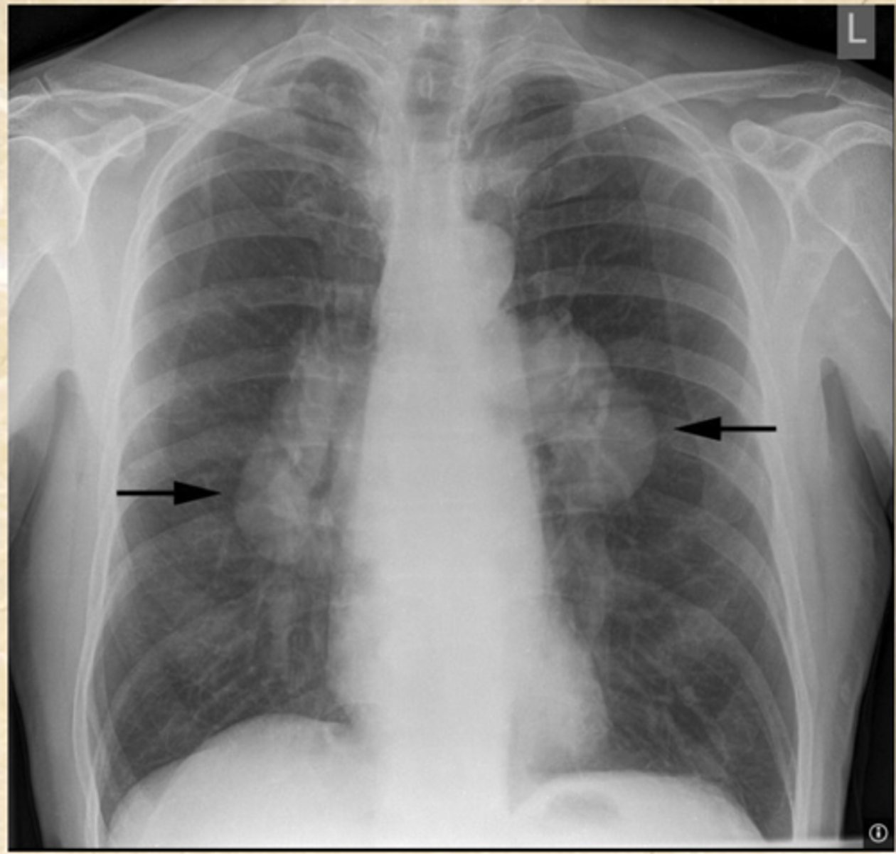
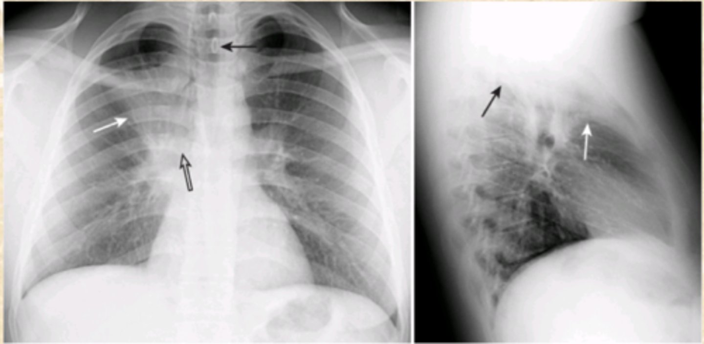
Hilar adenopathy
What does this CXR show?
Scarcoidosis
TQ: If you see bilateral hilar adenopathy; what condition should you think?
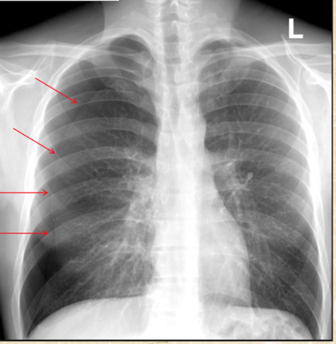
Hilar adenopathy
What does this CXR show?
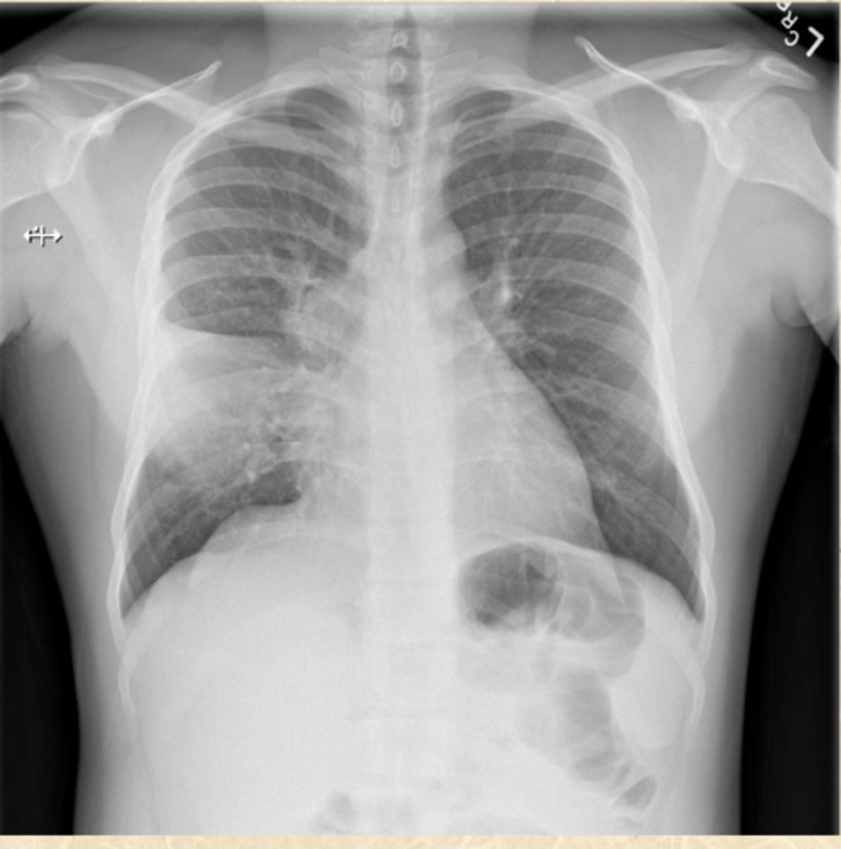
Free air under the diaphragm
What does this CXR show?
Free air under the diaphragm
What does this CXR show?
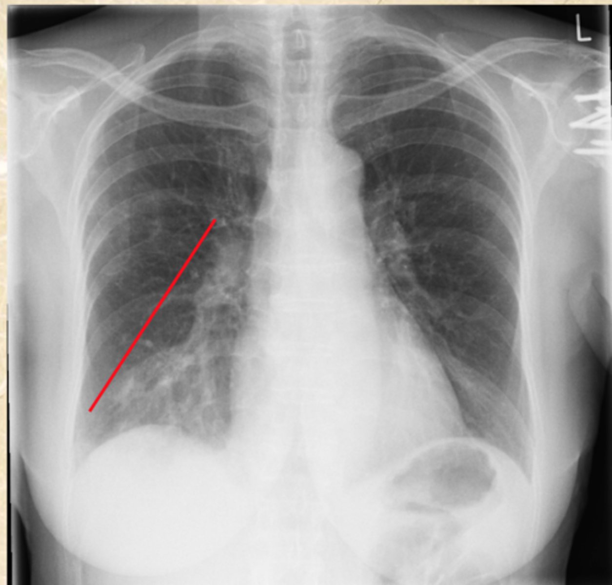
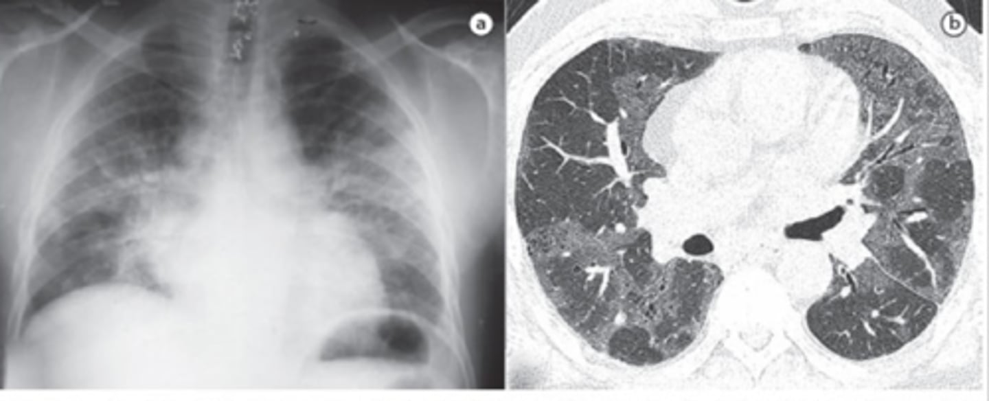
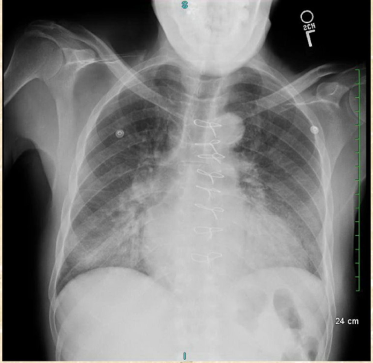
RML consolidation
Gastric bubble
What does this CXR show?
RLL consolidation
What does this CXR show?
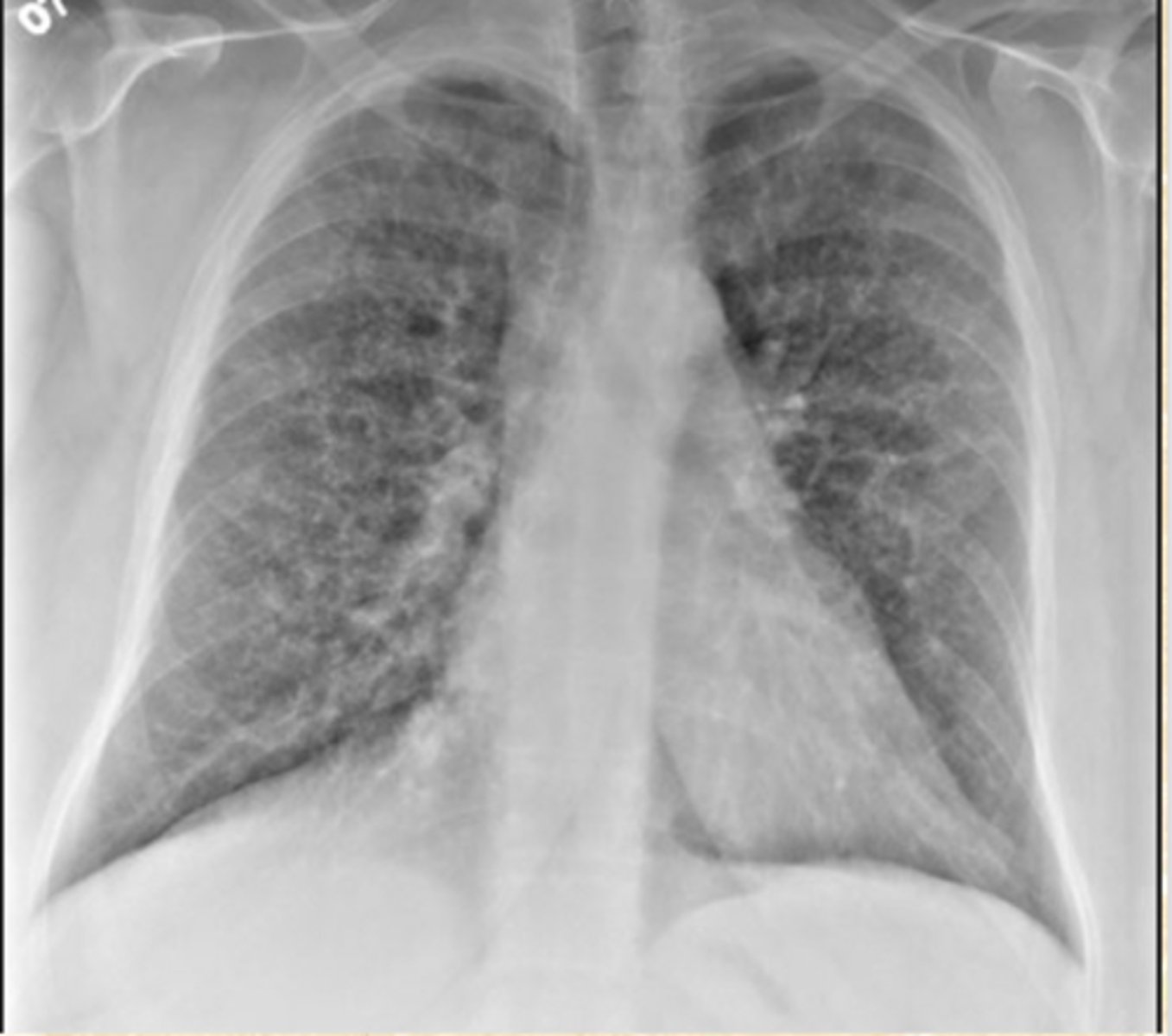
Interstitial Pulmonary Fibrosis
What does this CXR show?
Interstitial Lung Disease.
Reticular
What does this CXR show?
What term describes the pattern?
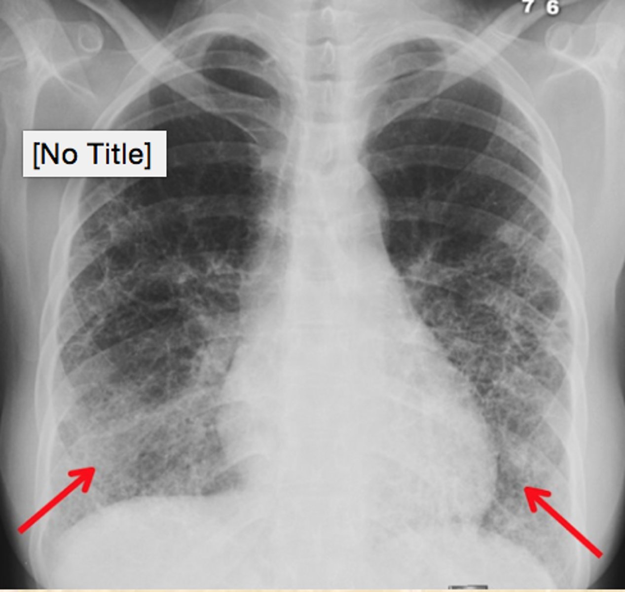
Interstitial Lung Disease
Reticular, Ground glass appearance
What does this CXR show?
What are the key words that describe the pattern seen in this CXR?
Atelectasis
Collapse of alveoli
RML Atelectasis
What does this CXR show?
RUL Atelectasis
What does this CXR show?
Full Atelectasis of the Left Lung
Lack of incentive spirometry post-op
What does this CXR show?
What is common cause of this condition?
LUL Consolidation
Its not atelectasis because you can see the bronchus through the white
What does this CXR show? How do you know?
Diffused Pulmonary Edema in BOTH lungs
What does this CXR show?
Pulmonary edema
Bat Wing Edema
What does this CXR show?
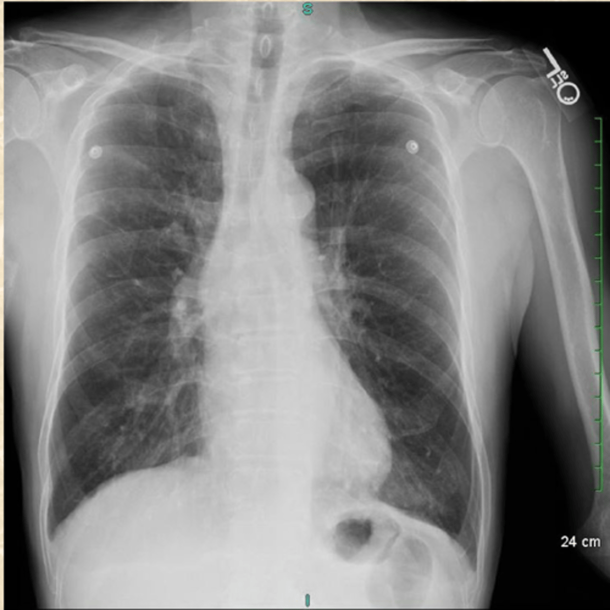
Cardiomegaly
What does this CXR show?
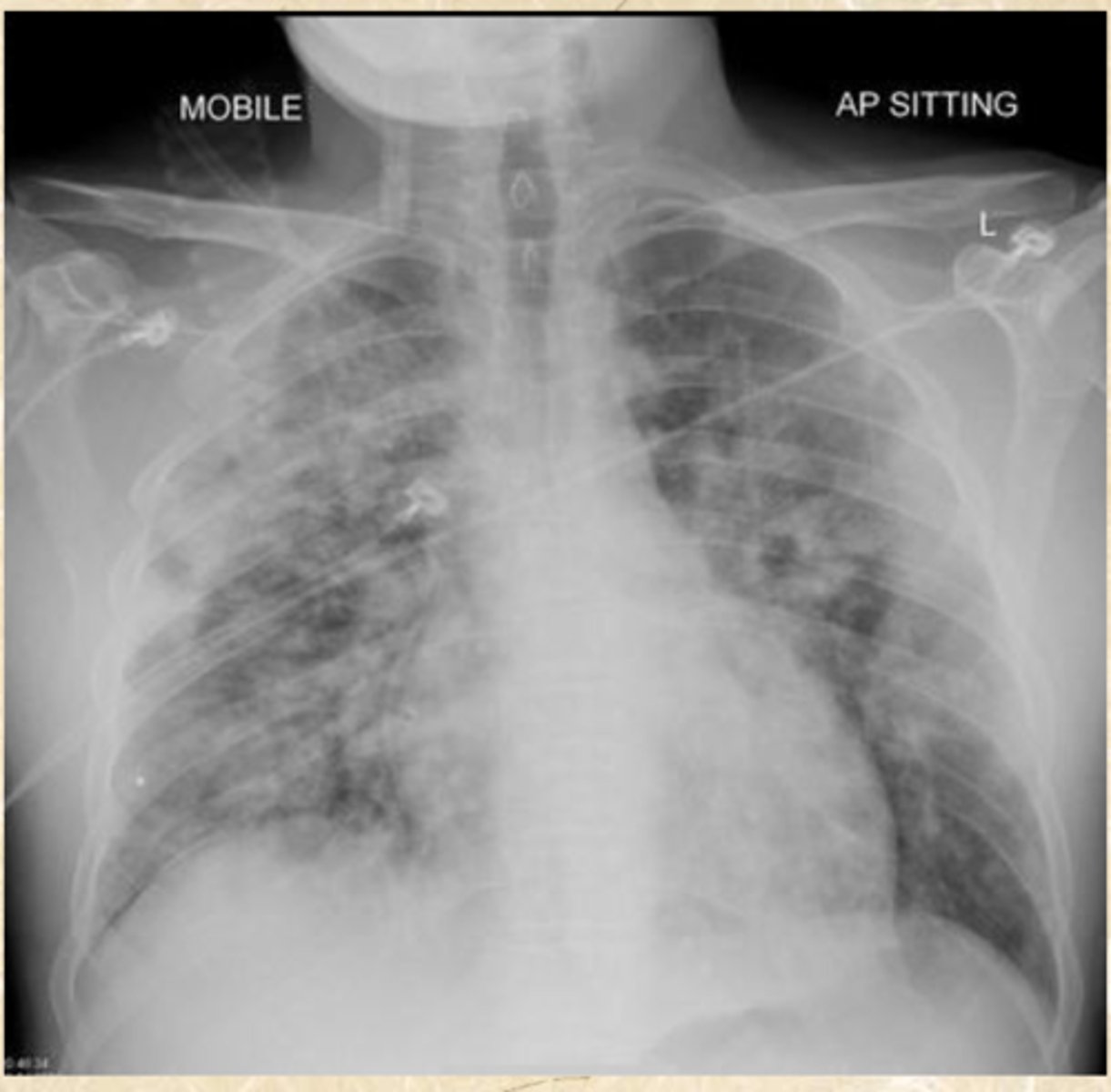
Bilateral Pulmonary Edema and Cardiomyopathy
What does this CXR show (2 things)?
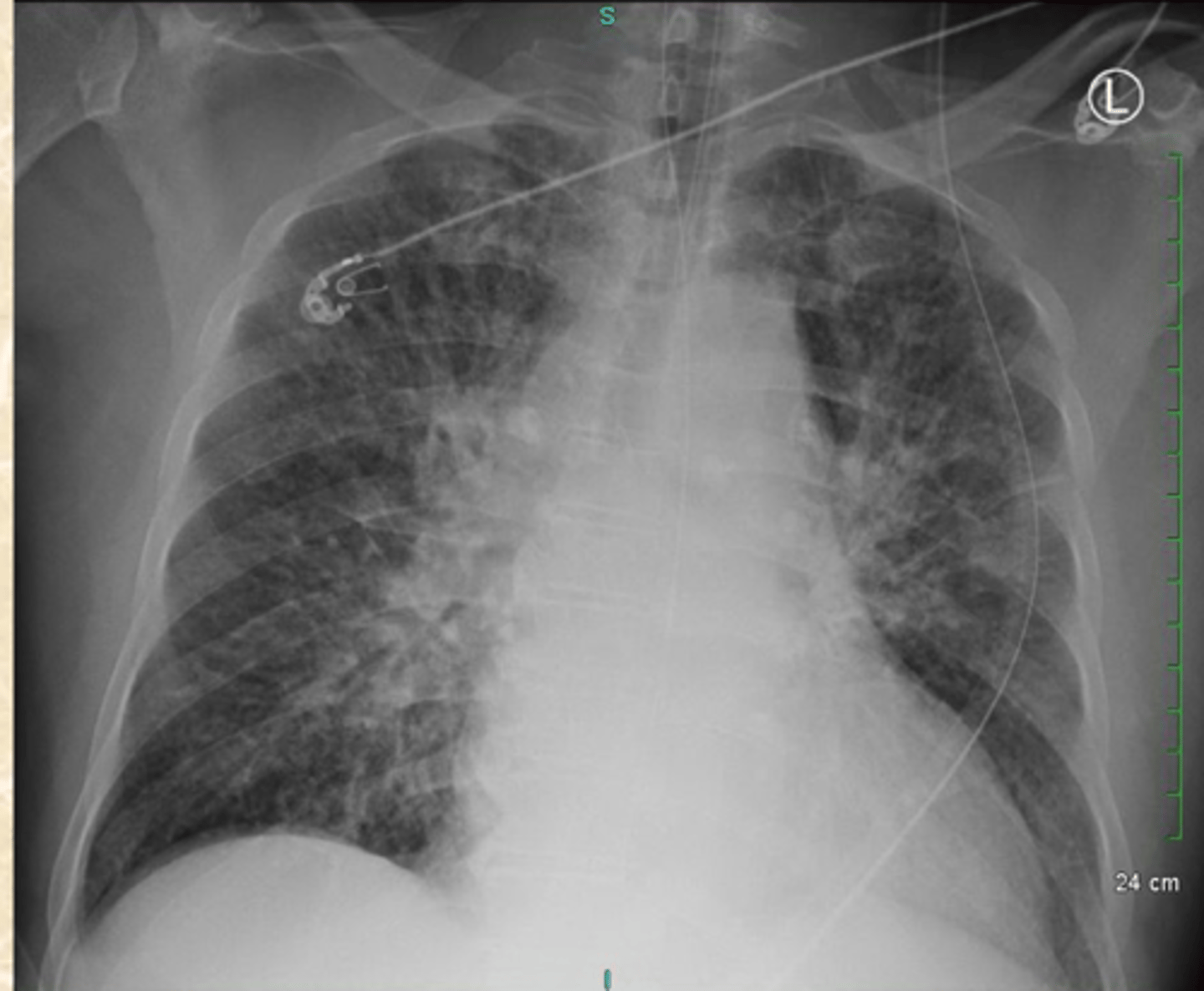
Bilateral Pulmonary Edema
What does this CXR show?
COPD
Large Lung Volume
Small Heart
Flattened Diaphragm
What do you believe this patient is suffering from?
What CXR findings are typical in this patients Dx?
COPD
Small Heart
Large Lung Volume
Flat Diaphragm
What is this Patient's Dx?
How do you know?
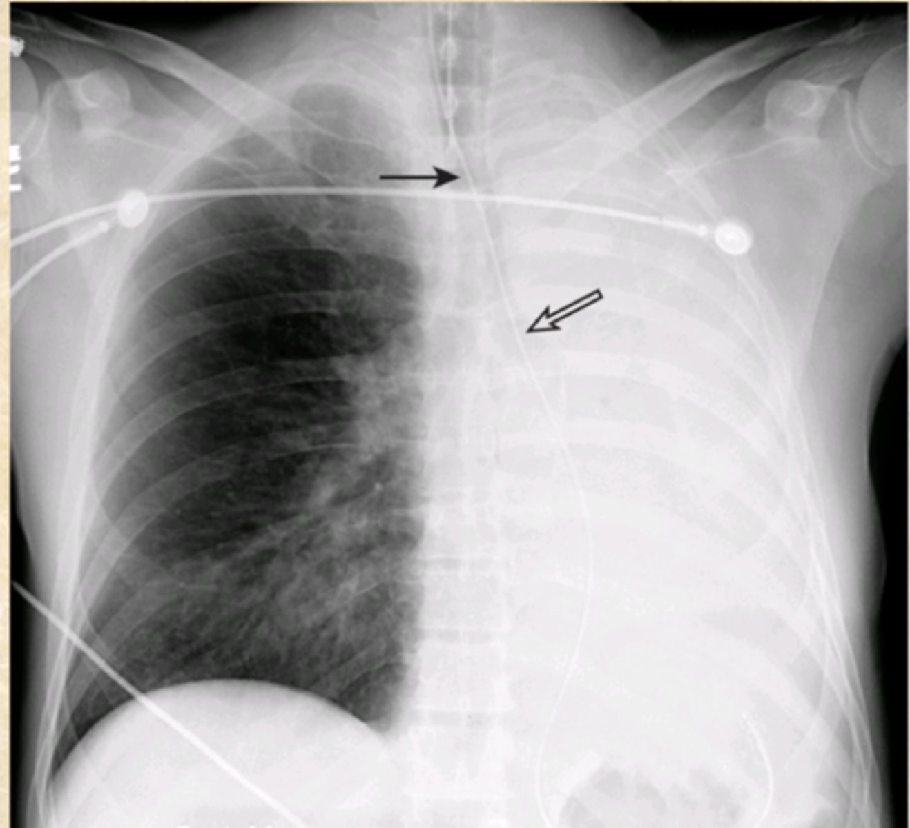
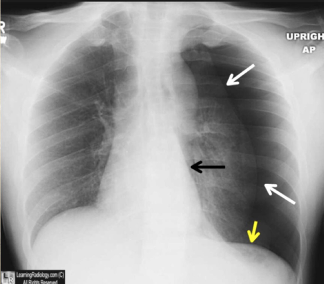
Tension Pneumo
What does this CXR show?

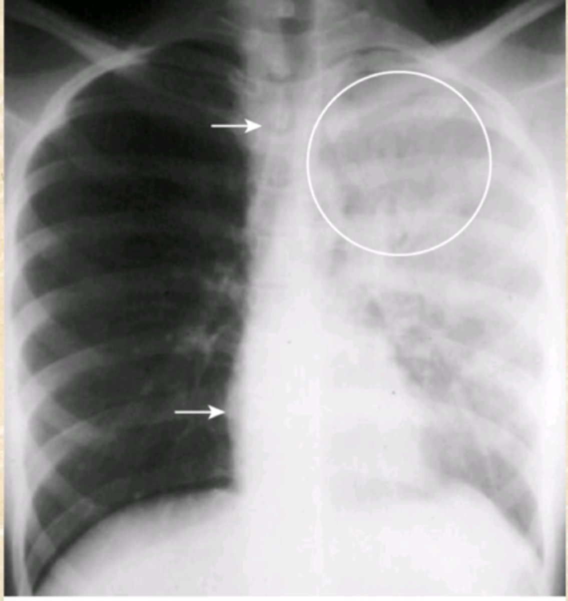
Pneumothorax
What does this CXR show?
Pneumothorax
What does this CXR show?
Tension Pneumo
What does this CXR show?
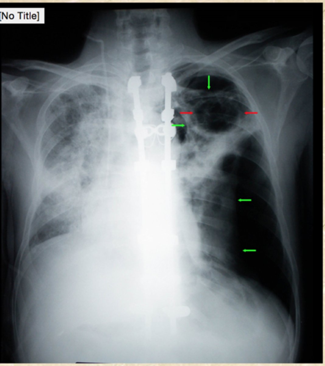
Right lung Pneumothorax
What does this CXR show?
wide mediastinum
What does this CXR show?
retrosternal and retrocardiac
What areas should have more lucency on lateral view?
Airway, Bone, Cardiac
ABC of chest X-ray
Diaphragm, Extra-pulmonary, Field
DEF of chest X-ray
Gastric Bubble, Hilum
GH of chest X-Ray