Anatomy Lab PA: Anterior Abdominal Wall
 Call Kai
 Learn
Learn Practice Test
 Spaced Repetition
 Match
Match Flashcards
 Knowt Play
1/45

There's no tags or description
Looks like no tags are added yet.
Name | Mastery | Learn | Test | Matching | Spaced | Call with Kai | Chat |
|---|
No analytics yet
Send a link to your students to track their progress
46 Terms
superiorficial fascia is composed of two layers: the fatty, outer layer is called:
Camper’s fascia
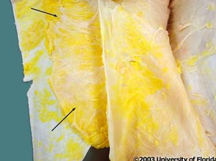
what layer of fascia are the arrows pointing to
Camper’s fascia
superiorficial fascia is composed of two layers: the membranous inner layer is called:
Scarpa’s fascia
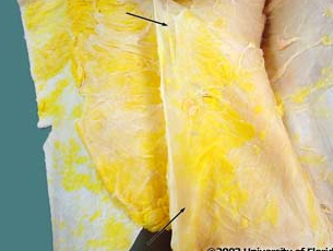
what layer of fascia are the arrows pointing to
Scarpa’s fascia
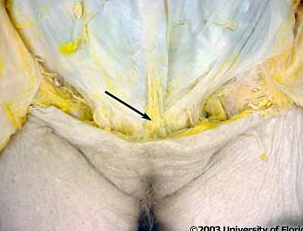
what ligament is shown in the image
fundiform ligament of the penis
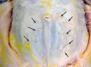
anterior cutaneous branches of what nerve is shown in the image?
thoracoabdominal nerve
_________ nerve is the functional name used to descive each of the lowest five intercostal nerves becuase the supply portions of both the thoracic and abdominal wall
thoracoabdominal
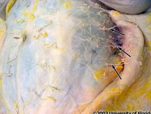
what vessels are indicated in the image; these vessels pierce the external abdominal oblique muscle and overlying deep investing fascia along the midaxillary line and pass anteriorly in the plane of superficial fascia
lateral cutaneous branches of throracoabdominal nerves and vessels
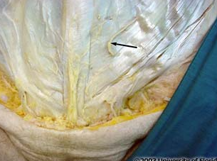
what nerve is being indicated to in the image
iliohypogastric nerve (L1)
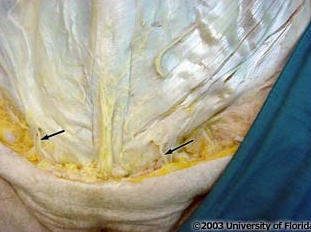
what nerve is being indicated to in the image
ilioinguinal nerve (L1)
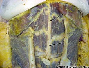
what is the name of the white line that runs vertically from the xiphoid process to the public symphysis called (pictured here)
aponeurotic linea alba
what is the name of the line that represents the midline convergence of aponeuroses of 3 abdominal muscles at the midlne?
aponeurotic linea alba
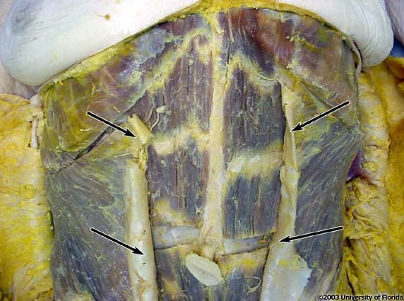
what structure is being indicted to in the image
linea semilunaris
what is the name of the paired and slightly curved vertical lines that represent the lateral border of the rectus sheath which encloses the rectus abdominis muscle on each side?
linea semilunaris
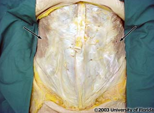
what muscle are the arrows pointing to?
external abdominal oblique muscle
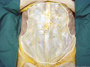
what structure is highlighted in the image
aponeurosis of the external abdominal oblique muscle
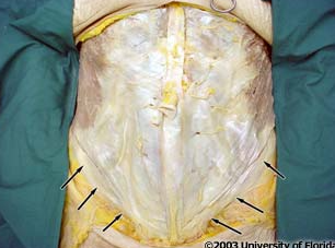
what ligament is being indicated to in the image?
inguinal ligament
what ligament is considered the inferior thickened border of the aponeurosis of the external abdominal oblique muscle?
inguinal ligament
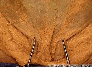
what structure is being indicated to with both probes (one structure on each side)
superficial inguinal ring
what is the name of the opening in the aponeurosis of the external abdominal oblique muscle through which the sermatic cord of the male passes to the scrotum or the round ligament of the female passes to the labia majora?
superfical inguinal ring
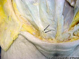
what structure is being indicated to in the image
medial crus
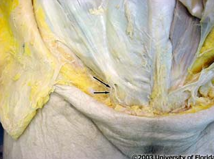
what structure is being indicated to in the image
lateral crus
_____ are thickenings of the external abdominal oblique aponeurosis around the superfical inguinal ring
crura
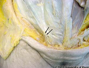
what is the name of the structure that is located superior and lateral to the superfical inguinal ring (shown in image)
intercrural fibers
what fibers reinforce the anterior abdominal wall in the area around the superficial inguinal ring and help prevent the crura from pulling apart?
intercrural fibers
what innervates the rectus abdominus muscle?
thoracoabdominal nerves
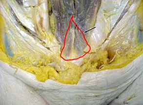
what msucle is highlighted in the image; it lies ANTERIOR to the lower part of the rectus abdominus muscle
pyramidalis
what two branches supplies the rectus abdominis muscle?
superior and inferior epigastric arteries
what two veins help drain the rectus abdominis muscle?
superior and inferior epigastric veins
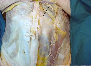
what artery is being indicated to in the image
superior epigastric
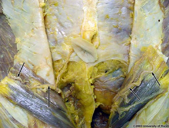
what artery is being indicated to in the image
inferior epigastric
the _____ ____ artery is a branch of the external iliac artery
inferior epigastric
the _____ ____ artery is a terminal branch of the internal thoracic artery
what is the slightly curved, inferior margin of the thickened posterior portion of the rectus sheath called?
arcuate line
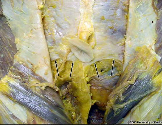
what boundary/margin is indicated in the image (context: on posterior surface of rectus sheath)
arcuate line
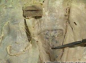
what is the name of the fascia that is inferior to the arcuate line (as pictured here)?
transversalis fascia
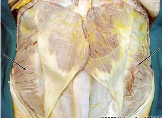
what muscle is being indicated to in the image
internal abdominal oblique
the fibers of the internal abdominal oblique run at ____ angles to those of the external oblique muscle
right
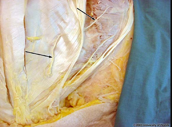
what nerve is being indicated to in the image; context: this is the deep surface of the external abdominal oblique along with the surface of the internal abdominal oblique muscle
iliohyogastric
what 2 nerves travel a portion of their course in the fascial plane between external abdominal oblique muscle and internal oblique muscle?
iliohypogastric and ilioinguinal
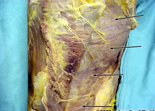
what muscle is being indicated in the image?
transversus abdominis muscle
what kind of muscle fiber orientation does the transversus abdominis muscle has that the external and internal abdominal oblique muscles do not have?
transverse
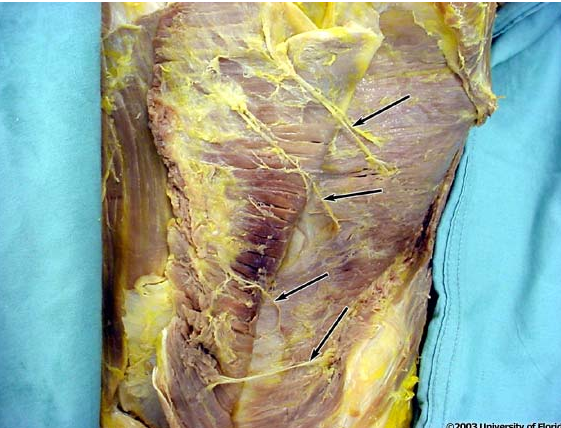
what kind of nerves/vessels are being indicated in the image?
thoracoabdominal nerves and vessels
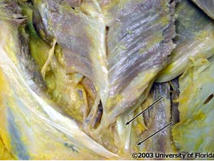
what tendon is being indicated to by the probe in the image; context: transversus abdominis muscle has been reflected
conjoint tendon
what portion of the aponeurosis joins the lowermost fibers of the aponeurosis of the internal abdominal oblique muscle and passes ANTERIOR to the rectus sheath to insert them directly into the pecten of the pubis?
conjoint tendon
the aponeurosis of the transversis abdominis muscle contributes to the _____ layer of the rectus sheath
posterior