lecture 15- accessory digestive organs, topography and blood supply
1/67
Earn XP
Description and Tags
sparks
Name | Mastery | Learn | Test | Matching | Spaced | Call with Kai | Chat |
|---|
No analytics yet
Send a link to your students to track their progress
68 Terms
where is the liver primarily located
to the right of median plane
what is special about the liver
no gallbladder
cranial extent of abdominal cavity
diaphragm
caudal extent of thoracic cavity
line of pleural reflection
basal border of lungs
ribs 6, 11, and 16
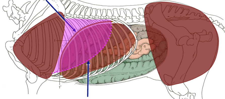
what is the top blue arrow
dome of diaphragm on midline

what is the bottom blue arrow
basal border of the lungs
what is liver cover by
ribs and diaphragm
what is special about the liver locationally
it does not extend to the abdominal floor as in the dogs

what is A
crura of diaphragm
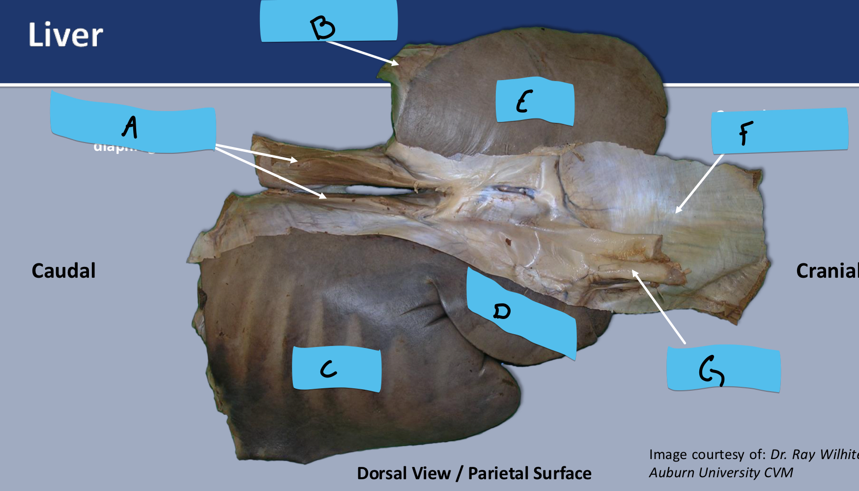
what is B
left triangular ligament of liver

what is C
right lobe

what is D
quadrate lobe

what is E
left lobe

what is F
central tendon of diaphragm

what is G
caudal vena cava
what is the opening into the omental bursa
epiploic foramen
what is the epiploic foramen bounded by
caudal vena cava and caudate lobe of liver dorsally
hepatic portal vein and pancreas ventrally
where can the small intestines become entrapped
epiploic foramen
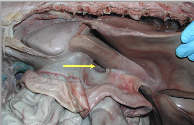
what is this
epiploic foramen
what is the most immobile abdominal organ
pancreas w
where does the dorsal surface of the pancreas attach
to liver and right kidney
what is the ventral attachment of the pancreas
base of cecum and right dorsal colon
where is the body of the pancreas found
extends into the cranial duodenal flexure
where is the right lobe of the pancreas found
contacts descending duodenum
where is the left lobe of the pancreas found
extends toward left kidney, contacts spleen and fundus of stomach
what does the pancreas form
a ring (anuls pancreatis) around the hepatic portal vein
we are performing a liver biopsy where would this be performed
on the right side of the horse between 12th and 14th intercostal space
you decide that you horse client needs a biopsy performed. During this we know that our landmarks are to go in around the 14th intercostal space on the right side. What all structures will our needle pass through
▪ Skin
▪ Intercostal muscles
▪ Costal pleura
▪ Pleural cavity
▪ Diaphragmatic pleura
▪ Diaphragm
▪ Peritoneal cavity
▪ Liver
what is the caudodorsal border of the liver
stomach
topography of left side of liver
6th-11/12 rib
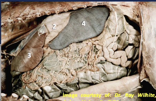
what is 2 3 and 4
2 stomach
3 liver
4 spleen
what part of the spleen might be found in the flank
caudal part
where is the parietal surface of the spleen against
internal aspect of diaphragm
what does the apex of the spleen reach to
10th or 9th rib
where is the gastrosplenic ligament found
greater curvature of stomach to spleen
where is the phrenicosplenic ligament found
attaches base of spleen to the left crus of the diaphragm
where is the renosplenic ligament found
extends from base of spleen to kidney
where is the large colon typically entrapped
in the space created between renosplenic ligament and abdominal wall
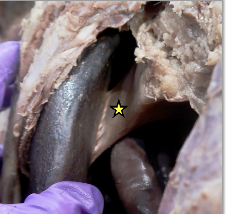
what is the star
space between renosplenic ligament and abdominal wall
supplies cranial abdominal organs
celiac artery
supplies distal portion of descending duodenum through proximal part of descending colon
cranial mesenteric artery
supplies distal part of descending colon and proximal (oral) part of rectum)
caudal mesenteric artery

what is A
celiac artery
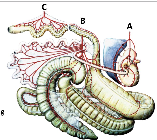
what is B
cranial mesenteric artery

what is C
caudal mesenteric artery
supplies lesser curvature of stomach and distal esophagus
left gastric a.
supplies liver, stomach, duodenum and pancreas
hepatic a.
what are the 2 main parts of hepatic a.
right gastric a.
gastroduodenal a.
what are the parts of gastroduodenal a.
cranial pancreaticoduodenal a.
right gastroepiploic a.
what supplies the lesser curvature of stomach
right gastric a.
what supplies the duodenum and pancreas
cranial pancreaticoduodenal a.
what supplies the greater curvature of stomach
right gastroepiploic a.
what supplies the spleen and stomach
splenic a.
what is the branch off splenic a.
left gastroepiploic a.
what supplies the greater curvature of stomach
left gastroepiploic a.
branches of cranial mesenteric a.
▪ Caudal pancreaticoduodenal a.
▪ Jejunal aa.
▪ Ileal a.
▪ Middle colic a.
▪ Right colic a.
▪ Ileocolic a
what supplies the small intestine
▪ Caudal pancreaticoduodenal a.
▪ Jejunal aa.
▪ Ileal a.
what arise from common trunk and supply colon
middle colic a.
right colic a.
caudal mesenteric a. branches
left colic a.
cranial rectal a.

what is A
right colic a.

what is B
colic branch of ileocolic a.

what is C
lateral and medial cecal arteries

what is D
ileocolic a.

what is E
common trunk

what is G
transition at pelvic flexure
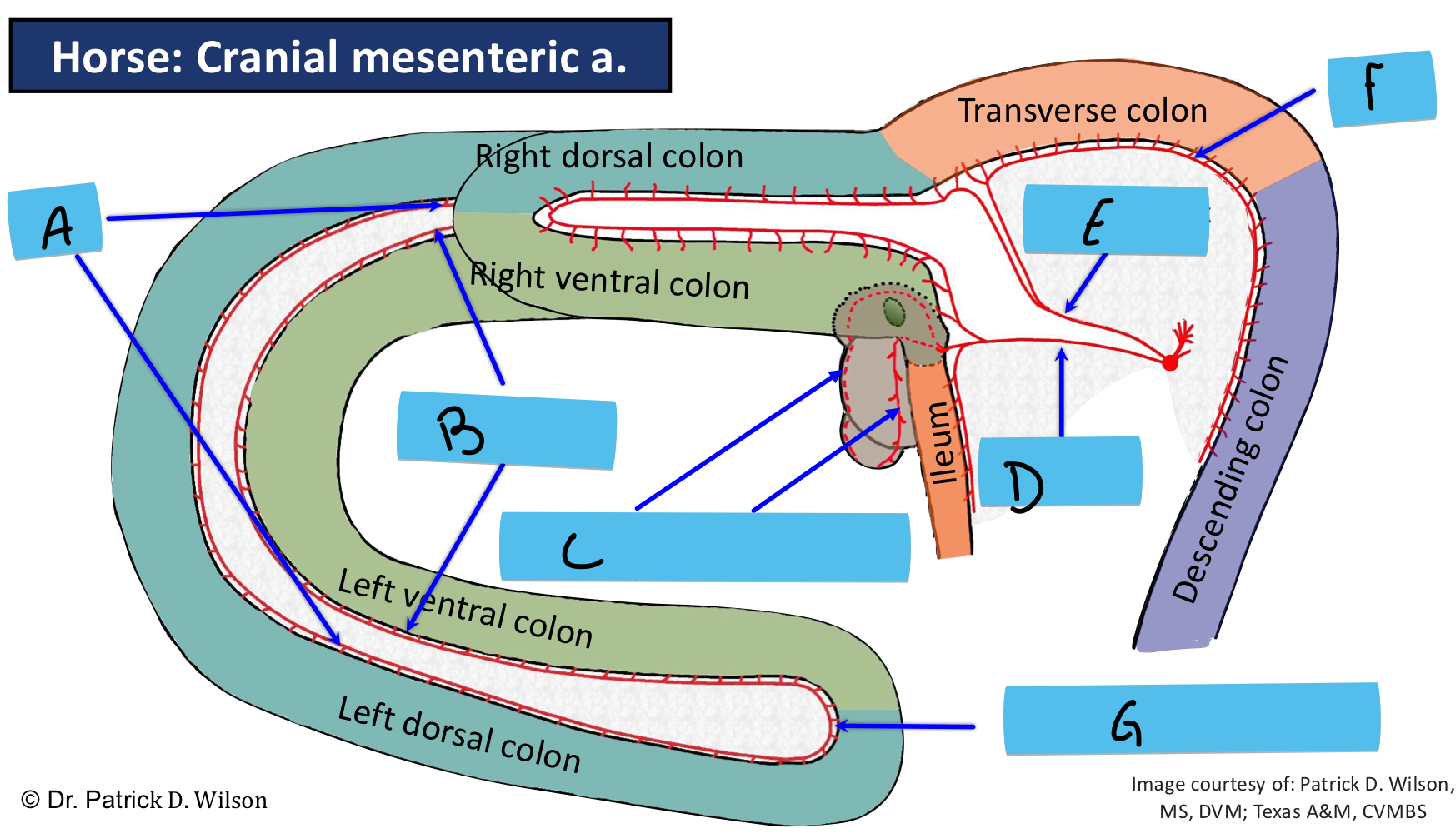
what is F
middle colic a.
what should you use for landmarks for rectal palpation
root of mesentery at the level fo the first lumbar vertebra