Locomotor Anatomy 4: Hindlimb Anatomy Demo
1/37
Earn XP
Description and Tags
Name | Mastery | Learn | Test | Matching | Spaced |
|---|
No study sessions yet.
38 Terms
BONES SECTION
BONES SECTION
IDENTIFY and CLASSIFY these hindlimb bones
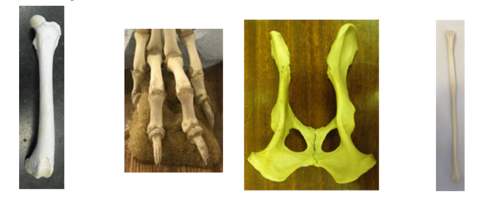
Left to Right:
Femur - long bone
Phalanges - long bones
Pelvis - Flat bone
Fibula - Long bone
IDENTIFY and CLASSIFY these hindlimb bones
Left to Right:
Tarsal bones - short bones
Tibia - long bone
Metatarsals - long bones
Patella and Fabellae - sesamoid bones
Note the following features using the picture:
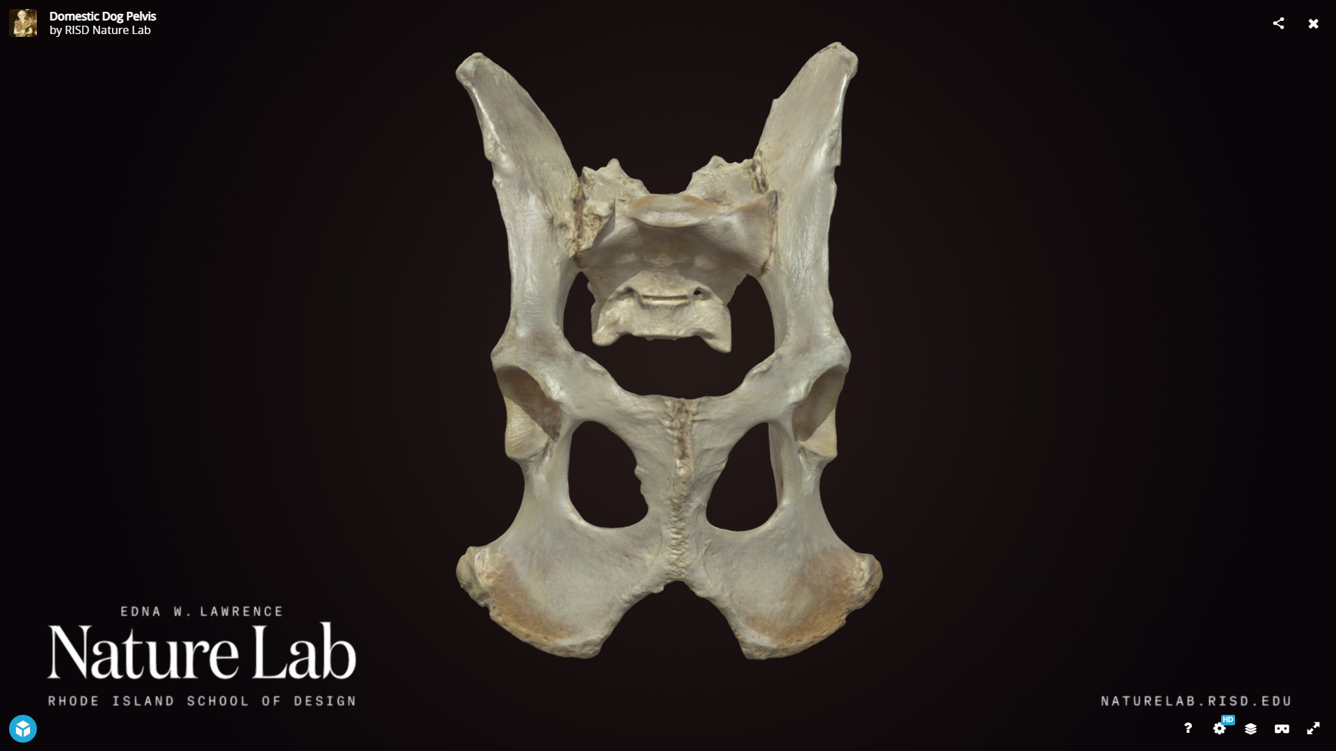
- Wing of ilium
- Body of ilium
- Dorsal iliac crest/spine
- Pubis
- Ischium
- Tuber ischium
- Obturator foramen
- Acetabulum
- Pelvic symphysis
- Ischiatic arch
*add picture with all of these labelled
Which of these features is palpable?
- Wing of ilium
- Body of ilium
- Dorsal iliac crest/spine
- Pubis
- Ischium
- Tuber ischium
- Obturator foramen
- Acetabulum
- Pelvic symphysis
- Ischiatic arch
Tuber Ischium
Dorsal iliac crest
What passes through the obturator foramen in the live animal?
Obturator Nerve
True or False: The obturator nerve provides supply to abductor muscles of the hindlimb?
False
Which structures originate on the tuber ischium?
A. Caudal thigh muscles
B. Cranial thigh muscles
C. Caudal tibial muscles
D. Cranial tibial muscles
Caudal thigh muscles
Which nerve supplies the caudal thigh muscle?
A. Femoral nerve
B. Obturator nerve
C. Radial nerve
D. Sciatic Nerve
Sciatic Nerve
Note the following features using the picture:
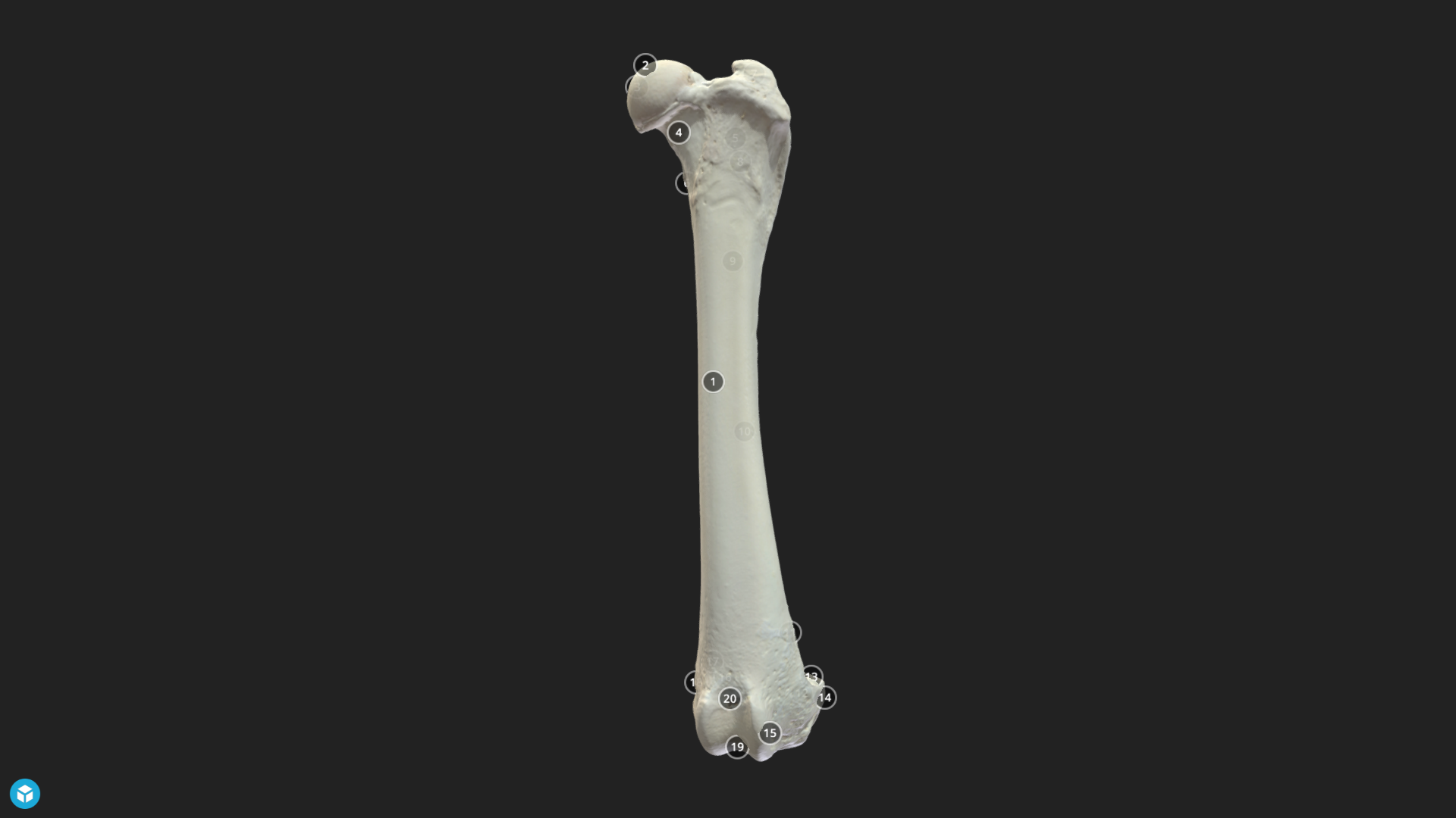
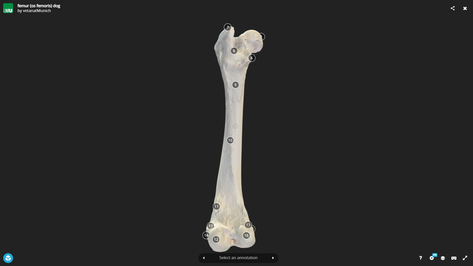
- Head
- Neck
- Greater trochanter
- Body
- Medial trochlear ridge
- Lateral trochlear ridge
- Trochlear groove
- Medial condyle
- Lateral condyle
- Intercondylar fossa
Put picture with labels
Which of these features are palpable?
- Head
- Neck
- Greater trochanter
- Body
- Medial trochlear ridge
- Lateral trochlear ridge
- Trochlear groove
- Medial condyle
- Lateral condyle
- Intercondylar fossa
Greater trochanter
Note the following features using the picture:
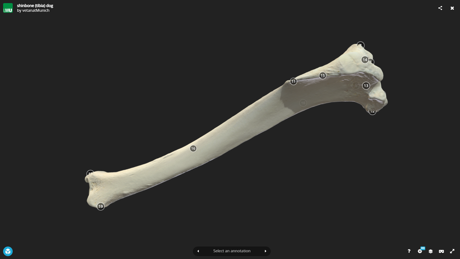
Tibia:
- Plateau
- Medial condyle
- Lateral condyle
- Intercondylar ridge
- Tibial crest
- Tibial tuberosity
- Medial malleolus
put labeled pictured
Which of these features are palpable on the Tibia?
Medial Malleolus
Tibial Tuberosity
Tibial Crest
Note the following features using the picture:
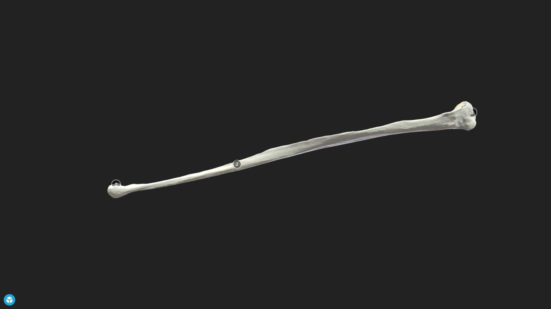
Fibula:
- Head
- Shaft
- Lateral malleolus
put labeled picture
Which of these features are palpable on the Fibula?
- Head
- Shaft
- Lateral malleolus
Lateral Malleolus
What attaches to the tibial tuberosity?
A. Biceps femoris
B. Gluteal muscles
C. Gracillis muscle
D. Quadriceps tendon of insertion
Quadriceps tendon of insertion
What is the function of the tendon of insertion of the quadriceps (aka patellar ligament)?
A. Extension of stifle
B. Flexion of stifle
C. Limb protraction
D. Limb retraction
Extension of stifle
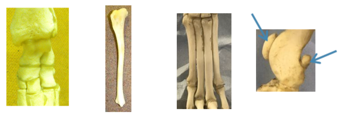
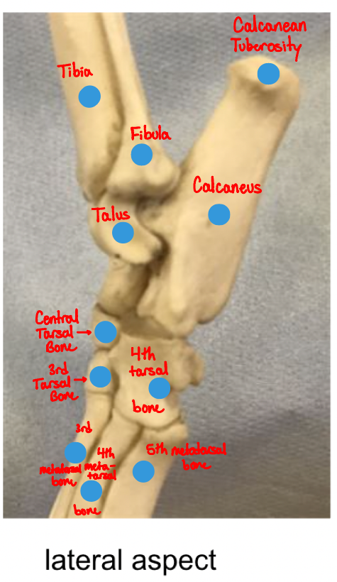
Name all of the hotspots of the bones of the canine tarsal and metatarsal bones.
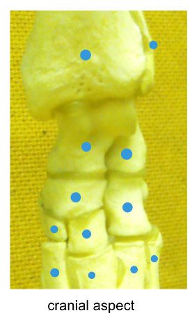
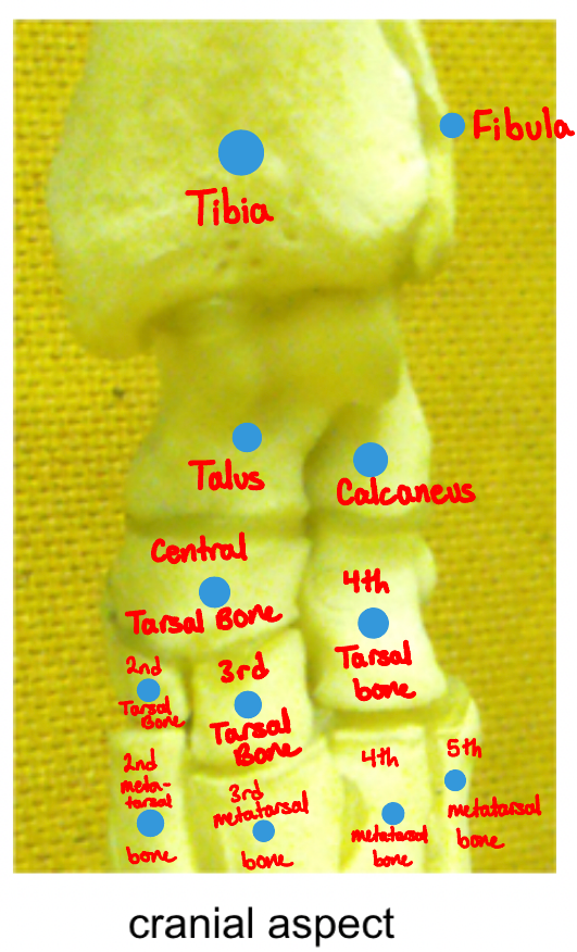
Name all of the hotspots of the bones of the canine tarsal and metatarsal bones.

Which important structure inserts onto the calcanean tuberosity?
Common calcanean tendon (Achilles tendon)
JOINTS SECTION
JOINTS SECTION
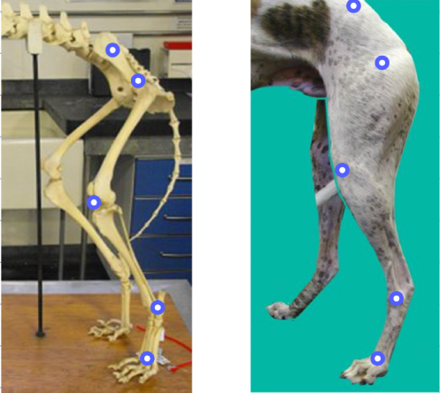
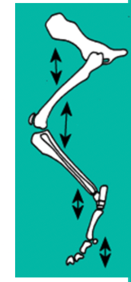
Locate the hindlimb joints on the images

Which aspect of each joint is the flexor angle on?
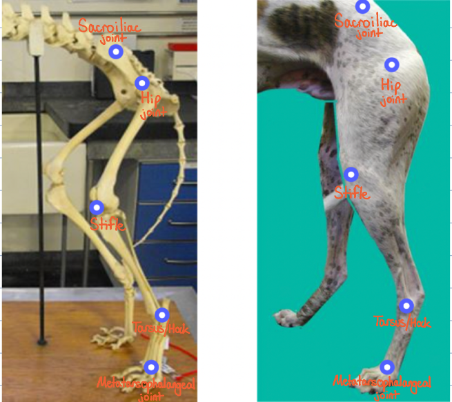
Hip Joint - cranial
Stifle - caudal
Tarsus/Hock - cranial
Metatarsophalangeal joint - caudal
Why does flexion of the hip result in protraction of the limb and extension of the hip result in retraction?
The sacroiliac joint is fixed therefore the hip is the first joint in the hindlimb where movement is possible.
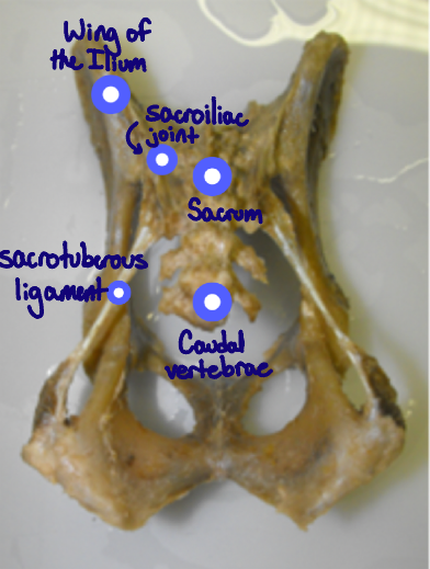
Note the hotspots on the pelvis and sacrum
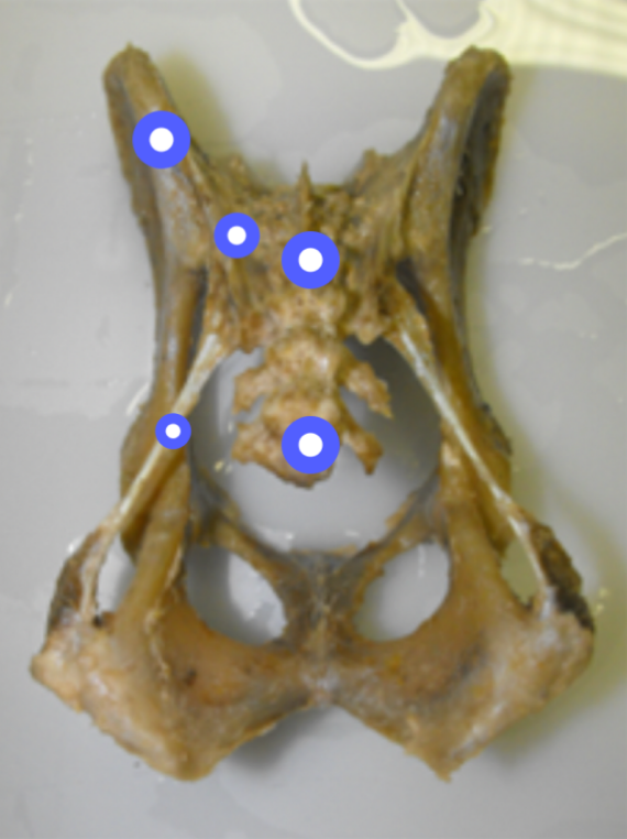
What range of movement is possible at the sacroiliac joint?
None - it is a fixed joint
Which structures does the sacrotuberous ligament run between?
Sacrum (S3) and tuber ischium
What type of joint is the sacroiliac joint?
A. Cartilagenous
B. Fibrous
C. Synovial
Fibrous
How many sacral vertebrae are there in a dog?
3
The hip is a ball and socket joint. Use the hotspots to locate major features of the hip articulation.
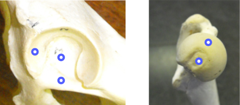
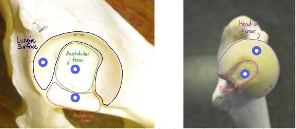
Which movement of these structures articulate together to allow movement at the hip joint?
lunate surface and femoral head
What attaches around the edge of the lunate surface in the live animal?
Labrum (cartilage lip extending around the acetabulum to better enclose the head of the femur in the acetabulum)
What occupies the acetabular notch in life?
a transverse ligament completes the lunate surface ventrally
What range of movement is possible at the hip joint of the dog?
Flexion
Extension
Abduction
Adduction
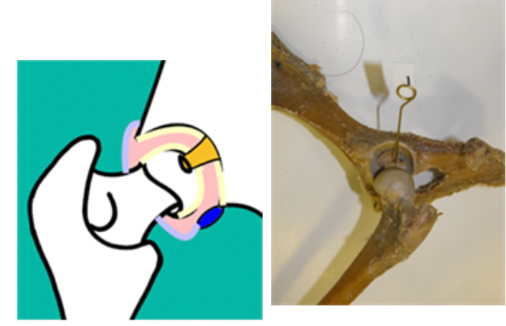
True or False: The hip joint is a synovial joint with no collateral ligaments
True
What provides stability to the hip joint in the absence of collateral ligaments?
large surrounding muscle mass
What other structure helps retain the femoral head in the acetabulum? What structures does this attach to?
Ligament of the head of the femur - runs from the acetabular fossa to the fovea of the head of the femur
This image indicates the boney landmarks that we can use to determine if a hip joint is dislocated
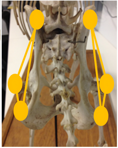
If normal, the greater trochanter, dorsal iliac crest and tuber ischium should form a symmetrical triangle on both sides of the pelvis