Lecture 22: Central Pathways
1/23
There's no tags or description
Looks like no tags are added yet.
Name | Mastery | Learn | Test | Matching | Spaced |
|---|
No study sessions yet.
24 Terms
How do retinal ganglion cell axons form the optic nerve + what happens at the optic chiasm?
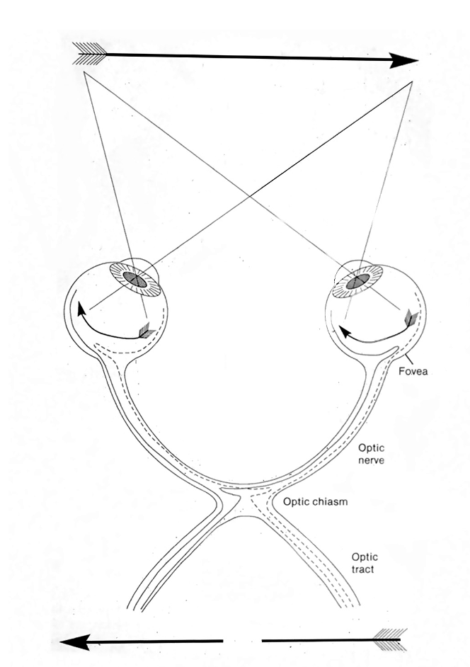
All retinal ganglion cell axons leave the eye as the optic nerve.
At the optic chiasm, axons from the nasal half of each retina cross to the opposite side, while axons from the temporal retina stay ipsilateral (partial decussation).
What is the function of the optic chiasm?
The optic chiasm ensures that information from each visual hemifield is sent to the contralateral hemisphere.
Right half of VF is seen in left hemisphere (vice versa).
What do the vertical and horizontal meridians do in the retina + why does the vertical one matter more?
Vertical meridian = splits the retina into nasal + temporal halves, which is crucial because nasal retinal axons cross at the optic chiasm, determining which hemisphere processes each visual hemifield.
Horizontal meridian = splits the retina into upper and lower halves, but this has relatively little impact on axon routing to the brain.
What are the major targets of optic tract axons?
Superior colliculus + lateral geniculate nucleus
How is the visual field represented in the brain when an object is straight ahead of you?
An object straight ahead is seen by both eyes.
Information from the right visual field falls on the nasal retina of the right eye and temporal retina of the left eye.
Nasal retinal axons cross at the optic chiasm, so the right visual field is processed in the left hemisphere.
How is eye input organized in the LGN?
The LGN has 6 layers and is monocular:
Contralateral eye → layers 1, 4, 6
Ipsilateral eye → layers 2, 3, 5
What are the three LGN pathways and what do they carry?
3 parallel pathways:
Magnocellular (layers 1–2): motion, luminance (parasol RGCs)
Parvocellular (layers 3–6): fine detail, red–green color (midget RGCs)
Koniocellular (between layers): blue–yellow color (small bistratified RGCs)
Neurons preserve the receptive fields of the layers donating.
Why do we even have an LGN (why isn’t it just a passive relay)?
The LGN modulates visual signals based on brain state (arousal, sleep) and attention, not just retinal input.
What are the two firing modes of LGN neurons and when do they occur?
Tonic mode: normal, alert state → LGN output closely tracks retinal input
Burst mode: drowsy/unfocused state → salient stimuli trigger bursts to “wake up” cortex
What are the functions of V1?
Begin synthesis by putting together elements of the visual scene into a more complex package.
Send right information to right targets.
What does retinotopy in V1 mean?
Spatial layout of the retina (visual field) is preserved in primary visual cortex (V1).
Where are foveal vs peripheral visual fields represented in V1?
Fovea → most caudal V1 (occipital pole)
Peripheral vision → more rostral V1
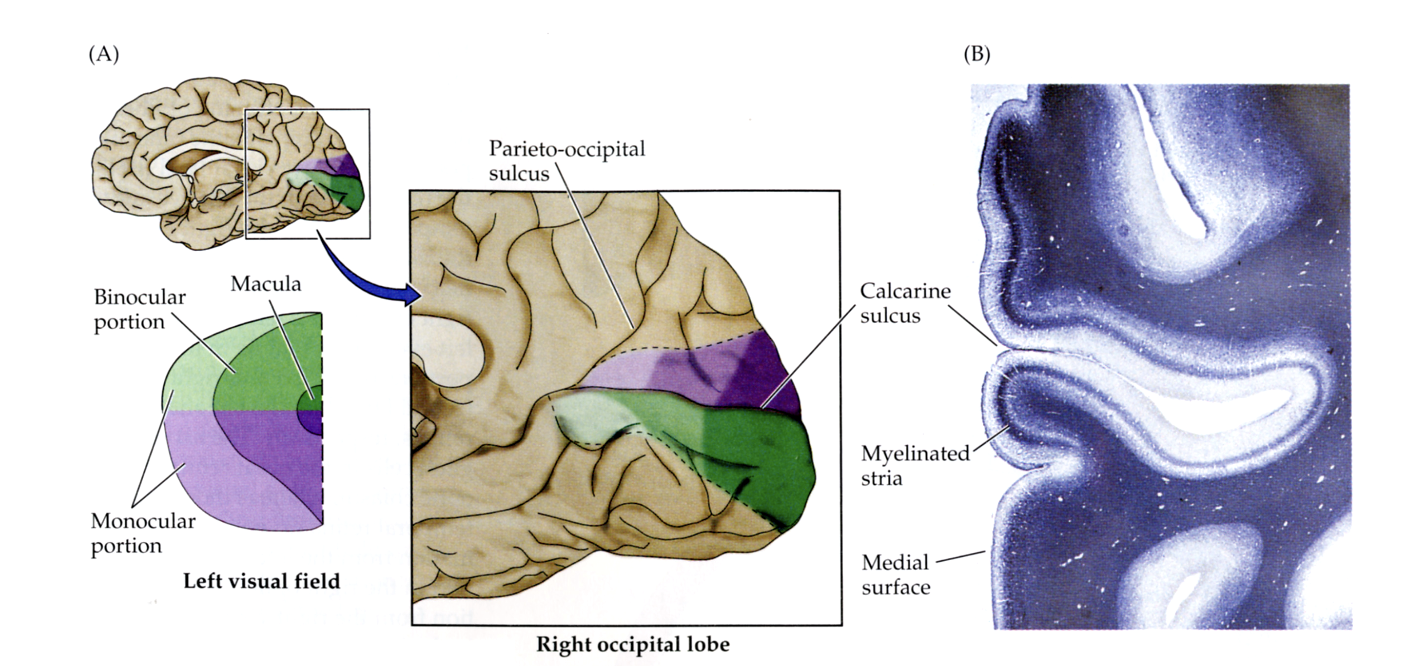
Why does the fovea occupy such a large portion of V1?
Because the fovea has a much higher density of retinal ganglion cells → cortical magnification.
How do simple cells in V1 differ from LGN neurons in what they respond to?
LGN neurons respond best to spots of light with center–surround contrast.
Simple cells in V1 combine LGN inputs to respond to oriented bars/edges.
What is orientation selectivity in V1 simple cells + why is it important?
Simple cells fire strongly only for a specific bar orientation (e.g., vertical but not horizontal).
This is the first step in reconstructing object shape in the visual system.
How do V1 simple cells become selective for the orientation of a bar?
By receiving convergent input from multiple LGN neurons whose center–surround receptive fields are spatially aligned.
When a bar of the correct orientation activates these LGN inputs together, their summed excitation drives the simple cell.
How do complex cells in V1 differ from simple cells in what they respond to?
Complex cells are tuned to a specific orientation + a specific direction (upward/downward) of motion of a bar.
Display greater specificity for a visual stimulus than simple cells.
How do receptive fields differ between retinal/LGN neurons and V1 simple cells?
Retinal ganglion cells and LGN neurons have center–surround receptive fields.
V1 simple cells have elongated receptive fields that respond best to oriented bars or edges.
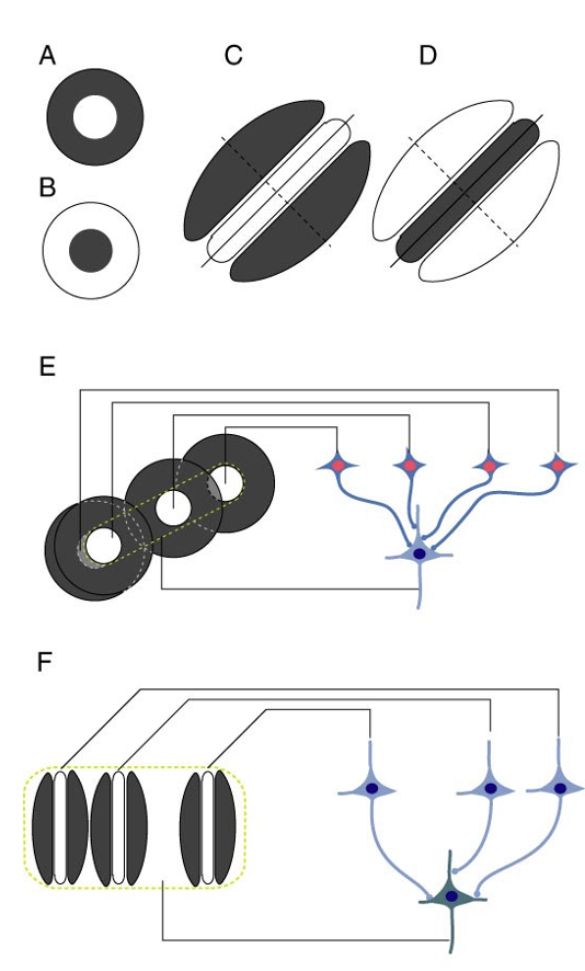
How do complex cells differ from simple cells in V1?
Complex cells arise from the nonlinear convergence of multiple simple cells, respond to oriented edges regardless of exact position, and are often direction-selective for motion.
What is the hypercomplex (end-stopped) property of some V1 neurons?
Hypercomplex cells are orientation-selective but have inhibitory flanks—their response is suppressed when a bar extends beyond a certain length.
This makes them sensitive to line endings, corners, and angles, not just long bars.
What is the non–image-forming visual pathway, and which retinal cells are involved?
It is a pathway where certain retinal ganglion cells do not project to the LGN.
Instead, they project to targets like the SCN (suprachiasmatic nucleus) and OPN (olivary pretectal nucleus) and are involved in circadian rhythms, pupil reflexes, and arousal, not visual perception.
What are intrinsically photosensitive retinal ganglion cells (ipRGCs)?
ipRGCs are a small population of retinal ganglion cells that are directly light-sensitive because they express their own opsin, melanopsin.
They have large dendritic fields, respond best to blue light, and project mainly to non–image-forming targets (e.g., SCN for circadian rhythms, OPN for pupil reflex), though they also receive rod and cone input.
How does light entrain (reset) circadian rhythms in the brain?
Light activates intrinsically photosensitive retinal ganglion cells (ipRGCs), which send signals via the retinohypothalamic tract to the suprachiasmatic nucleus (SCN).
The SCN is the brain’s circadian pacemaker, and daily input from ipRGCs resets the clock to align internal rhythms with the light–dark cycle.
How do ipRGCs mediate the pupillary light reflex?
ipRGCs send light signals to the olivary pretectal nucleus (OPN) in the midbrain.
Axons of OPN neurons send their own axons to parasympathetic neurons on both sides of the brain.
Output of these neurons (by ciliary ganglion neurons) cause pupils to shrink by contracting the iris sphincter muscle.