Session 4: Ossification and Disorders of Bone
 Call Kai
 Learn
Learn Practice Test
 Spaced Repetition
 Match
Match Flashcards
 Knowt Play
1/85

There's no tags or description
Looks like no tags are added yet.
Name | Mastery | Learn | Test | Matching | Spaced | Call with Kai | Chat |
|---|
No analytics yet
Send a link to your students to track their progress
86 Terms
What disorder of bone results from genetic abnormalities in type I collagen synthesis?
Osteogenesis Imperfecta
Name the principle collagen type found in bone?
Collagen type I
From what progenitor are osteoblasts derived?
Osteoprogenitors
What is the osteoid
the organic part of the matrix of bone secreted by osteoblasts
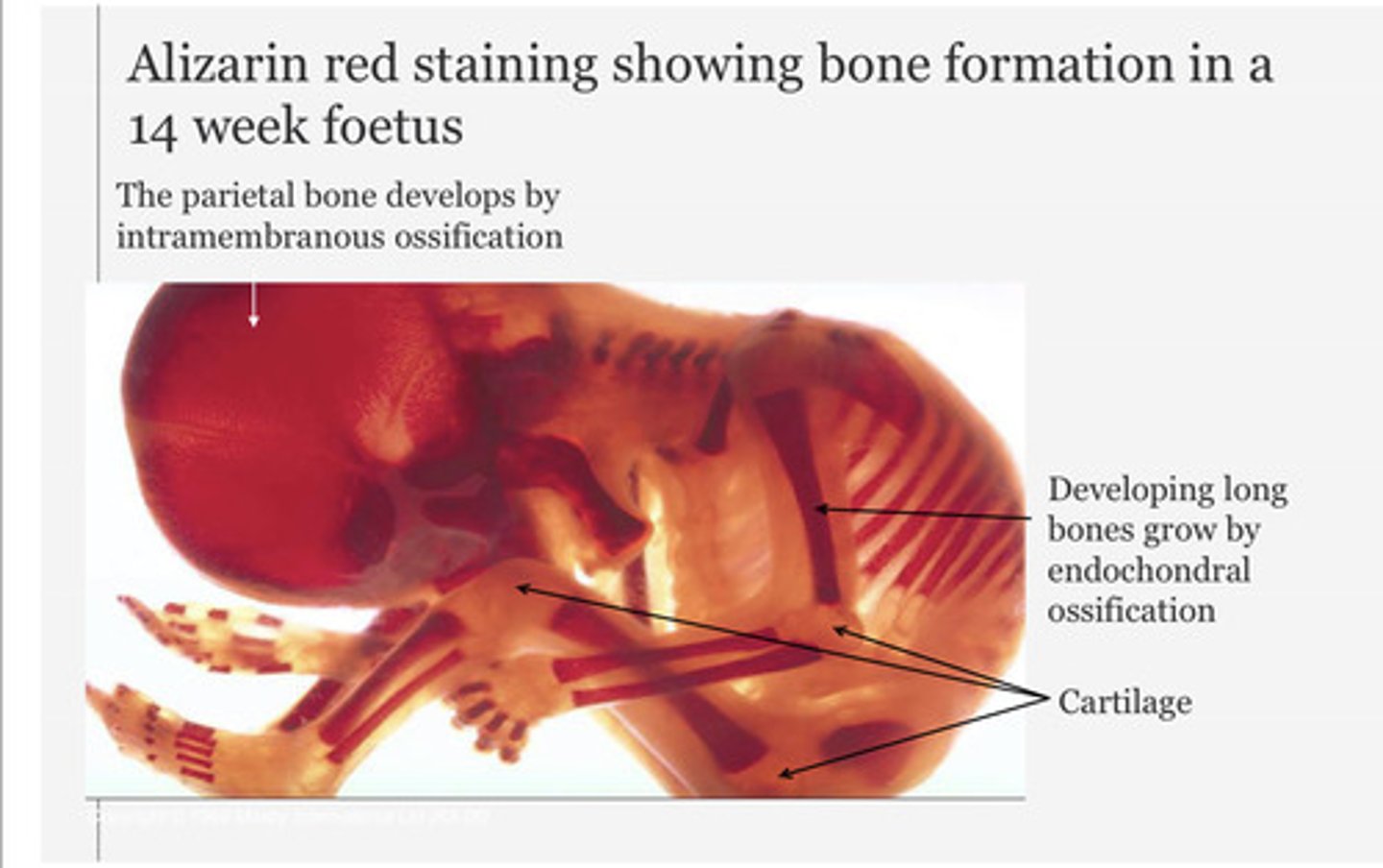
By what mechanism does the parietal bone develop in the foetus?
Intramembranous ossification
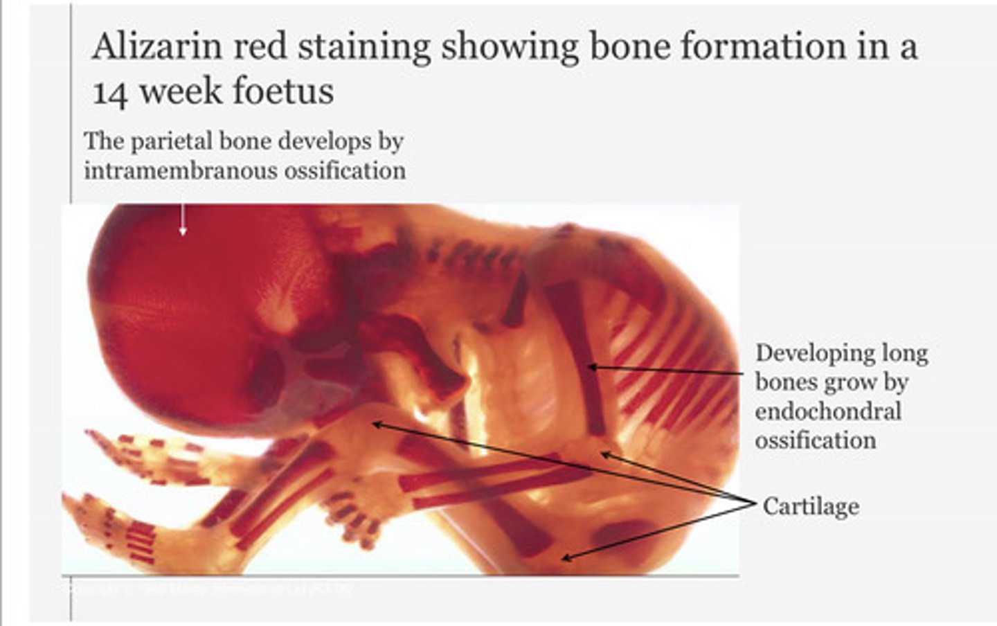
By what mechanism does the long bones develop in the foetus?
Endochondral ossification
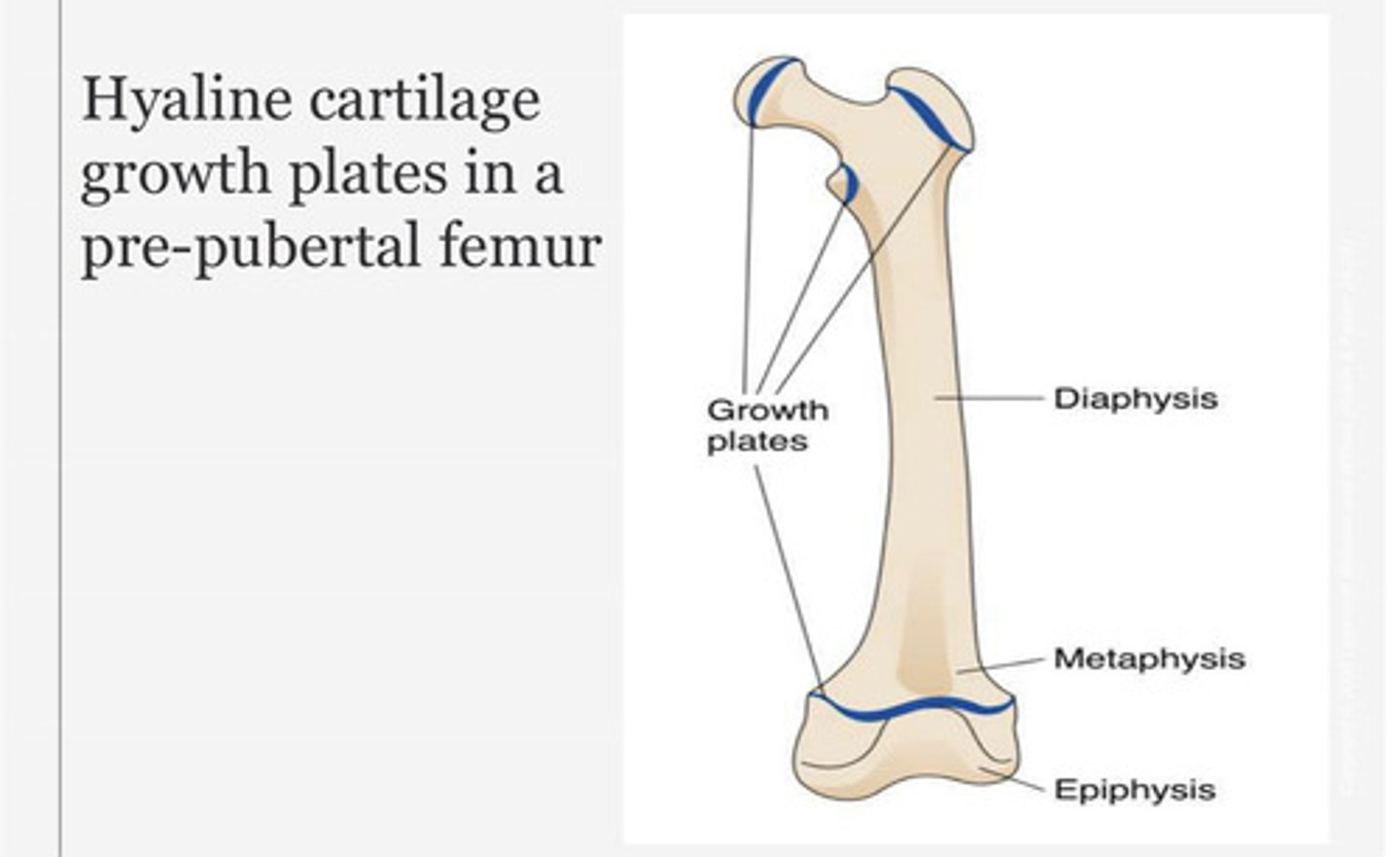
What is the highlighted part of the hyaline cartilage referring to?
Growth plates
Describe endochondral ossification process
1) Fetal hyaline cartilage model develops
2) Periosteal bone collar forms around diaphysis with degeneration of underlying cartilage
3) Invasion of capullaries and osteoprogenitors create primary ossification centre in diaphysis.
Osteoid undergoes calcification to form woven bone.
4) Secondary ossification centres appear in epiphyses around time of birth
5) In childhood - primary and secondary ossification centres are separated by epiphyseal growth plates that allow elongation
6) Growth plates close at end of puberty (no more elongation can occur). But osteoblast reservoir in periosteum allows for thickening (appositional growth)
At what stage in growth do the growth plates close?
At the end of puberty
How do flat bones develop?
Intramembranous ossification
Cranial bones of the skull (e.g., parietal, occipital, temporal and frontal).
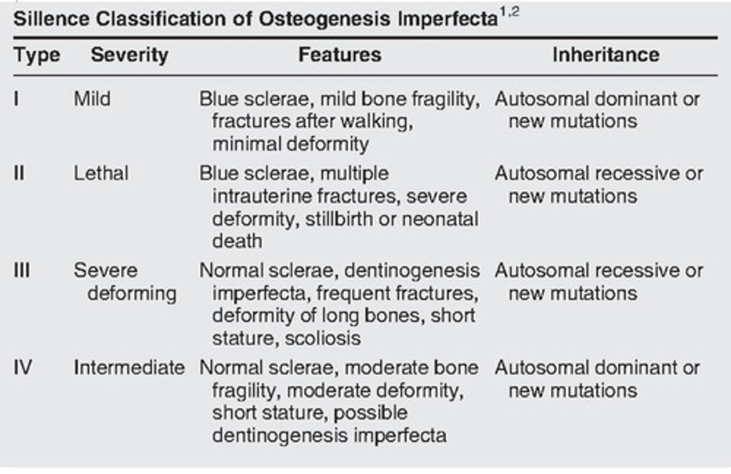
What mutation causes osteogenesis imperfecta (OI)?
Types I-IV arising from mutations in one of the genes encoding type I collagen (COL1A1 or COL1A2).
What structures does osteogenesis imperfecta affect?
Skeleton (tibial fractures; deformity of long bones), joints, ears, ligaments, teeth, sclarae (blue sclerae) and skin
What is the name of the growth hormone (GH) stored in the anterior pituitary?
Somatotropin
What is the role of somatotropin (growth hormone; GH)?
Promote the growth of epiphyseal cartilage
How do you distinguish adult and child x-ray?
Presence of growth plates
How can disorders of the growth plate arise from excessive somatotropin (growth hormone; GH) BEFORE puberty?
Before puberty - excess GH can cause gigantism through promotion of epiphyseal growth plate activity. Insufficient GH can affect epiphyseal cartilage and lead to pituitary dwarfism.
What is acromegaly? When does it occur?
Rare condition resulting from excess secretion of growth hormone (GH).
Acromegaly is the condition that results when there is excess GH AFTER puberty (when the epiphyseal growth plates close).
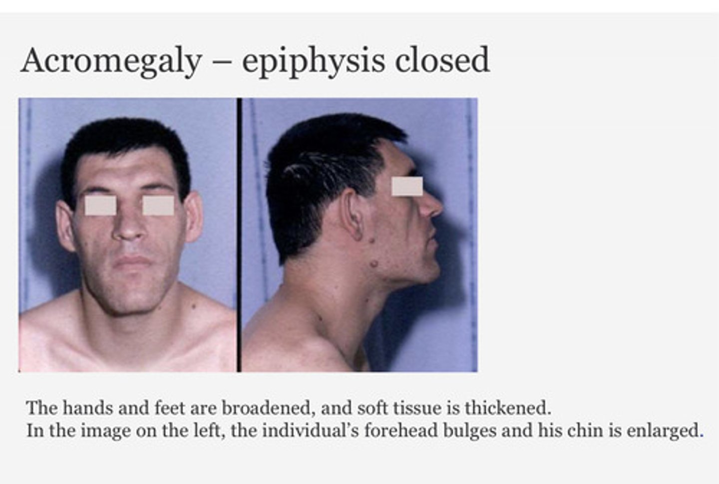
What are the symptoms of acromegaly?
Hands and feet are broadened, soft tissues are thickened.
What does the Fibroblast Growth Factor Receptor 3 (FGFR3) mutation lead to?
The mutation leads to a REDUCTION in:
1) Reduced proliferation of chondrocytes in growth plate cartilage
2) Reduced cartilage matrix production
3) Reduced chondrocyte hypertrophy
Achondroplasia
What are the signs/symptoms of Achondroplasia?
- Normal intelligence
- Large head: frontal bossing
- Small midface
- Proximal long bone shortening
- Short fingers/toes
- Trident hand
- Spinal stenosis
- Lumbar lordosis
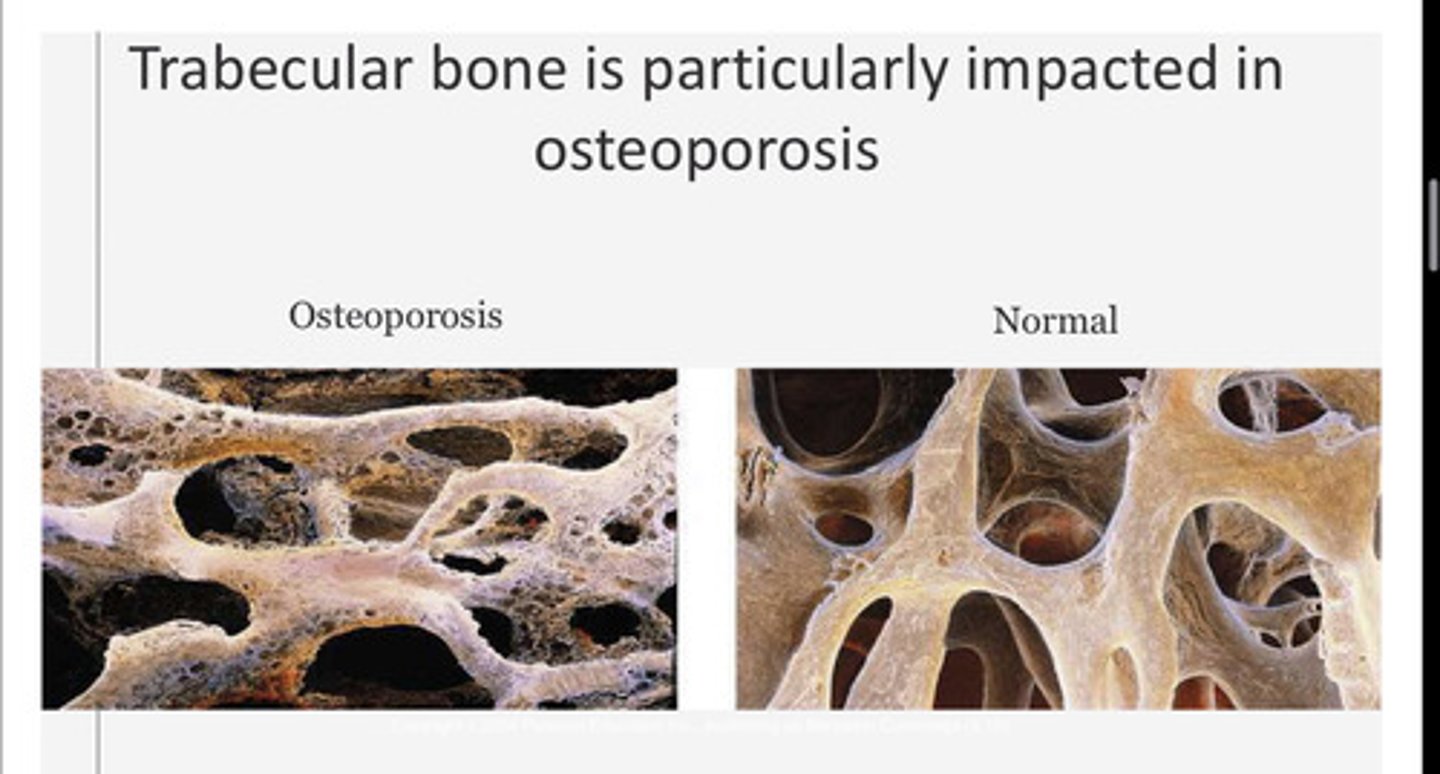
What is osteoporosis?
The loss of cancellous (spongy) bone associated with reduced mechanical strength and increased susceptibility to fractures (common in wrists, hips and spine).
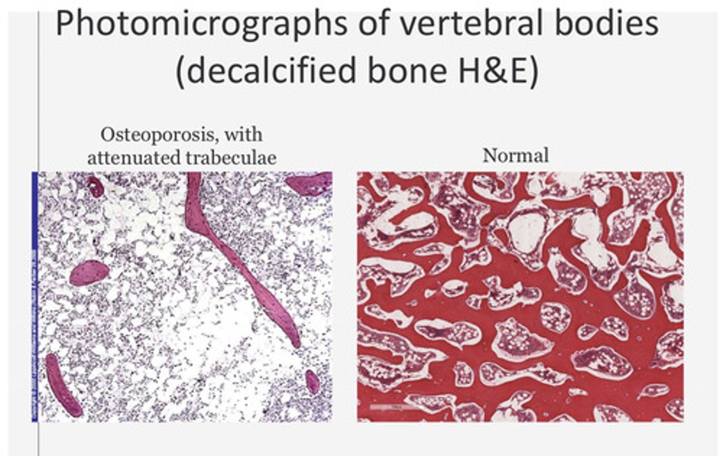
Decalcified bone (H&E) due to osteoporosis
Which sex hormones can influence the growth plate?
Androgens and oestrogens
Give an example of a condition arising from deficient thyroid hormones in neonates resulting in short stature and intellectual impairment
Neonatal hypothyroidism results in intellectual impairment and short stature.
It can be diagnosed with the heel prick screening test
At what age does bone mass peak?
What age does bone mass begin to decline?
Bone mass peaks between age 25-35 years
Bone mass begins to decline from around age of 40
What is the standard pharmacological treatment for osteoporosis?
Bisphosphonates - these adhere to bone and suppress osteoclast activity
What are the two types of primary osteoporosis? When do they occur?
Primary osteoporosis is the most common form of osteoporosis
Type 1
Post-menopausal women = increase in osteoclasts number due to oestrogen withdrawal
Type 2
Both sexes - from age of >70 (senile osteoporosis) = reduced osteoblast function
What are some non-modifiable risk factors for osteoporosis?
1. Genetics: family history, ethnicity (black populations have higher bone density)
2. Age/gender
3. Small frame
4. Previous fractures
5. Other disorders e.g., rheumatoid arthritis, hyperthyroidism
What causes rickets?
Failure of osteoid to calcify in children due to lack of calcium.
What is the effect of rickets?
Long bones become soft and deformed (bowing of legs)
Frontal bone bossing, enlargement of costochondral junctions of the ribs (rachitic or rickety rosary)
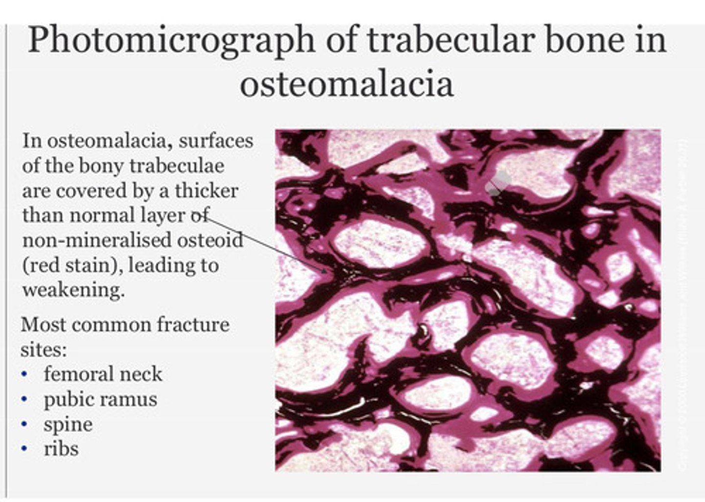
What does the photomicrograph show?
Surfaces of the bone trabeculae covered by thicker than normal layer of non-mineralised osteoid (red stain) leading to weakening.
What is osteomalacia?
Adult counterpart of rickets - caused by significant calcium deficiency or lack of vitamin D (poor diet, lack of sunshine, cultural, liver or kidney disease)
What are common symptoms of osteomalacia?
Bone pain, back ache, muscle weakness, increased risk of fracture
What is the most common form of short-limbed dwarfism
Achondroplasia
What is the name of the autosomal dominant point mutation leading to Achondroplasia?
Fibroblast growth factor receptor-3 gene (FGFR3) impacts endochondral ossification and leads to short-limbed dwarfism.
What is exostoses and when do they form?
Exostoses is an extra growth of bone that extends outward from existing bone.
These can form when there are abnormalities in heparan sulphate (HS) synthesis which affect chondrogenesis signalling pathways.
This results in defects in chondrocyte differentiation - leads to exostoses formation along border of the growth plates
What gene mutation leads to abnormalities in the heparan sulphate (HS) synthesis - which leads to defects in chondrogenesis/chondrocyte differentiation leading to the formation of exostoses along border of growth plates?
Autosomal dominant mutations in the exostosin-1 and exostosin-2 (Ext-1 and Ext-2) genes.
How is vitamin D obtained?
Some vitamin D is dietary (e.g. from fish). Most vitamin D is produced in the skin from 7-dehydrocholesterol by the action of UV light (sun).
Where is vitamin D hydroxylated?
Vitamin D is hydroxylated in the liver and then the kidney to form the ACTIVE 1,25 dihydroxy vitamin D (calcitriol).
What is the role of calcitriol?
Increase calcium absorption in the small bowel
Promote bone mineralisation (promotes bone production)
What are the signs of rickets?
Long bones become soft and deformed, frontal bone bossing, enlargement of the costochondral junctions of the ribs (rachitic or rickety rosary).
What is the pathogenesis of osteomalacia in trabecular bone?
In osteomalacia - surfaces of the bony trabeculae are covered by a thicker than normal layer of non-mineralised osteoid (red stain) leading to weakening.
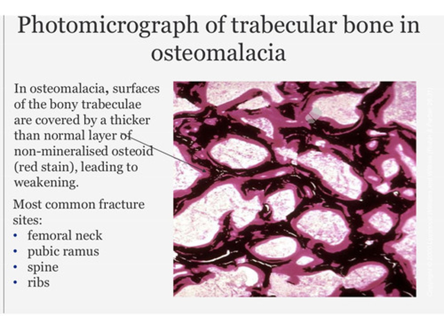
What are the most common fracture sites in osteomalacia?
Femoral neck, pubic ramus, spine and ribs
What disorder of bone is this:
Short-limbed dwarfism resulting from an activating mutation in the FGFR-3 gene leading to reduced chondrocyte activity at the epiphyseal growth plate, and early closure.
Achondroplasia
What disorder of bone is this:
Loss of bone density due to overactive osteoclasts following menopause
Type I osteoporosis
What disorder of bone is this:
Increased stature as a consequence of enhanced activity at epiphyseal growth plates stimulated by an over-active pituitary gland before puberty
Gigantism
What disorder of bone is this:
Brittle bones form due to defects in type I collagen production
Osteogenesis imperfecta types I-IV
What disorder of bone is this:
Diminished stature as a consequence of reduced activity at epiphyseal growth plates resulting from an under-active pituitary gland before puberty
Pituitary dwarfism
What disorder of bone is this:
Reduced mineralisation of osteoid leading to bone pain and muscle weakness in adults with a vitamin D deficiency
Osteomalacia
What disorder of bone is this:
Increase in appositional growth in adults resulting from an over-active pituitary gland
Acromegaly
What disorder of bone is this:
Loss of bone density due to diminished osteoblast function with age
Type II osteoporosis
What disorder of bone is this:
Bone softening in childhood as a consequence of a vitamin D deficiency
Rickets
Intramembranous ossification is the method by which most ___ bones are formed. It takes place within condensations of embryonic ___ cells at ossification centres. These cells differentiate into osteoprogenitor cells, which in turn proliferate to form a layer of ___. These cells secrete the ___ that calcifies to form woven bone. Neighbouring ossification centres fuse and undergo remodelling to form mature bone.
flat, mesenchymal cells, osteoblasts, osteoid that calcifies to form woven bone.
Which is a feature of osteoarthritis (OA), and which is a feature of rheumatoid arthritis (RA)?
Systemic symptoms include fatigue, malaise, and depression
Rheumatoid arthritis (RA)
Which is a feature of osteoarthritis (OA), and which is a feature of rheumatoid arthritis (RA)?
Can run in families
Rheumatoid arthritis (RA)
Which is a feature of osteoarthritis (OA), and which is a feature of rheumatoid arthritis (RA)?
Autoimmune disorder
Rheumatoid arthritis (RA)
Which is a feature of osteoarthritis (OA), and which is a feature of rheumatoid arthritis (RA)?
Degenerative disorder
Osteoarthritis (OA)
Which is a feature of osteoarthritis (OA), and which is a feature of rheumatoid arthritis (RA)?
Most commonly affects joints of the hand and wrist, and small joints of the feet
Rheumatoid arthritis (RA)
Which is a feature of osteoarthritis (OA), and which is a feature of rheumatoid arthritis (RA)?
Increased risk with obesity
Osteoarthritis (OA)
Which is a feature of osteoarthritis (OA), and which is a feature of rheumatoid arthritis (RA)?
Commonly involves knees and hips
Osteoarthritis (OA)
Which is a feature of osteoarthritis (OA), and which is a feature of rheumatoid arthritis (RA)?
Most common type of arthritis
Osteoarthritis (OA)
Endochondral ossification is the method by which most bones, particularly ___ bones are formed.
Endochondral ossification is the method by which most bones, particularly long bones are formed.
Late in the first trimester, a periosteal bone ___ surrounds the cartilaginous template, causing the cartilage to become ischaemic.
bone collar
When do the growth plates close?
Puberty
Case study: fractures/ dislocation.
A 70 year old female presents to the Emergency Department with a wrist fracture following a fall at home.
What type of bone is disproportionately impacted in osteoporosis?
Cancellous (spongy) bone
Case study: fractures/ dislocation.
A 70 year old female presents to the Emergency Department with a wrist fracture following a fall at home.
Apart from the wrist, which two bones/ sites are most commonly fractured in osteoporosis?
Vertebrae
Case study: fractures/ dislocation.
A 70 year old female presents to the Emergency Department with a wrist fracture following a fall at home.
At what age range does bone mass density reach its peak?
25-35 years
Which type of osteoporosis?
Loss of bone density resulting from disease (e.g., multiple myeloma) or long-term steroid usage
Secondary osteoporosis
Which type of osteoporosis?
Affects both men and women, and results from an age-related decrease in osteoblast activity
Primary osteoporosis type 2
Which type of osteoporosis?
Affects post-menopausal women, and results from an increase in osteoclast activity
Primary osteoporosis, type 1
Identify features in the section of ground bone
A = Osteocytes in lacunae
B = Volkmann's (perforating) canal
C = Haversian (central) canal
D = Canaliculi
E = Interstital lamella
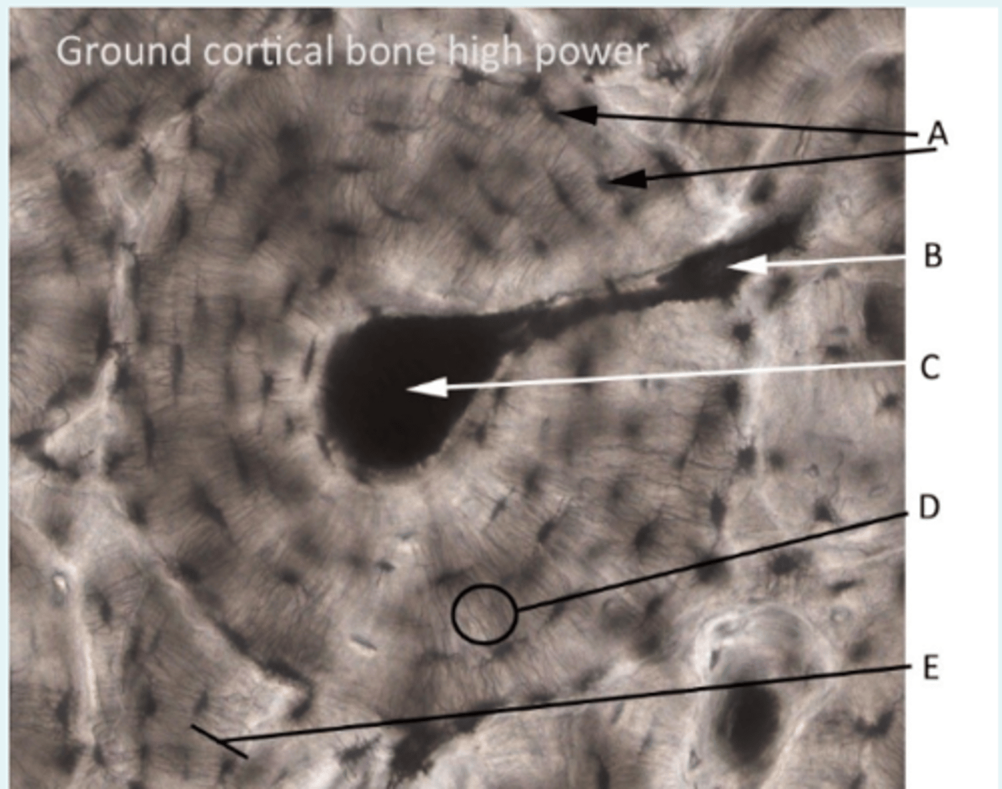
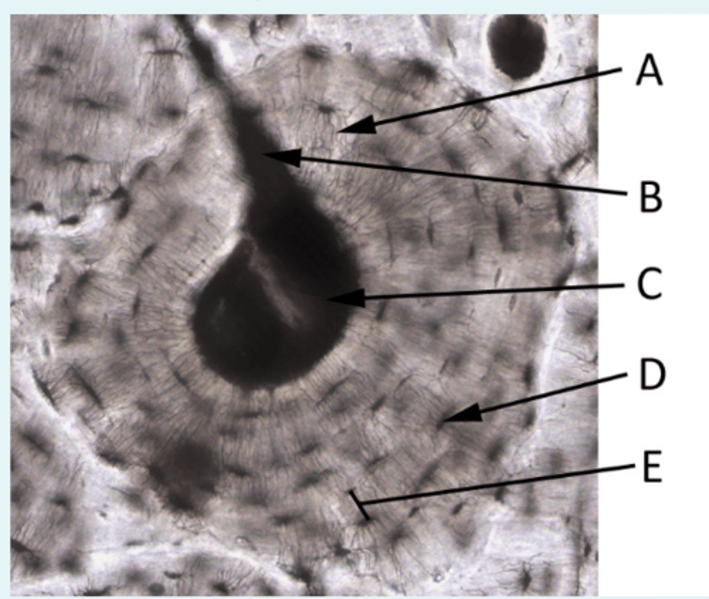
In the following diagram of ground bone, which letters indicate a feature that provides channels for osteocyte communication?
A = Canaliculi are small channels which connect lacunae
Which hormone is primarily responsible for the release of calcium stored in bone?
Parathyroid hormone
How many genes contribute to the development of osteopororis?
Many genes contribute to the phenotype (i.e., it's polygenic)
Bisphosphonates have two phosphate groups that bind ___ ions, allowing the drug to accumulate at bony surfaces. Bisphosphonates are taken up by ___ and reduce bone loss by reducing their action.
Bisphosphonates have two phosphate groups that bind calcium ions, allowing the drug to accumulate at bony surfaces. Bisphosphonates are taken up by osteoclasts and reduce bone loss by reducing their action.
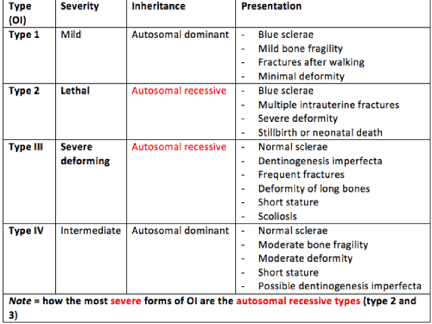
Describe Types I, II, III and IV of Osteogenesis Imperfecta
- Severity
- Inheritance pattern
- Features/Presentation
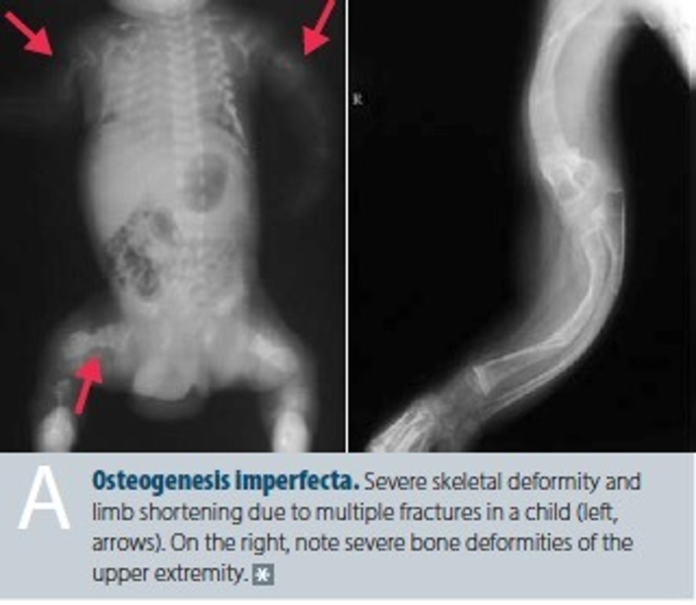
Name the locations of some deformities and fractures/callouses that are typically presented in Osteogenesis Imperfecta (OI)?
Deformities:
- Tibia
- Carpal bone
Fractures and callouses:
- Femurs
Forearms bones are typically shortened, thinned and bowed as well
Achondroplasia is an autosomal dominant disorder, and lethal in the homozygous form. What percentage of offspring will suffer from achondroplasia assuming one affected parent?
There is a 50% chance that the offspring might suffer from achondroplasia (punnett square)
A lack of growth hormone prior to puberty will also impact the growth of epiphyseal cartilage. What disorder results from this?
Pituitary dwarfism
A) Why would an excess of growth hormone post-puberty not lead to gigantism?
B) Why would result instead?
A) Because the growth plates close usually near the end of puberty.
B) Acromegaly would result instead.
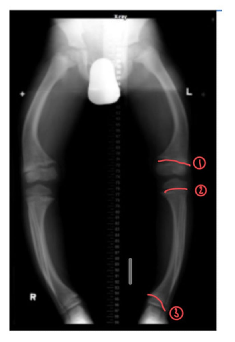
The following x-ray shows a child diagnosed with rickets. Identify the three areas where you can see the epiphyseal growth plate
The femur (patellar surface) and the tibia (proximal and distal head)
In osteoporosis, bone ___ exceeds bone formation, ___ bone mineral density.
Loss of ___ bone is associated with reduced mechanical strength and increased susceptibility to fracture.
In osteoporosis, bone resorption exceeds bone formation, reducing bone mineral density.
Loss of cancellous bone is associated with reduced mechanical strength and increased susceptibility to fracture.
Common sites of fracture in osteopororis other than the hips include the ___ and ___
Wrists and spine
Which one of the following statements is TRUE regarding osteoporosis?
A) Primary osteoporosis is 2nd most common form of osteoporosis
B) Type 1 osteoporosis occurs in postmenopausal women
C) Type 2 osteoporosis is more common in women and reflects increased osteoclast activity
D) Bisphosphonate treatment should not be initiated for people with normal Calcium and Vitamin D levels
B) Type 1 osteoporosis occurs in postmenopausal women
Type 2 osteoporosis (senile) occurs in both women and men, particularly from the age of 70 and reflects reduced ___ function
Osteoblast
Two non-modifiable risk factors for osteoporosis
- Family history
- Increasing age
- Female sex
- Post-menopause
- History of fractures