3.6.1 Stimuli, both internal and external, are detected and lead to a response
1/110
Earn XP
Description and Tags
.
Name | Mastery | Learn | Test | Matching | Spaced | Call with Kai |
|---|
No analytics yet
Send a link to your students to track their progress
111 Terms
How do organisms increase their chance of survival? (1)
By responding to changes in their environment
what is a stimulus
a change in the external environment (i.e. temperature, light intensity and pressure)
What are taxes and kineses? (1)
Simple, innate responses that can maintain a mobile organism in a favourable environment
What is a taxes (tactic response) ? (2)
- Directional movement
- Towards (positive) or away (negative) from a stimulus
What is a common example of taxes? (1)
Woodlice moving away from a light source to avoid predators
What is a kineses (kinetic response) ? (2)
- Non-directional (random) response to a stimulus
- Resulting in organisms changing their speed or rate of direction
- to stay in/move to a favourable environment
What is an example of a non-directional stimulus? (1)
The conditions of an environment
What is a common example of kinesis? (3)
- Woodlice move faster when in drier environment
- To increase chances of moving to an environment with a higher humidity
- To prevent drying out
What is tropism? (1)
the growth response of a plant to a directional stimulus (light or gravity)
What is positive tropism? (1)
growth TOWARDS a stimulus
What is negative tropism? (1)
growth AWAY from a stimulus
What is photo tropism? (1)
growth of a plant in response to light
Describe phototropism in plant shoots and roots (6)
1. Cells in the tip of the shoot or root produce IAA
2. IAA diffuses down the shoot or root
3. IAA moves to shaded side of the shoot or root
3. In shoots, this stimulates cell elongation
4. Whereas in roots, it inhibits cell elongation
5. So shoots bend towards light
6. Whereas, roots bend away from light
Describe gravitropism in plant shoots and roots (7)
1. Cells in the tips of the shoot or root produce IAA
2. IAA diffuses down the shoot or root
3. IAA moves to the lower side of the shoot or root
4. In shoots, this stimulates cell elongation
5. In roots, this inhibits cell elongation
6. So shoots bend away from gravity
7. Whereas, roots bend towards gravity
What is the role of growth factors in flowering plants? (3)
1. Hormone like chemicals that allow the plant to respond to directional stimuli by speeding up or slowing down plant growth
2. Move from growing regions, e.g., shoot and root tips (where they are produced)
3. To other tissues where they regulate growth in response to directional stimuli
Describe auxins in flowering plants (4)
produced in the tips of shoots
diffuse backwards to stimulate the cell just behind the tips to elongate
If the tip of a shoot is removed, no auxin will be available and the shoot stops growing.
Auxins stimulate growth in shoots but high concentrations inhibit growth in roots.
what are other types of growth factors apart from auxins and their functions in plants
Gibberellins stimulate seed germination and flowering.
Abscisic acid (ABA) helps plants respond to environmental stress and is involved in stomatal closure.
Cytokinins stimulate cell division and cell differentiation.
Ethene stimulates flowering and fruit ripening.
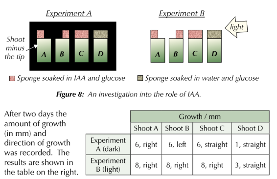
EXAMPLE - An experiment was carried out to investigate the role of IAA in shoot growth.
Eight shoots, equal in height and mass, had their tips removed.
Sponges soaked in glucose and either IAA or water were then placed where the tip should be.
Four shoots were then placed in the dark (experiment A) and the other four shoots were exposed to a light source, directed at them from the right (experiment B) — see Figure 8.
After two days the amount of growth (in mm) and direction of growth was recorded. The results are shown in the table on the right.
a) Explain the data.
b) suggest why this experimental design was good
c) why were all plants soaked in glucose
a)
The results show how the movement of IAA controls phototropism in plant shoots.
In experiment A shoot A, the IAA diffused straight down from the sponge into the left-hand side of the shoot. This stimulated the cells on this side to elongate, so the shoot grew towards the right.
In shoot B, the opposite occurred, making the shoot grow towards the left.
In shoot C, equal amounts of IAA diffused down both sides, making all the cells elongate at the same rate.
In experiment B, the shoots were exposed to a light source. The IAA diffused into the shoot and accumulated on the shaded side (left-hand side) regardless of where the sponge was placed.
Shoots A, B and C all grew towards the right because most IAA accumulated on the left, stimulating cell elongation there.
b) In this experiment the negative control treatment was the sponge soaked in water (and glucose) which was included to show that it was the IAA causing the observed effects and nothing else.
c) to provide energy for growth of shoots.
Photosynthesis can’t take place in the dark so the growth of seedlings in experiment A might have been limited if they weren’t provided with glucose (an external energy source).
What is Indoleacetic acid (IAA)
an important auxin that’s produced in the tips of shoots and roots in flowering plants.
It’s moved around the plant to control tropisms — it moves by diffusion and active transport over short distances, and via the phloem over long distances.
This results in different parts of the plant having different concentrations of IAA.
The uneven distribution of IAA means there’s uneven growth of the plant.

EXAMPLE
Q1 Thigmotropism is a plant growth response to touch.
a) In the diagram on the right, does the shoot display positive or negative thigmotropism?
b) Is the concentration of auxins, such as IAA, likely to be highest at the point labelled X or Y? Explain why.
a) positive. The shoot is bending towards the stimulus.
b) Y because this is where cell elongation is taking place, causing the shoot to bend towards the opposite side.
what is the role of a receptor
detect stimuli — they can be cells, or proteins on cell surface membranes.
receptors are specific to one type of stimulus
example of a receptor
e.g. baroreceptors are a type of receptor that detect changes in blood pressure,
what is the role of an effector
cells that bring about a response to a stimulus, to produce an effect.
example of an effector
muscle cells and cells in glands
how do receptors and effectors communicate
the nervous system or the hormonal system, or sometimes using both.
what are the 3 types of neurones
Sensory neurones
Motor neurones
Relay neurones
what is the role of a sensory neurone
transmit electrical impulses from receptors to the central nervous system (CNS)
what is the role of a relay neurone
transmit electrical impulses between sensory neurones and motor neurones.
what is the role of motor neurones
transmit electrical impulses from the CNS to effectors.
what is the role of neurotransmitters
take the information across the gap (called a synapse) to the next neurone, where another electrical impulse is generated
what is the role of the CNS
processes the information from the sensory neurone and sends impulses along motor neurones to an effector
what is a coordinator giving an example
- an organ that receives messages from receptors and uses the message to coordinate the activities in the body.
- For example, the brain or spinal cord (CNS)
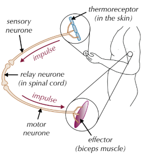
What is the order of the protective effect of a simple reflex arc? (7)
1. Stimulus
2. Receptor
3. Sensory neurone
4. Relay neurone
5. Motor neurone
6. Effector
7. Response
What are the important features of the protective effect of a simple reflex arc? (3)
- Rapid (only 3 neurones and few synapses are involved)
- Automatic (It doesn't have to be learnt) - goes through the spinal cord but not conscious parts of the brain
- Offers protection from harmful stimuli - as the response happens so rapidly
what is a simple reflex
a rapid, involuntary response to a stimulus.
where are relay neurons located
in CNS
what part of the CNS do reflexes pass to
only spinal cord as faster
draw a simple reflex arc
Explain why nervous communication leads to a localised and short-lived response.
localised → neurotransmitters are secreted directly onto cells
short-lived → neurotransmitters are quickly removed once they have done their job.
EXAMPLE The knee-jerk reflex involves lightly tapping a person on the patellar tendon (just below the knee-cap) with a tendon hammer. When this happens, the quadriceps muscle (in the thigh) immediately contracts, causing the person’s lower leg to jerk forward.
a) This response is a reflex. Suggest one way in which the response would differ if it was not a reflex.
b) Name the stimulus and the effector in the knee-jerk reflex.
c) The knee-jerk reflex is unusual because the sensory neurone synapses directly onto the motor neurone in the spinal cord.
i) Describe how this differs from a simple reflex, such as the hand-withdrawal response to heat.
ii) Suggest what effect tapping the patellar tendon might have in someone with a spinal cord injury. Explain your answer.
a) Any one from, e.g. the response would be slower /
the response would be voluntary.
b) Stimulus — light tap/touch.
Effector — quadriceps muscle.
c) i) The knee-jerk reflex doesn’t involve a relay neurone
in the spinal cord. / There are usually three neurones
involved in a simple reflex.
ii) E.g. the quadriceps muscle may not contract/there may be no response. If the spinal cord is damaged then the sensory neurone may not be able to transmit nervous impulses to the motor neurone / the motor neurone may not be able to transmit nervous impulses to the leg muscle.
EXAMPLE
Many nociceptors (pain receptors) are located in the skin.
a) Describe the pathway of nervous communication that would take place in a healthy person if they pricked their finger with a pin.
Congenital insensitivity to pain is a condition where the body does not feel physical pain. The condition is a result of non-functional nociceptors. The ability of sufferers to feel a light touch is usually normal.
b) Suggest why people with this condition are able to feel a light touch even though they’re unable to feel pain.
c) Suggest why it’s beneficial to an organism to be able to detect and respond to pain.
a) The nociceptors detect the stimulus and impulses are passed to a sensory neurone. This passes the electrical impulses to a relay neurone in the spinal cord/CNS which carries the impulse to a motor neurone. The motor neurone carries impulses to an effector (e.g. a biceps muscle).
b) Particular receptors are specific to a particular stimulus. This means that it’s possible that while their pain receptors aren’t functional (so pain isn’t felt), their touch receptors are functional allowing light touches to be felt.
c) It helps to protect the body by reacting to situations/environments that could cause the body harm.
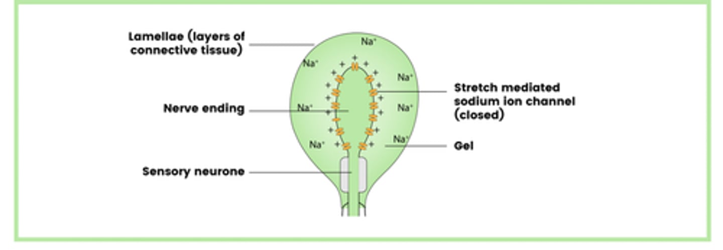
What is a pacinian corpuscle? (2)
mechanoreceptors in the skin
contain the end of a sensory neurone, (sensory nerve ending) wrapped in loads of connective tissue called lamellae
what type of stimuli do Pacinian corpuscles respond to
mechanical stimuli (i.e. pressure and vibrations), not to any other type of stimulus as receptors only respond to specific stimuli.
Draw the basic structure of a Pacinian corpuscle
- Lamellae
- Nerve ending
- Sensory neurone
- Stretch-mediate sodium ion channels
- Gel
What does the pacinian corpuscle illustrate? (3)
- Receptors respond only to a specific stimuli - a mechanical pressure
- Stimulation of a receptor leads to the establishment of a generator potential
- When the threshold is reached, action potential is sent (all-or-nothing principle)
what is a generator potential
Electrical signal generated in sensory receptors
created when sodium ions (Na+)
move across the axon membrane of Pacinian corpuscles in response to mechanical pressure.
Describe how a generator potential is established in a pacinian corpuscle (5)
1. A mechanical stimulus (e.g., pressure) deforms the lamellae and causes the sensory neurons cell membrane to stretch deforming the stretch-mediated sodium (Na+) channels
2. This causes stretch mediated Na+ channels in the membrane to open, allowing Na+ to diffuse into the sensory neuron
3. Greater pressure causes more Na+ channels to open, allowing more Na+ to enter
4. This influx of Na+ causes depolarisation, leading to a generator potential
5. If the generator potential reaches the threshold, it triggers an action potential
what is depolarisation
when a cell's membrane potential becomes less negative
leading to the generation of an action potential
What is meant by resting potential of receptors and how is it maintained
the inside of the cell is more negatively charged than the outside
The resting potential is generated by ion pumps and ion channels
What leads to a larger generator potential in receptors
A bigger stimulus excites the cell membrane more, making it more permeable causing a bigger movement of ions into and out of the cell and a bigger change in potential difference
action potential in receptors
If the generator potential is big enough to go over the threshold it’ll trigger an action potential — an electrical impulse along a neurone.
The strength of the stimulus is measured by the frequency of action potentials (the number of action potentials triggered during a certain time period).
If the stimulus is too weak the generator potential won’t reach the threshold, so there’s no action potential (all or nothing principle).
EXAMPLE Suggest how a person’s perception of touch might be affected by drugs that block stretch-mediated sodium ion channels in cell membranes.
pressure from touch would normally deform the stretch‐mediated sodium ion channels in Pacinian corpuscles, leading to a generator potential. However, by blocking sodium ion channels the drug would stop sodium ions from diffusing into the cell and generating an action potential. This would mean the person wouldn’t be able to perceive that they were being touched.
what are photoreceptors
receptors in your eye that detect light.
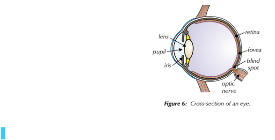
what is the pathway of light that enters the eye
Pupil (amount of light that enters is controlled by the muscles of the iris) → lens → retina → optic nerve
what is the forvea
an area of the retina where there are lots of photoreceptors.
where are no photoreceptors located in the eye
in the blind spot - where the optic nerve leaves the eye
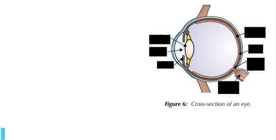
label the eye
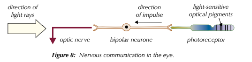
How photoreceptors work
Light enters the eye, hits the photoreceptors and is absorbed by light-sensitive optical pigments.
Light bleaches the pigments, altering the membrane permeability to sodium ions, producing a generator potential.
if it reaches the threshold, an action potential is produced which is sent along a bipolar (sensory) neurone.
Bipolar neurones connect photoreceptors to the optic nerve, which takes impulses to the brain
Draw a diagram to show how photoreceptors work
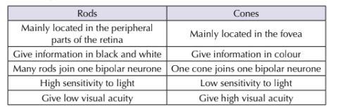
What are the two photoreceptor cells found in the retina of the eye? (2)
- Rods
- Cones
where are most rod cells found
the peripheral parts of the retina,
where are most cone cells found
fovea
What is the differences in colour vision for rods and cones? (2)
- Rods only allow monochromatic vision (black and white)
- Cones allow coloured vision
Explain how there are differences in colour vision for rods and cones? (1)
Rods and cones contain different optical pigments making them sensitive to different wavelengths of light.
How do cones allow for coloured vision? (4)
- Three different types of cones
- With different optical pigments
- So will absorb different wavelengths
- Stimulation of different combinations / proportions of cones gives a range of colour perception
What are the three different types of cones? (3)
- Red Sensitive
- Blue sensitive
- Green sensitive
Why are rods more sensitive to light than cones? (3)
- Several rods are connected to a single bipolar (sensory) neurone
- many weak generator potentials combine (via spatial summation) to reach/overcome the threshold
- and trigger an action potential.
Why are cones less sensitive to light than rods? (2)
- Each cone connected to a single bipolar (sensory) neurone
- There is no spatial summation occurring
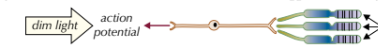
is this diagram pointing to rods or cones
rods
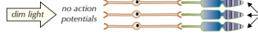
is this diagram pointing to rods or cones
cones
What is visual acuity? (1)
The clarity or sharpness of vision
low visual acuity = blurry vision
What is the difference in visual acuity between rods and cones? (1)
Cones give a higher visual acuity than rods
Why do cones give a higher visual acuity? (3)
- Each cone is connected to a single bipolar (sensory) neurone
- Cones send separate sets of impulses to the brain
- So they can distinguish between 2 separate sources of light
Why do rods give a lower visual acuity? (3)
- Several rods are connected to a single bipolar (sensory) neurone
- Several rods send a single set of impulses to the brain
- Therefore, it cannot distinguish between separate sources of light
Why do rods only allow for monochromatic vision? (2)
- There is only one type of rod
- That only contains one pigment
what type of photo receptor works best in dim light and why
rods - contain light sensitive pigment called rhodopsin
what type of photo receptor works best in bright light and why
cones - contain light sensitive pigments called photopsins that allow them to detect different wavelengths of light
summarise the differences between a rods and cones
mneumonic to remember where rods and cones are located
cones are packed closely together in the fovea.
what are the 2 divisions of the nervous system
Central
Peripheral
what are the 2 divisions of the peripheral nervous system
autonomic
somatic
what are the 2 divisions of the autonomic nervous system
parasympathetic
sympathetic
what is the CNS
is made up of the brain and spinal cord,
what is the PNS
made up of the neurones that connect the CNS to the rest of the body.
what is the autonomic nervous system
controls unconscious activities inside the body i.e. digestion
what is the somatic nervous system
system controls conscious activities and processes sensory information from the environment.
what is the sympathetic nervous system
‘fight or flight’ system that gets the body ready for action.
what is the parasympathetic nervous system
the ‘rest and digest’ system that calms the body down.
what part of the nervous system is involved in the control of heart rate
autonomic
what is myogenic stimulation
the process by which heart beat is controlled (via contracting and relaxing without receiving signals from the nerves)
EXAM TIP REGARDING AVN
there’s a delay before the AVN reacts. Don’t write in the exam that there is a delay in the wave of electrical activity reaching the AVN.
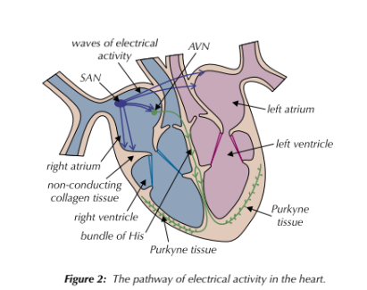
what is the sinoatrial node (SAN)
a small mass of tissue in the wall of the right atrium, that acts as a pacemaker — it sets the rhythm of the heartbeat by sending out regular waves of electrical activity to the atrial walls.
what is the bundle of His and Purkyne tissue
a group of muscle fibres responsible for conducting the waves of electrical activity between the ventricles to the apex (bottom) of the heart.
The bundle splits into finer muscle fibres in the right and left ventricle walls, called the Purkyne tissue.
Describe the myogenic stimulation of the heart
1. The sinoatrial node (SAN) acts as a pacemaker, sending regular waves of electrical activity across the atria, causing them to contract simultaneously
2. Non-conducting tissue between the atria and ventricles prevents the impulse from passing directly to the ventricles, preventing immediate contraction of the ventricles
3. Waves of electrical activity reach the atrioventricular node (AVN), which delays the impulse, allowing the atria to fully contract and empty before the ventricles contract
4. The AVN sends a wave of electrical activity down the bundle of His, which branches into Purkyne tissue which carries the waves of activity into the walls of the right and left ventricles causing them to contract simultaneously from the base up.
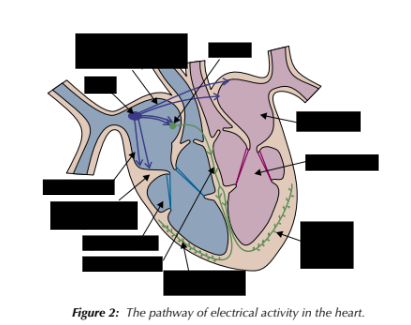
label the areas of the heart needed for myogenic stimulation
what is the rate at which the SAN fires caused by
unconsciously caused by the medulla in the brain
why may animals need to alter their heart rate
to respond to internal stimuli, e.g. to prevent fainting due to low blood pressure or to make sure the heart rate is high enough to supply the body with enough oxygen.
how are internal stimuli detected in the body
by pressure receptors (baroreceptors) and chemical receptors (chemoreceptors)
where are baroreceptors located and what are they stimulated by
pressure receptors
in the aorta and carotid arteries.
Stimulated by high and low blood pressure.
where are chemoreceptors located and what are they stimulated by
Chemical receptors
In aorta, the carotid arteries and in the medulla.
Detect changes in the oxygen level in the blood and carbon dioxide and pH (which are indicators of O2 level).