DNT 104 Chapter 15 Dentition Overview Vocabulary Flashcards
 Call Kai
 Learn
Learn Practice Test
 Spaced Repetition
 Match
Match Flashcards
 Knowt Play
1/81

Earn XP
Description and Tags
Vocabulary flashcards for reviewing DNT 104 Chapter 15 Dentition Overview, covering dentition periods, tooth structures, tooth types, numbering systems, line/point angles, and anatomical relationships.
Name | Mastery | Learn | Test | Matching | Spaced | Call with Kai | Chat |
|---|
No analytics yet
Send a link to your students to track their progress
82 Terms
Primary Dentition Start period
6 months to 6 years of age, beginning with the eruption of the primary mandibular central incisor.
Primary Dentition end
Period usually ends when the first permanent tooth erupt: the permanent mandibular first molar at approximately 6 to 7 years of age
Mixed Dentition start period
approximately 6 to 12 years of age containing both primary and permanent teeth, beginning with the eruption of the permanent mandibular first molar.
Mixed Dentition end period
Shedding of the last primary tooth which usually occurs from age 11 to 12
Color differences, example: permanent teeth and Primary teeth
less opaque enamel, which makes the underlining yellow dentin more noticeable; Opaque- not able to see through; not transparent
Permanent Dentition Period
The final dentition period occurring after approximately 12 years of age, beginning with the shedding of the last primary tooth.
Deciduous Dentition
Another term for the primary dentition, consisting of 20 baby teeth that are shed and replaced by permanent teeth.
Succedaneous Tooth
A permanent tooth that succeeds or replaces a primary baby tooth. (teeth 4-13 and 20-29 represent the succedaneous teeth in the permanent dentition.)
Non-Succedaneous Permanent Teeth
The permanent molars (teeth 1, 2, 3, 14, 15, 16, 17, 18, 19, 30, 31, and 32), which erupt behind primary teeth without replacing any primary teeth.
Crown
covered by enamel, whether it is erupted or not
Anatomical Crown
The top of the crown covered by enamel, bounded apically by the Cementoenamel Junction (CEJ), which never changes throughout life.
Clinical Crown
The part of the tooth crown that is seen above the gingiva and not covered by the gingiva, which can change in length over time.
Why can clinical crown change overtime?
Before tooth eruption: tooth is still under the gum so you cannot see the crown
When the tooth erupts: tooth comes through the gum, so the crown is more visible
With gingival recession: the gum moves downward toward the root exposing more of the root of the teeth, making the clinical crown LONGER than the anatomical crown
Gingival inflammation/swelling: causes gums to rise up which covering portions of the anatomical crown and making the clinical crown SHORTER then the anatomical crown
Anatomical Root
The portion of the tooth root covered entirely by cementum, extending from the CEJ to the root apex, which remains constant.
Clinical Root
The portion of the tooth root embedded within and attached to the periodontium(surrounding gingiva and alveolar bone), which changes length in relation to gingival.
Enamel
The hardest tissue of the body
Thick over the tip of the crown and thin in the cervical area (where the root is)
Color will vary, depending on the thickness
Resistant to wear
Self cleaning abilities because it is smooth(normally)
Dentin
Forms the main portion of the tooth
Softer than enamel but harder than cementum. (It it covered by cementum so if the cementum is weak or broken it is cause sensitivity to the dentin bc the dentin is now exposed)
70% mineralized
Capable of producing secondary and tertiary dentin
Pulp Cavity
The central space of the tooth containing blood vessels, nerves, lymph nodes, and connective tissue that detects pain sensory input.
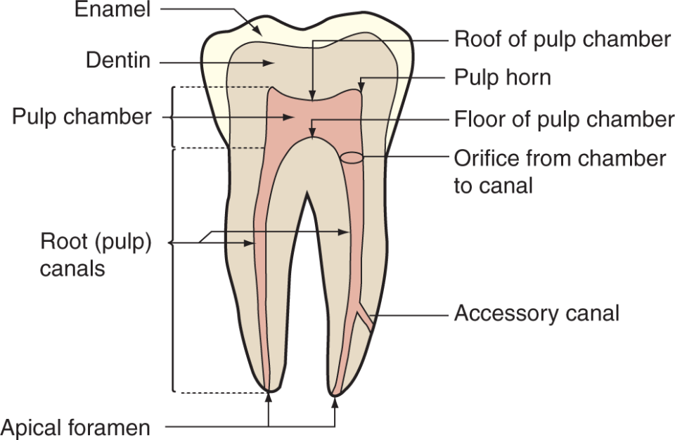
Pulp Horns
Horn-like coronal extensions of the pulp chamber projecting into the cusp tips of the tooth.
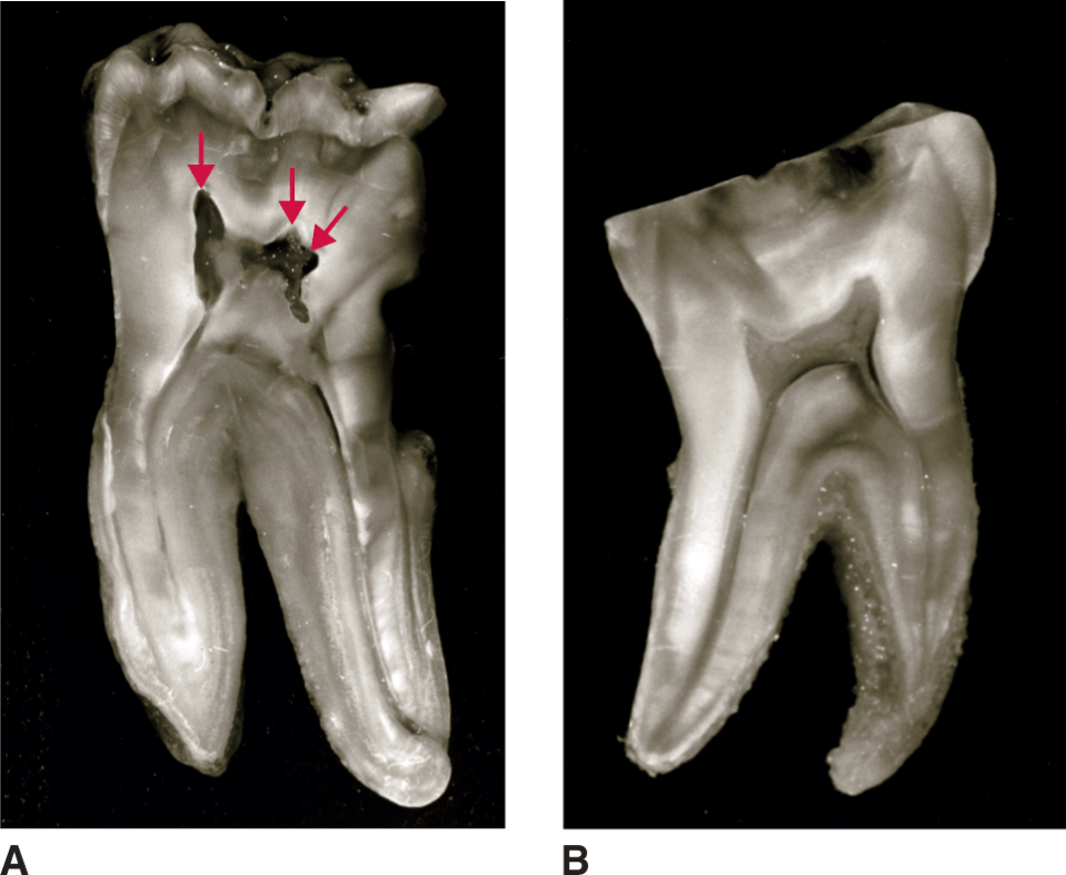
Pulp Canals
Elongated root canals extending from the pulp chamber down through the roots to the root apex.

Pulp Chamber
The large central portion of the pulp cavity located within the coronal section of the tooth.

Root Apex
The terminal end or tip of the tooth root.
Apical Foramen
The terminal opening at the root apex through which nerves, blood vessels, and lymphatics enter and exit the internal pulp cavity.
Furcation
The specific anatomical location where a multi-rooted tooth trunk divides into separate root branches.
What are the separate root branches of Furcation?
Bifurcation: splitting into 2 distinct roots
Trifurcation: Splitting into 3 distinct roots
Root Trunk
The undivided portion of the root extending from the Cementoenamel Junction (CEJ) to the point of furcation.
Which tooth, often exhibit short root trunks, rendering their furcations more susceptible to early clinical exposure during periodontal bone loss.
Mandibular molars
Cementum
A bone-like substance covering the root that is thinner at the CEJ and thicker at the root apex. also contains, alveolus, gingiva, PDL, occlusion
Amount of roots in molars
Maxillary - 3 roots (Trifurcation)
Mandibular- 2 roots (Bifurcation)
Amount of roots in premolars
Maxillary- 2 roots (Bifurcation)
Mandibular -1 root
Alveolus
An individual tooth socket consisting of the bone that surrounds and supports the tooth.
Gingiva
cover the alveolar process- bone that surrounds the teeth
Periodontal Ligament (PDL)
The tissue that attaches the tooth to the socket (alveolus), keeping teeth in place so they do not twist or break during mastication.
Occlusion
The contact relationship when the maxillary and mandibular teeth come together.
Cementoenamel Junction (CEJ)
The area where the enamel of the crown meets the cementum covering the root of the tooth, crucial for maintaining tooth stability.

CEJ Relationships Diagram
A diagram illustrating that cementum overlaps enamel in 60% of teeth which is ideal, cementum and enamel meet end-to-end in 30%, and a gap exposing dentin exists in 10% that can cause pain.
Incisors
8 total
used for biting or cutting
appearing triangular from the proximal aspect.
What are the numbers of the central incisors?
(Maxillary 8 and 9) (Mandibular 24 and 25)
What are the numbers of the lateral incisors
(Maxillary 7, 10) (Mandibular 23, 26)
Canines
4 total
helpful for piercing or tearing food
featuring a single cusp.
Numbers of canines
6, 11 , 22, 27
Premolars
Transitional posterior teeth
(8 total)
tear, crush, and grind food
featuring 2, 3, or 4 cusps.
Numbers of premolars
4,5,12,13,20,21,28,29
Molars
The largest and strongest teeth
(12 total)
used to grind food
featuring 2, 3, 4, or 5 defined cusps.
number of molars
1,2,3,14,15,16,17,18,19,30,31,32
Definition of sextants
breaks quadrants down further by focusing on a smaller section of teeth.
Posterior Sextant: premolar and pre molar
Maxillary left sextant( 12,13,14 and 15)
Anterior Sextant: incisor and canine. Has 6
DAQT System
A standardized sequence used to describe a tooth by stating its Dentition, Arch, Quadrant, and Tooth type.
Universal Numbering System
The primary numbering system used in the US, designating primary teeth with letters A through T and permanent teeth with numbers 1 through 32 clockwise.
International Numbering System (INS)
A tooth numbering system that uses numbers 1-4 for permanent teeth and 5-8 for primary teeth. children uses letters not numbers no more than 5 while adults can go from 1 to 8
Palmer Notation System
A tooth notation system used in orthodontics using numbers 1-8 for permanent teeth and letters a-e for primary teeth set inside a quadrant bracket symbol.
Proximal Surfaces
The surfaces of adjacent teeth that face and touch each other in the same arch, divided into mesial and distal surfaces.
Lingual(tongue side) / palatal (Maxillary) surfaces
the inner surface of the tongue
Occlusal surface and Incisal surface
mainly posterior teeth that sit right on top of each other; anterior teeth where incisor teeth at the bottom
Facial/labial surface and Buccal surface
anterior teeth; posterior teeth
Line Angle
The line formed where only 2 crown surfaces meet along a curve or corner.
How do you name Line Angles
Proximal surfaces(mesial or distal) should always be first. Then the Incisal or occlusion(anterior or posterior teeth) should be named last
Anterior Line Angles (6)= 2 crown surfaces meet
Distolabial
Distolingual
Mesiolabial
Mesiolingual
Labio-incisal
Linguo-incisal
Posterior Lines Angles (8)= 2 crown surfaces meet
Bucco-occlusal
Disto-Occlusal
Distolingual
Distobuccal
Linguo-occlusal
Mesio-occlusal
Mesiolingual
Mesiobuccal
Point Angle
The specific point formed where only 3 crown surfaces intersect.
How to name Point Angles
First term: must always be proximal surface(mesial or distal)
Second term: must be facial/labial/buccal or lingual/palatal surface
*Third term: must always be the chewing surface (incisal or occlusional. (New thing added)
Anterior Point Angles (4)= 3 surfaces meet
Distolinguo-incisal
Distolabio-incisal
Mesiolinguo-incisal
mesiolabio-incisal
Posterior Point Angles (4)= 3 surfaces meet
Distolinguo-occlusal
Distobucco-occlusal
Mesiolinguo-occlusal
Mesiobucco-occlusal
How are Crowns divided
into 3rds horizontally and vertically, creating a grid of six functional areas/boxes per surface
Horizontal divisions: incisal/occusal third, middle third, cervical third
Vertical divisions: mesial third, middle third, distal third
How are roots divided
only be divided into 3rds horizontally!
Typical third, middle third, cervical third
Used to precisely localized root carries or radiographic lesions, ex: lesion at the apical third of the mesial root
Cusp (mountains on the teeth on the chewing surface)
Help crush, tearing, and grind food
Help with occlusion (teeth coming together)
Mainly found on posterior teeth (premolars, and molars)
Canines have cups and it is the only anterior teeth that does
Height of Contour
crest of curvature
Buccal Bump identification: located on the buccal aspect of the posterior teeth near the CEJ
Viewing a tooth from the proximal aspect, the buccal side shows a distinct bulge/bump near the ECJ before tampering inward
The lingual side is smoother and a subtle curvature(not a big bump)
Contact Area (where floss goes)
A region on the proximal surface (mesial or distal) where a tooth touches its adjacent neighboring tooth in the same arch.
Contact Area (Anterior teeth)
more narrow and up higher, closer to incisal third
Contact Area (Posterior teeth)
contact area, shifts lower down the tooth, closer to the middle third.
Contact Point
The point on the occlusal surface of a tooth that touches another tooth in the opposing arch during occlusion. Dentist use this
Interproximal Space
A triangular space between two neighboring teeth filled in healthy periodontium by interdental papilla gingival tissue.
Helps prevent food entrapment and allow food to slide smoothly along the crown surfaces during mastication
Embrasures
Spaces between the teeth(area) that diverge, buccally, labially or lingually and occlusal/incisal to the contact areas
Open, continuous V-shaped spillway spaces surrounding contact areas that radiate outward to allow food to spillways during mastication.
CEJ curvature (Proximal surfaces(mesial and distal)
the CEJ curves coronally( towards the incisal edge or occlusal surface) towards the crown. Rules are the same for both maxillary and mandibular teeth
CEJ curvature Facial/labial/lingual surfaces
the CEJ curves apically(towards the root Apex). Rules are the same for both maxillary and mandibular teeth
CEJ curvature Mesial surface vs distal surface
demonstrates a higher more pronounced CEJ curvature than distal surfaces
The CEJ curvature becomes flatter when going towards the distal teeth
CEJ curvature Anterior teeth
higher CEJ curve because they are narrow and need more support/ stability
CEJ curvature Posterior teeth
flatter CEJ because they are wider and more stable bc of their multiple roots
** how to identify mesial versus distal aspect on anterior teeth, instead of inspecting the CEJ curvature variations.
Mesial aspect: incisal angle/outline forms nearly a sharp right angle
Distal aspect: incisal angle/outline is visibly more rounded
how to identify facial/buccal vs lingual aspects on teeth with landmarks.
Buccal prominence/height of contour: a distinct facial bulge located on the cervical third helps identify the buccal surface
Root configuration: multi rooted teeth, provide clear orientation markers; for instance, maxillary molars always feature a distinct lingual root alongside two buccal roots
Self Cleaning Qualities of countours
(both facial and lingual contour are designed to direct food off the teeth during mastication protecting the surrounding periodontium
What causes food impaction?
Open contacts (diastema) and Recession exposed root surfaces makes it hard for patient to clean
Diastema
An open contact space or gap between two adjacent teeth in the same arch, commonly found between teeth #8 and #9.