Integument flashcards
 Call Kai
 Learn
Learn Practice Test
 Spaced Repetition
 Match
Match Flashcards
 Knowt Play
1/100

There's no tags or description
Looks like no tags are added yet.
Name | Mastery | Learn | Test | Matching | Spaced | Call with Kai | Chat |
|---|
No analytics yet
Send a link to your students to track their progress
101 Terms
Physiologic functions of the Epidermis
Primary protection/physical barrier against microbes, chemicals, toxins, physical impacts via (formation of the cornified envelope, production of antimicrobial substances during this process, and permanent shedding of corneocytes)
Primary envelope to contain water, electrolytes, macromolecules (protein, carbohydrates, fat)
Vitamin D production
Contribution to temperature regulation via transepidermal water loss.
Main physiological functions of the dermis
Absorbs stress from movement
Maintains skin shape
Water storage
Immunologic surveillance
Thermoregulation/cooling via radiation/conduction/convection and blood vessels and sweat glands
Erection of hair
Cutaneous sensation
Main physiologic functions of the subcutis
Energy reserve (90% triglycerides, ‘fat’)
Thermoinsulation
Protective padding
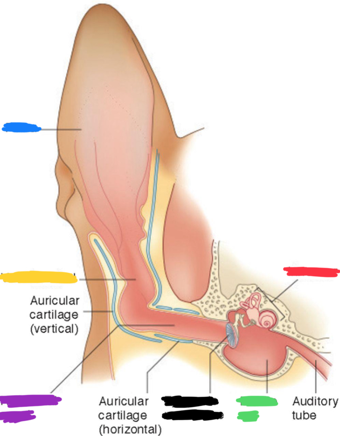
What is labeled in blue?
Pinna
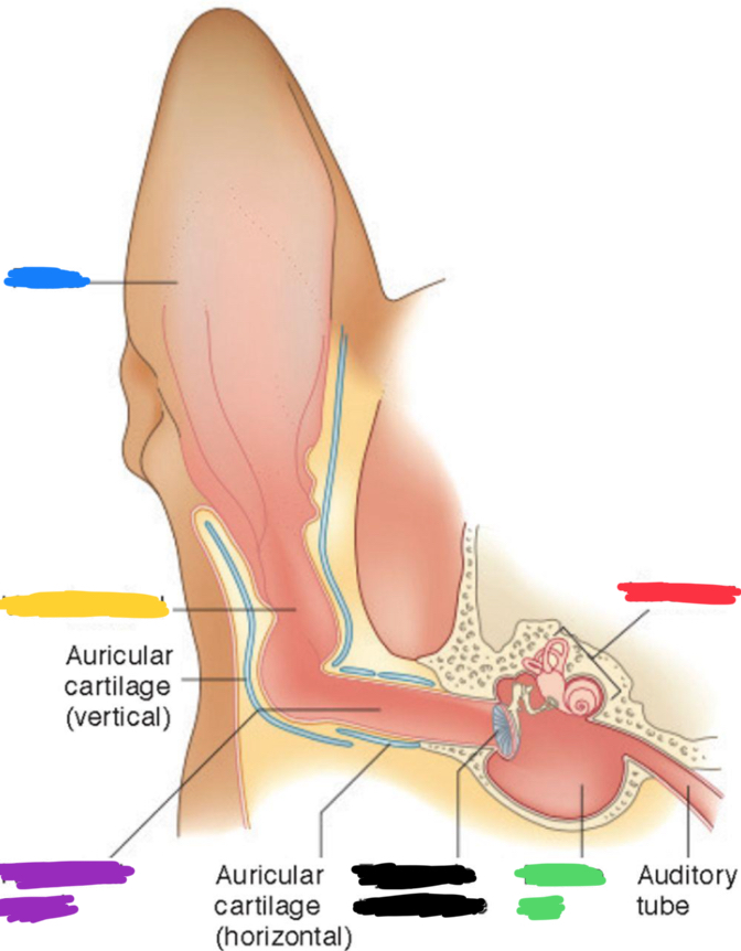
What is labeled in yellow?
Vertical canal
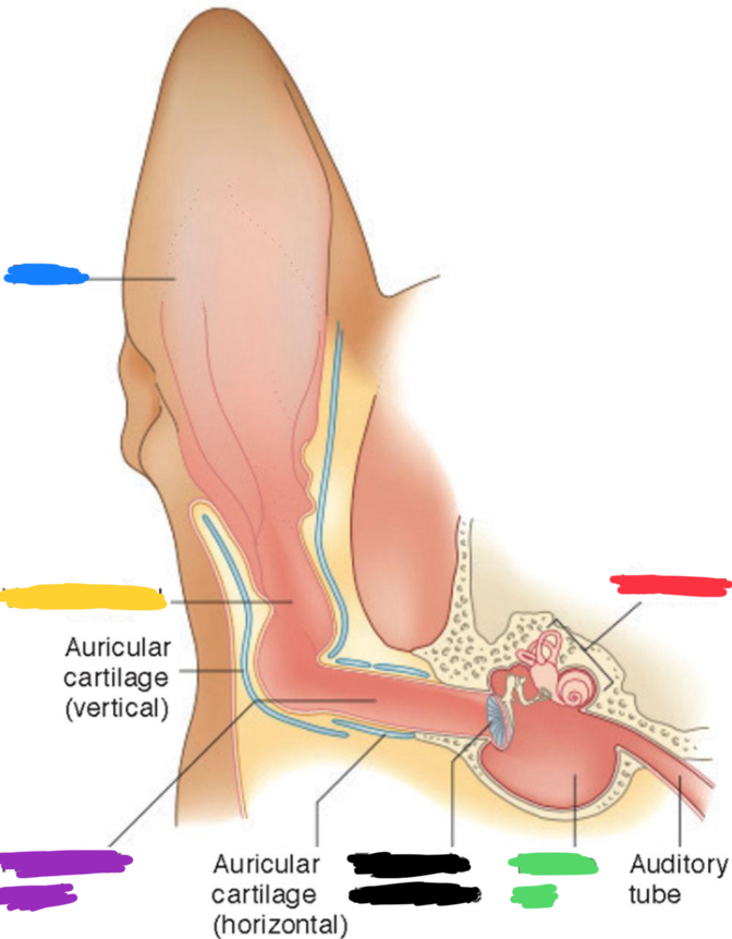
What is labeled in purple?
Horizontal canal
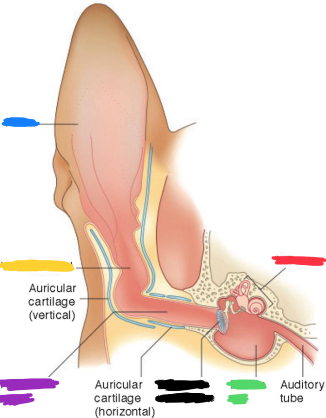
What is labeled in red?
Inner ear
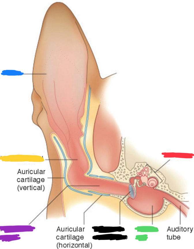
What is labeled in green?
Middle ear
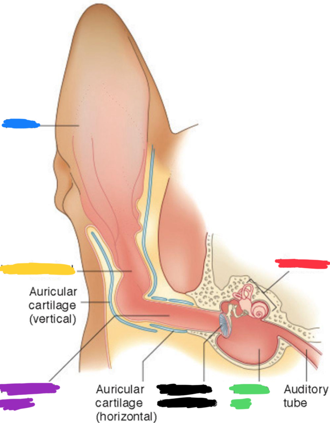
What is labeled in black?
Tympanic membrane
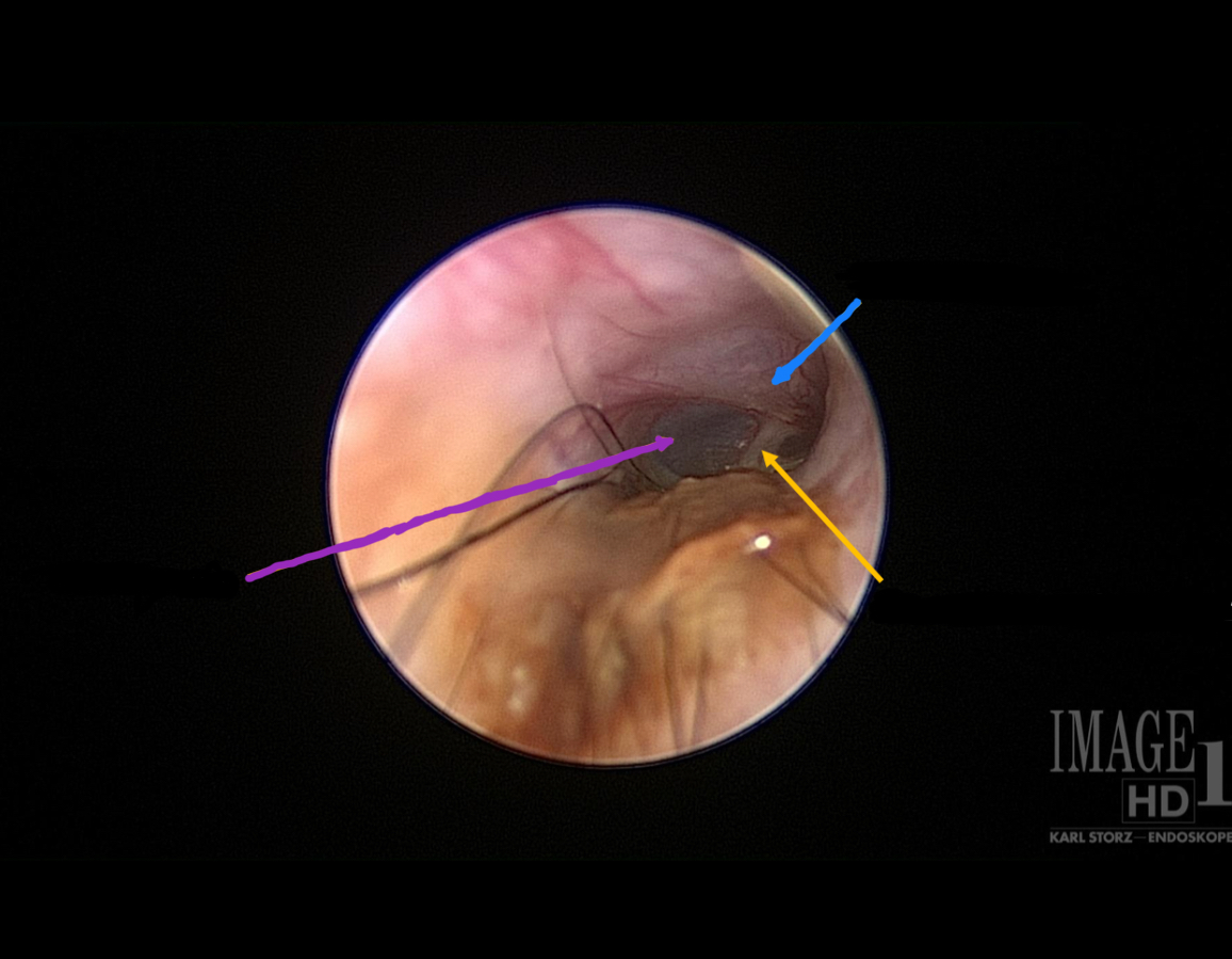
Yellow arrow
Manubrium of Malleus
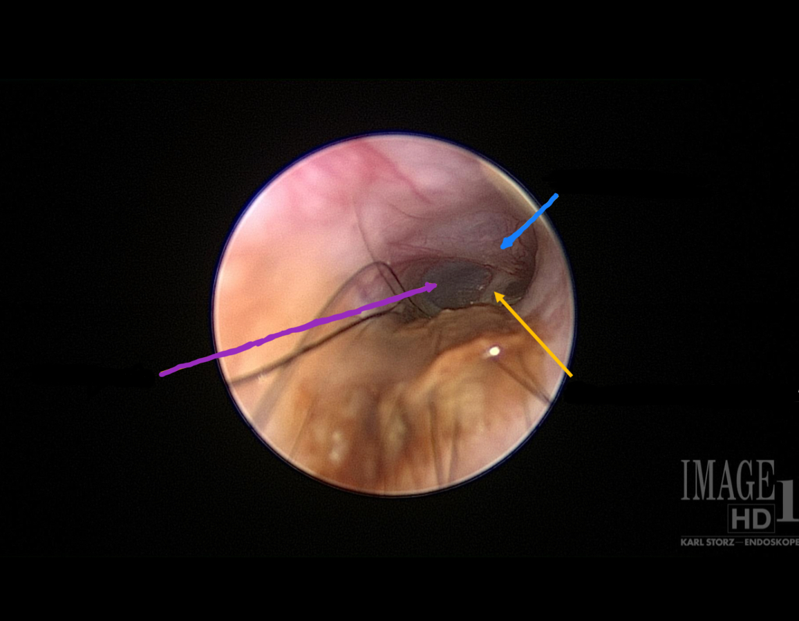
Blue arrow
Pars flaccida
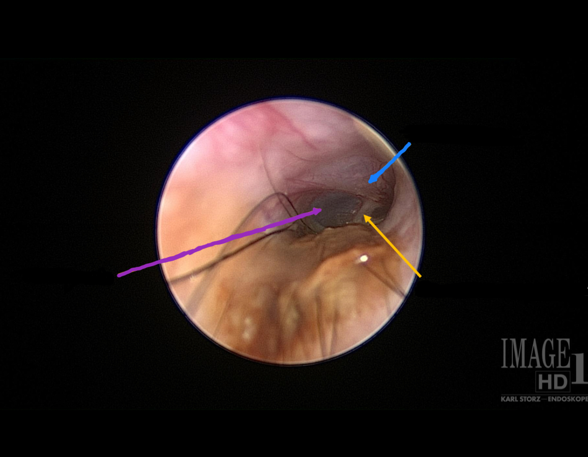
Purple arrow
Pars tensa
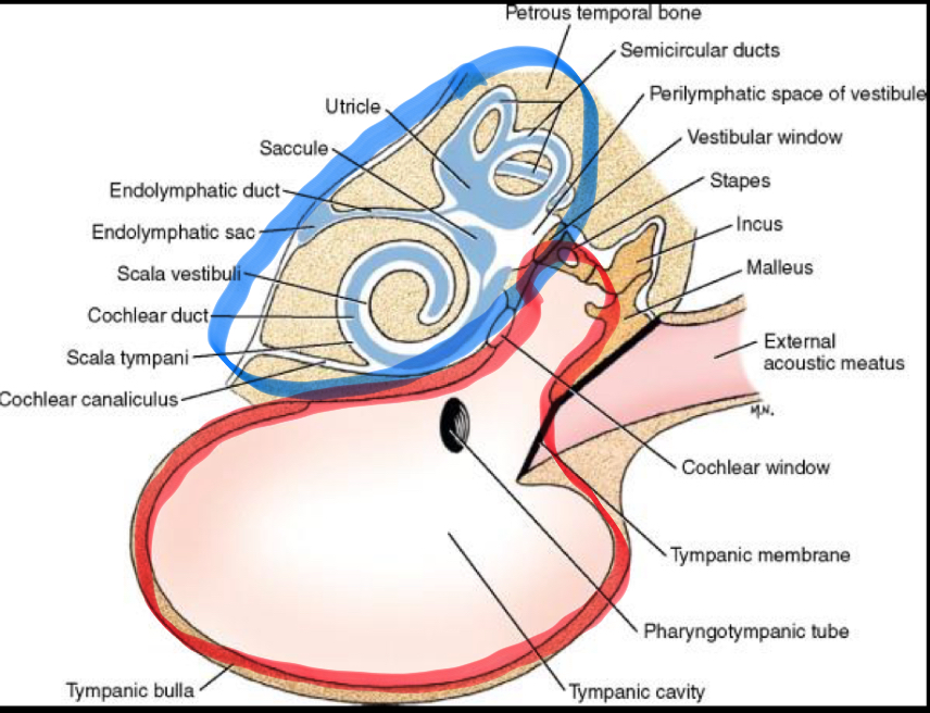
Encircled in blue
Inner ear
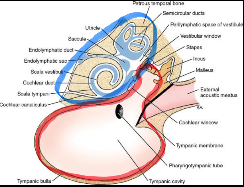
Encircled in red
Middle ear
Skin microbiome
Collection of microbes that naturally live on our bodies
It is in a constant state of flux
Its composition varies depending on species
Varies between individuals of the same species
Varies depending on body site in the same individual
Exposure to a varied microbiome is considered a key component in immune regulation
What did this study state regarding ears?
It demonstrated a shift in bacterial populations between allergic and healthy dogs. The observed results in the ear canal are similar to those reported on the skin and could explain the propensity of allergic dogs to develop bacterial otitis.
Internal impacts on skin
malnutrition/lack of essential fatty acids → epidermal lipid deficiency/disturbance → scaling, poor barrier function
External impacts on skin
excessive washing, chemicals, dry/wet environment → weakening of epidermal barrier, increasing scaling, increased transepidermal water loss
Epidermal keratinization definition
A highly orchestrated, genetically programmed process resulting in structural and metabolic changes
Epidermal keratinization steps
Proliferation/Reproduction
Firm adhesion (desmosomes ‘hands’, hemidesmosome ‘anchors’ connect intracellular keratin filament types K5/14 to neighboring keratinocytes)Production of additional keratin filaments K1/10
Beginning synthesis of lipid-containing lamellar bodies
(cholesterol, ceramides, free fatty acids & enzymes)The cornified envelopes are firmly connected via corneodesmosomes + tight junctions
Extrusion of lipid-containing lamellar bodies -> lipids fills intercorneocyte spaceProgressive flattening of cells, keratin filaments get progressively packed into large, insoluble polymers (= formation of the ‘cornified envelope’) with the help
of filaggrin (keratohyaline ‘granules’)Keratinocytes are terminally differentiated (disintegration of organelles, absence of nucleus), ‘coated’ by lipids (ceramides), separated by proteases, and are constantly shed
Hair follicle cycle order
Anagen, Catagen, Telogen, Anagen
What happens during the first Anagen phase in the hair follicle cycle?
This is considered the growth phase, the hair follicle grows
Increased levels = mitotic activity, melanin production, thyroid hormones, growth hormones
Discouragement of growth = glucocorticoids and estrogen
What happens during the Catagen phase of the hair follicle cycle?
This is considered the regression phase, it is where hair growth stops, the follicle shrinks, and the hair detaches from its blood supply.
Increased activity = Caspase activity and Keratinocyte apoptosis
Decreased = Melanin production
What happens during the Telogen phase of the hair follicle cycle?
This is also considered the resting phase, hair is not growing
Small dermal papilla
No mitotic activity
What happens during the second Anagen phase of the hair follicle cycle?
This is also considered the exogen phase, the shedding of the hair follicle and the new anagen phase commences
Epidermis
a layer of epithelium and this epithelium is keratinized stratified squamous.
Dermis
-network of collagen, elastic, reticular fibers within amorphous ground substance
- typically contains fibroblasts, macrophages, plasma cells, mast cells, fat, and scattered melanocytes
- houses hair follicles, sweat glands, and sebaceous glands
- plentiful blood and lymphatic vessels and nerves
- functions: anchors and supports the epidermis, gives rise to small vessels that nourish the epidermis via simple diffusion, immune surveillance highway, insulates and protects the organism
Hypodermis (subcutis)
-the superficial fascia (seen in gross anatomy) that anchors the dermis to the underlying muscle and bone
- location of varying amounts of adipose tissue (panniculus adiposus) and cutaneous muscles (panniculus carnosus)
What are the layers of the cutis?
Epidermis, Dermis, and Hypodermis (subcutis)
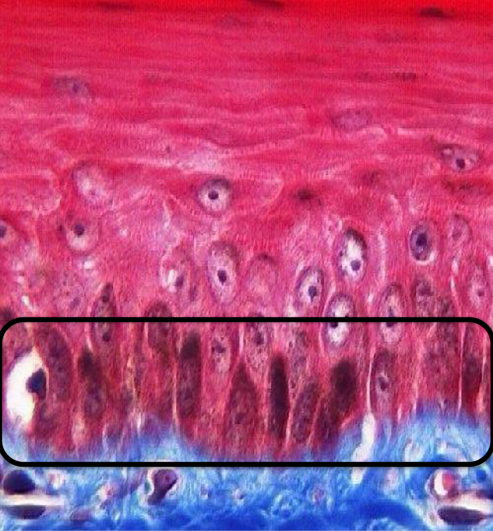
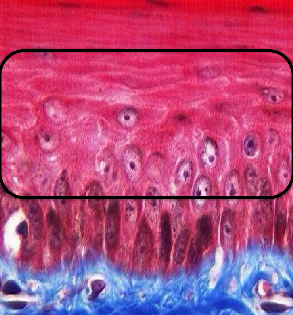
What layer of the epidermis is this?
Stratum basale
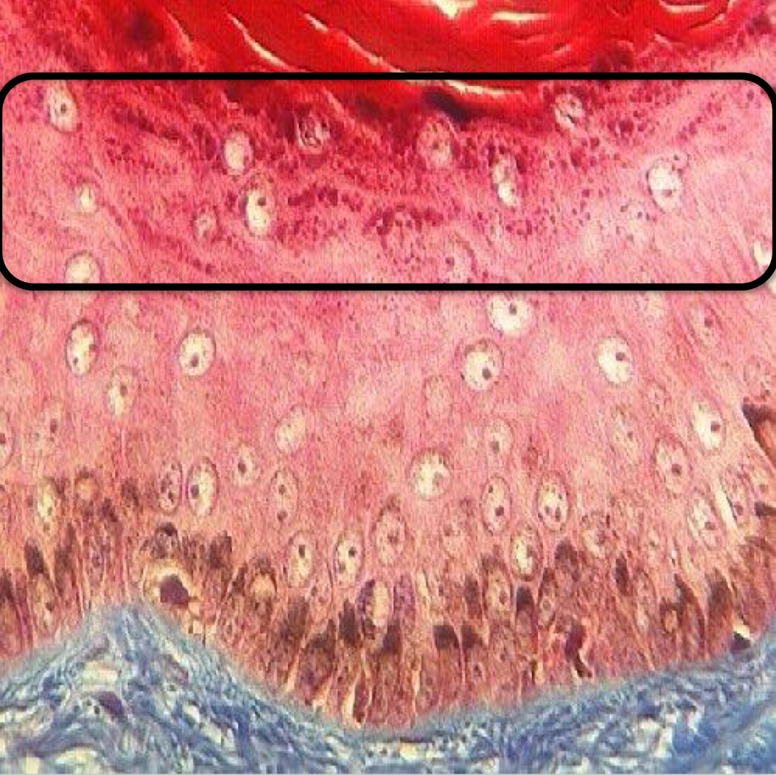
What layer of the epidermis is this?
Stratum spinosum
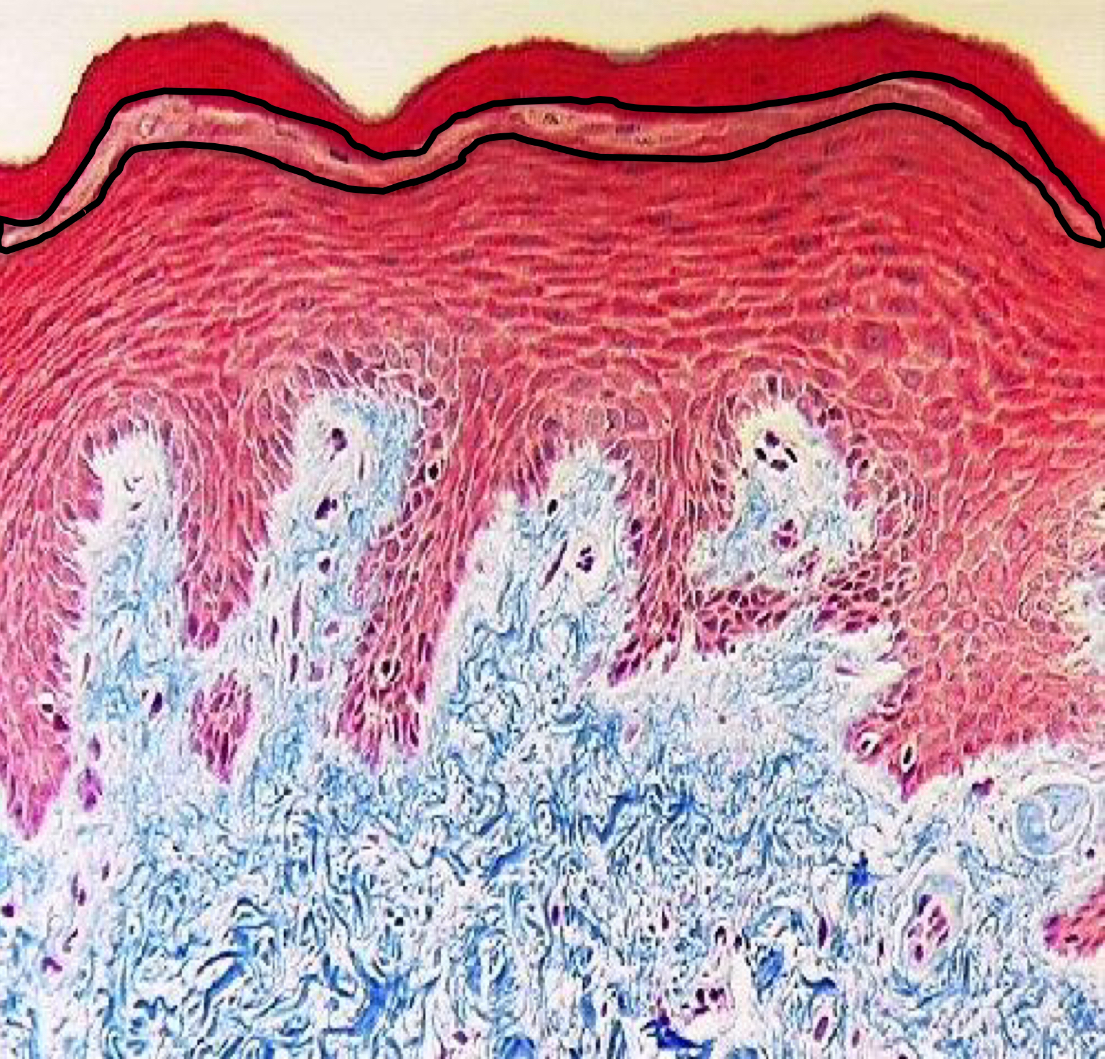
What layer of the epidermis is this?
Stratum granulosum
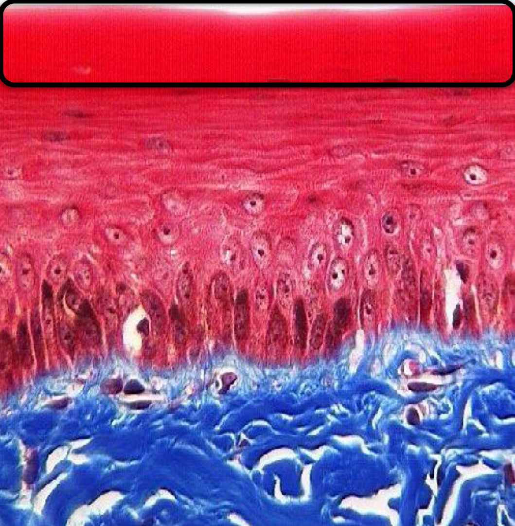
What layer of the epidermis is this?
Stratum lucidum
What layer of the epidermis is this?
Stratum corneum
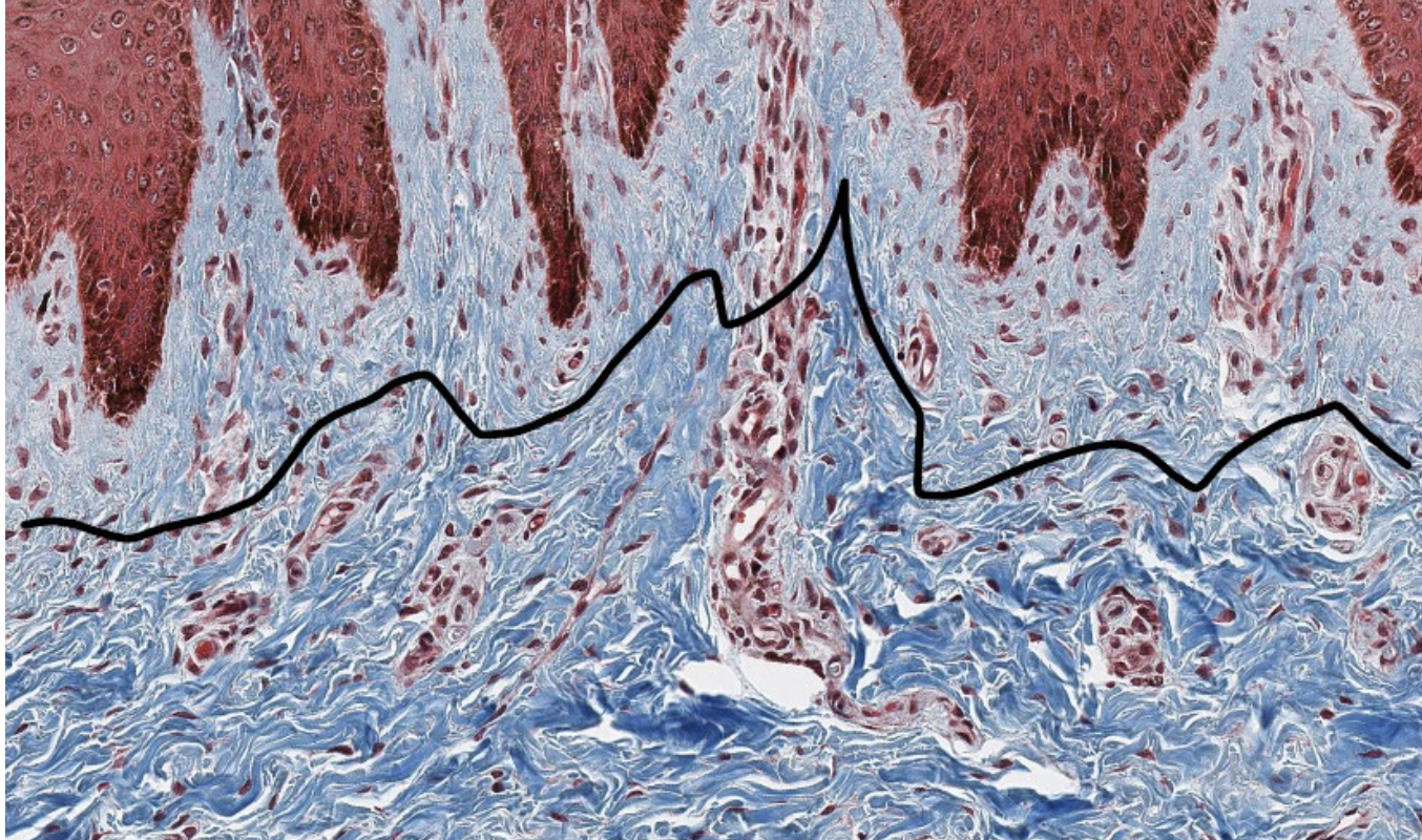
The top is the ______ layer of the dermis and the bottom is the _____ layer of the dermis.
Papillary; Reticular
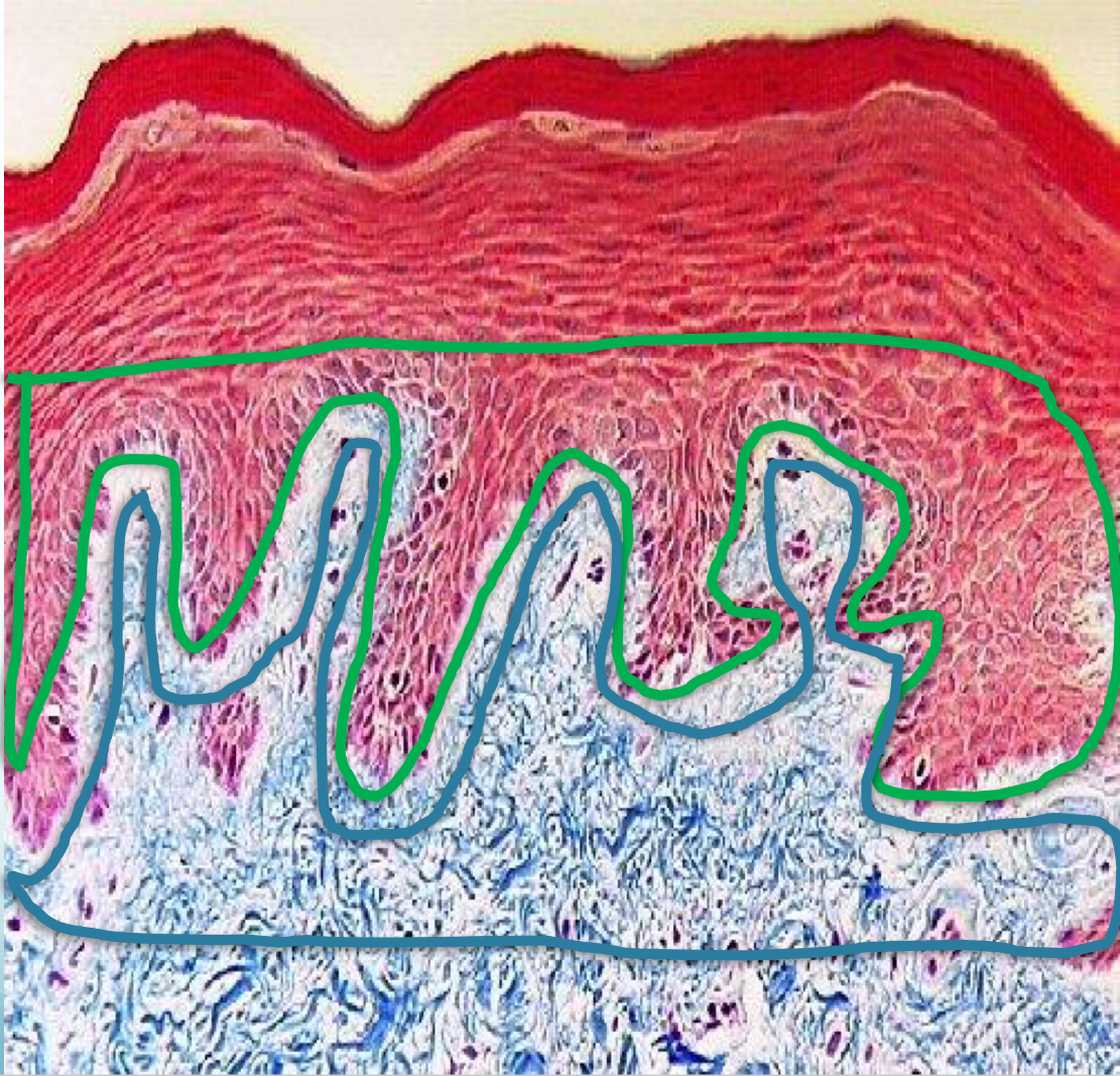
Green are the ______
Epidermal ridges
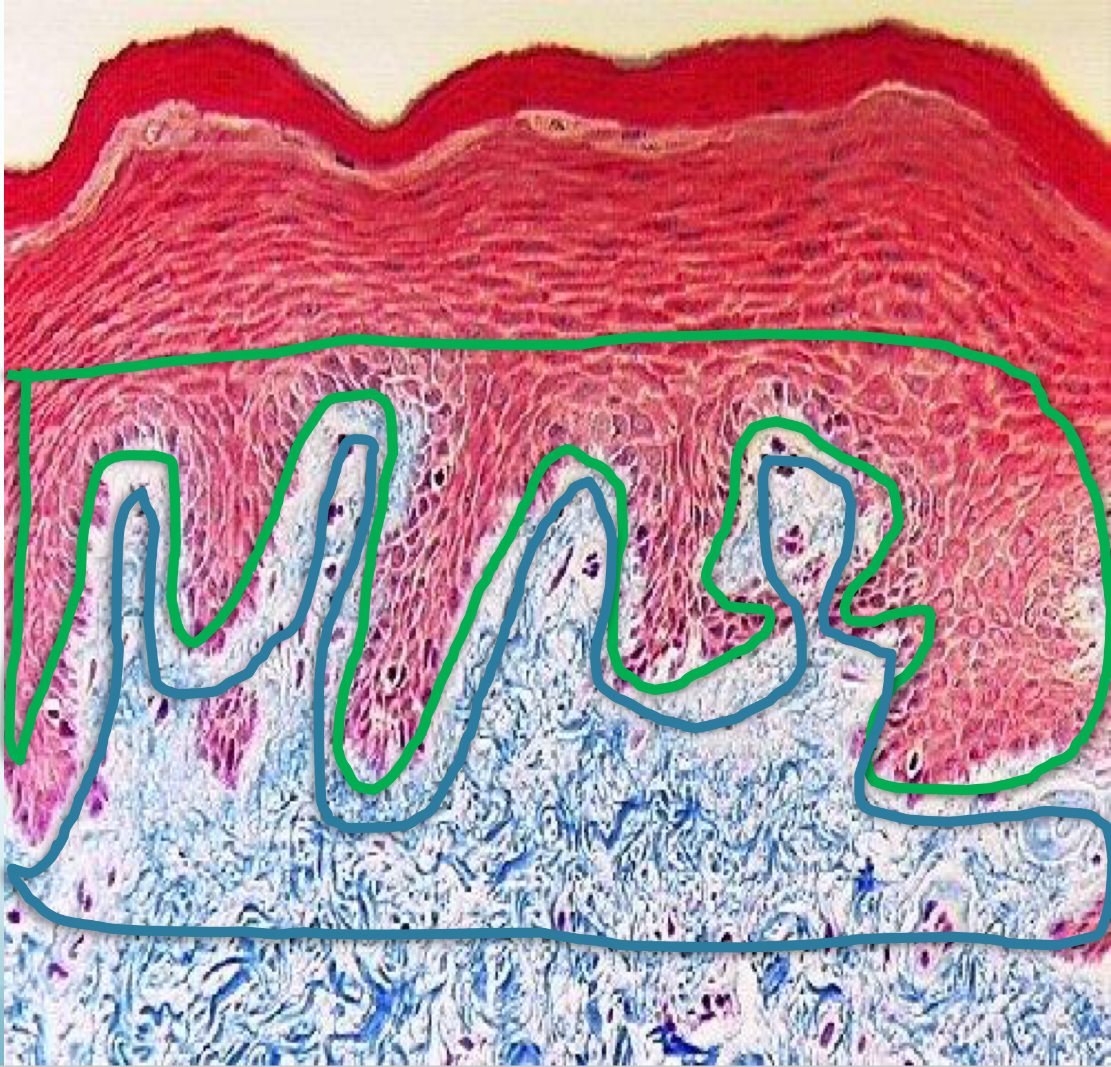
Blue are the ______
Dermal Papillae
Kerotinocytes
The cells that make up the layers of the epidermis
Non-keratinocytes
melanocytes, tactile cells, and antigen-presenting cells
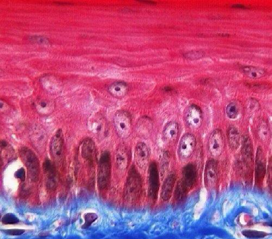
What is this? (kertinized/non-keratinized)
Keratinized (layers)
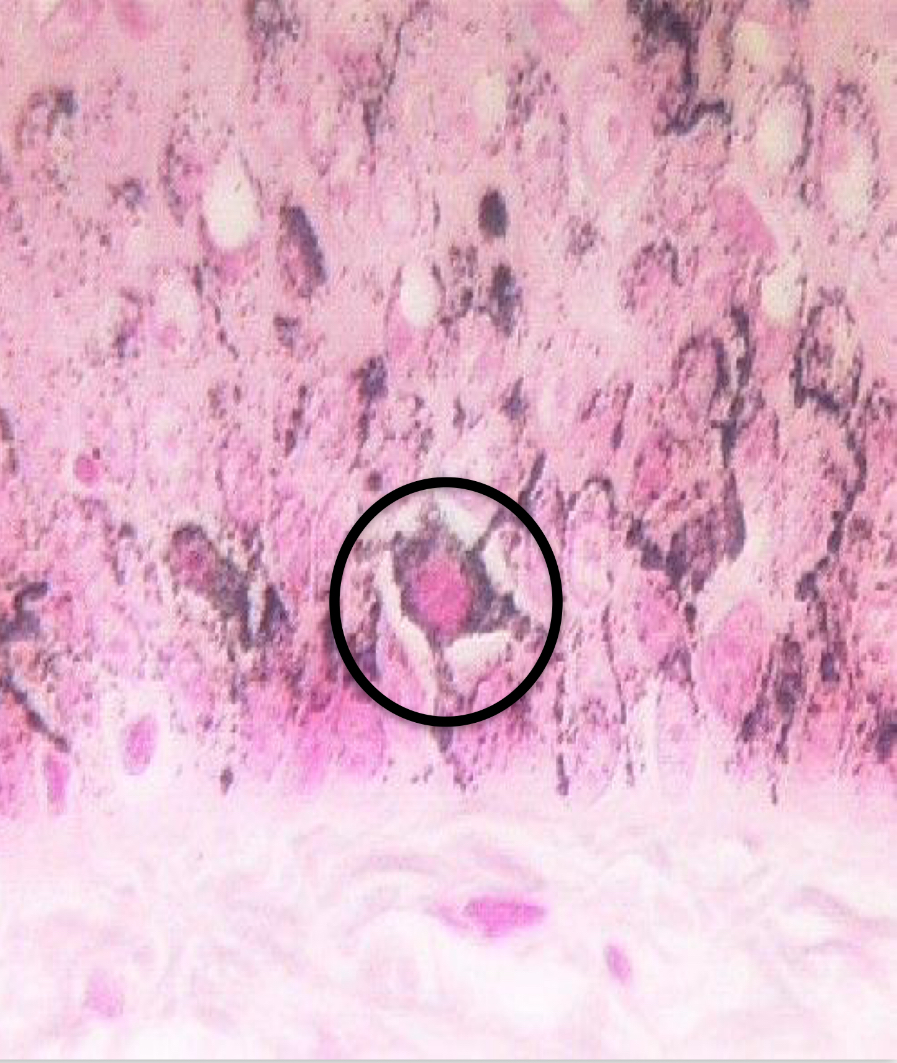
What cell is this? Keratinized or Non-keratinized?
Melanocyte; non-keratinized
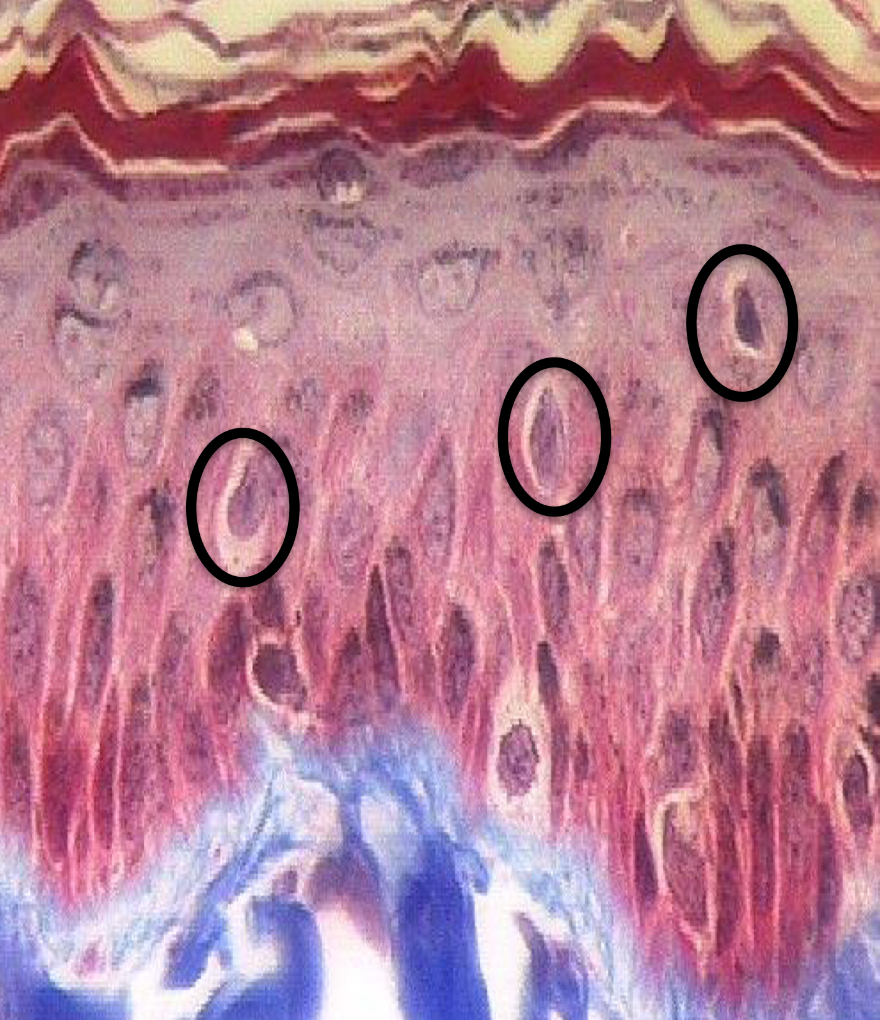
What cell are these? Keratinized or Non-keratinized?
Langerhan’s cells; Non-keratinized
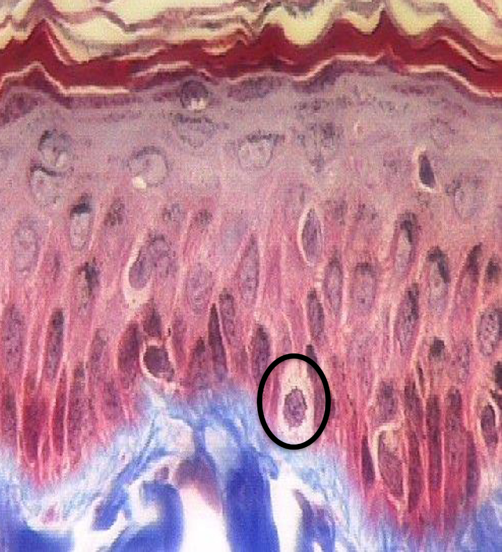
What cell is this? Keratinized or Non-keratinized?
Merkel cell; Non-keratinized
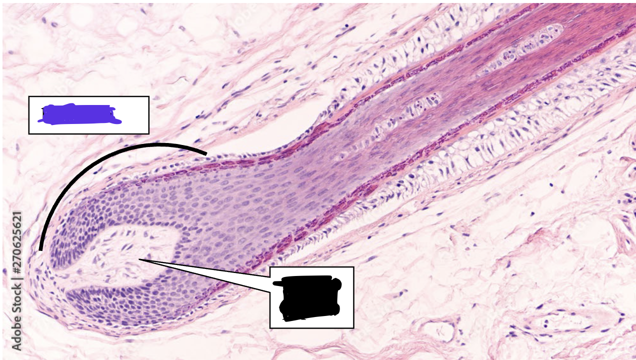
What part of hair is this? In purple
Hair bulb
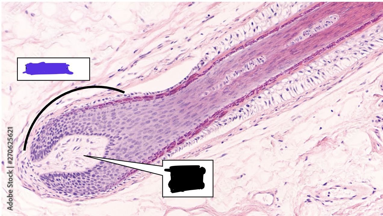
What is highlighted in black in this hair follicle?
Dermal Papilla
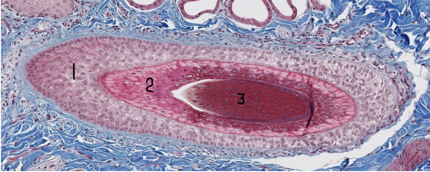
What is 1 in this hair follicle?
External root sheath
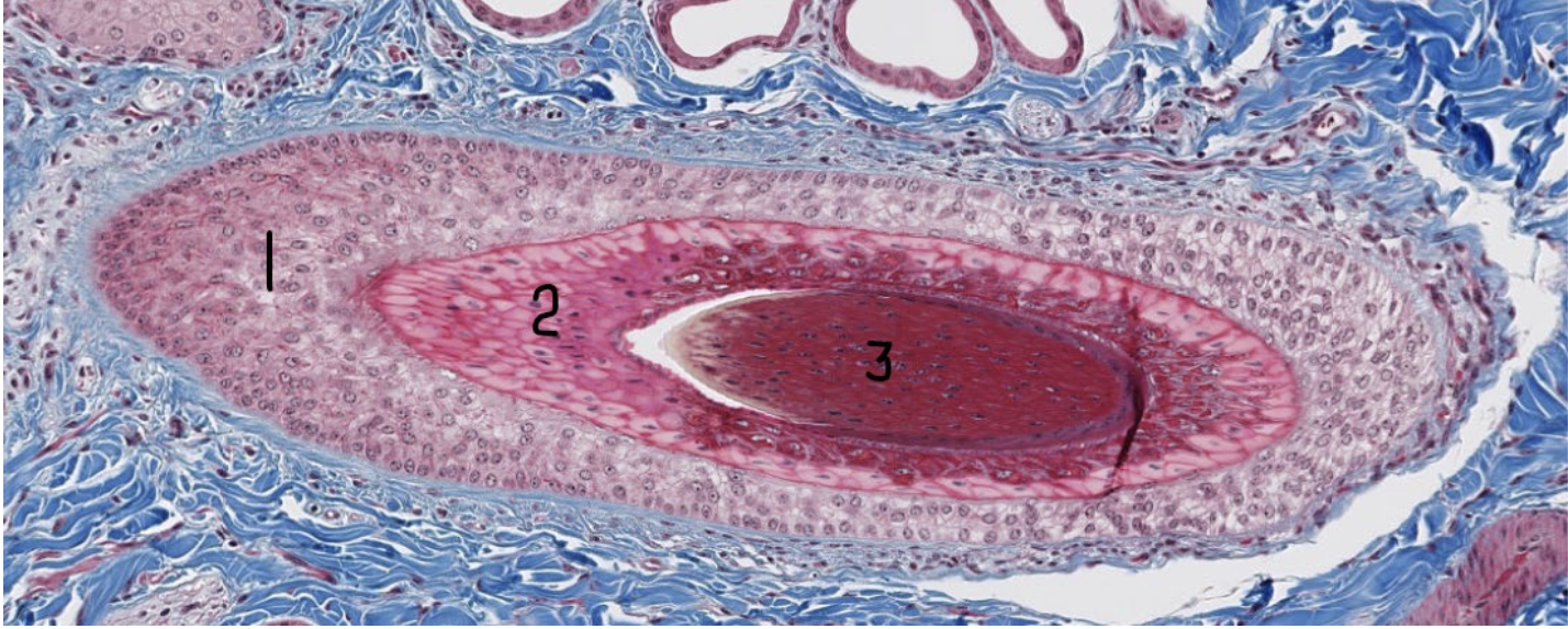
What is 2 in this hair follicle?
Internal root sheath
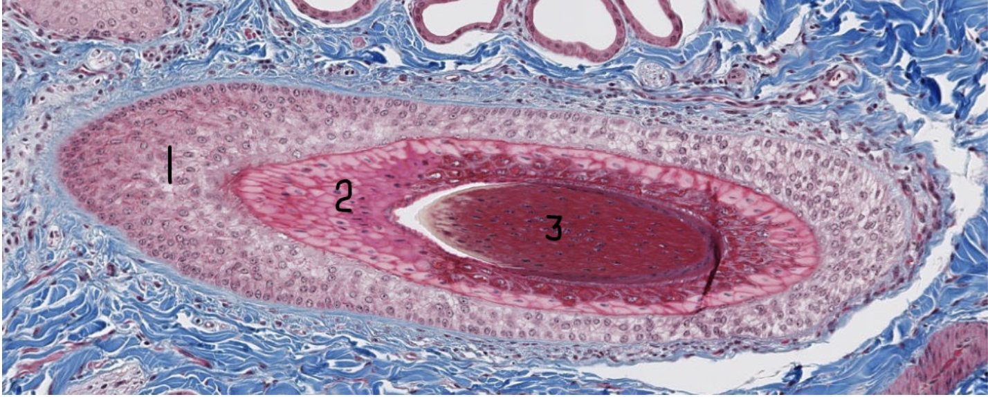
What is 3 in this hair follicle?
Hair root
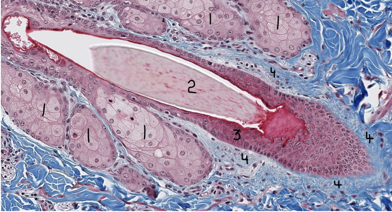
What are 1?
Sebaceous glands
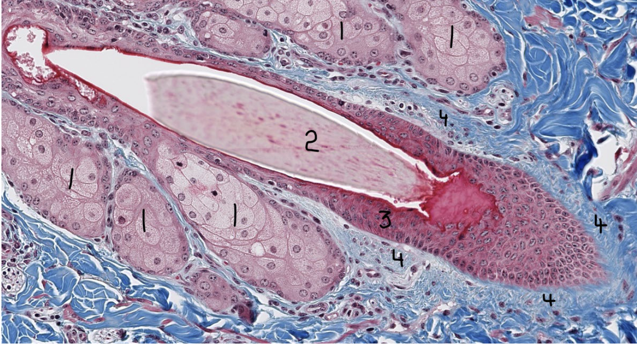
What is 2?
Hair root
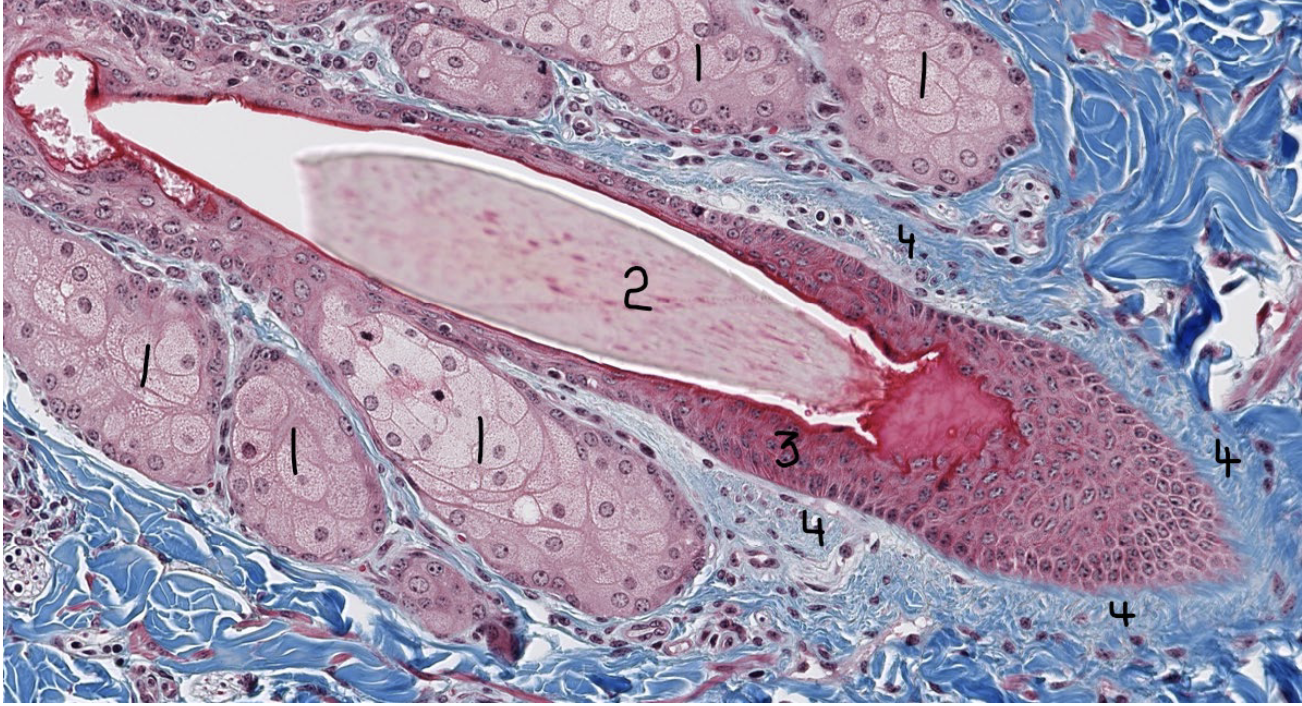
What is 3?
Hair follicle
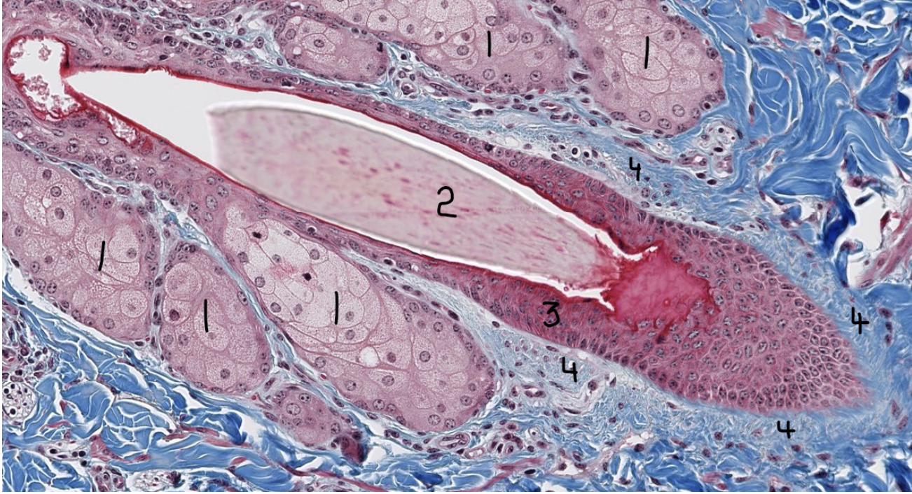
What is 4?
dermal root sheath
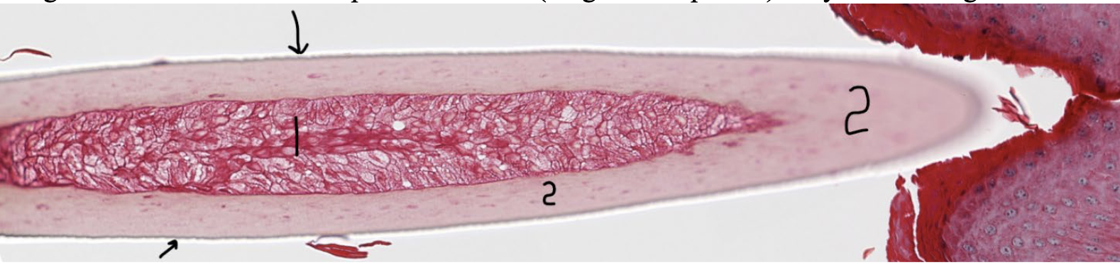
Close up of a hair shaft, what is 1?
Hair medulla
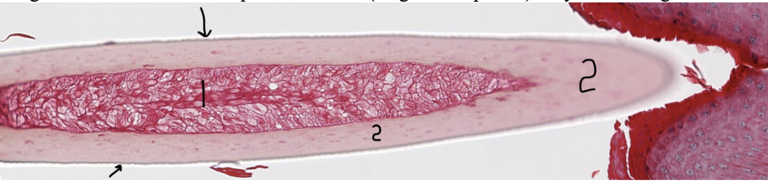
Close up of a hair shaft, what is 2?
Hair cortex
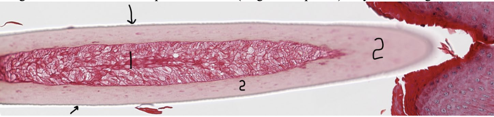
Close up of a hair shaft, what are the arrows pointing at?
Hair cuticle
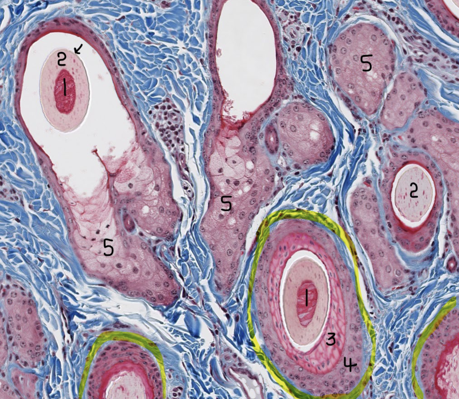
Image of haired skin, what is 1?
Hair Medulla
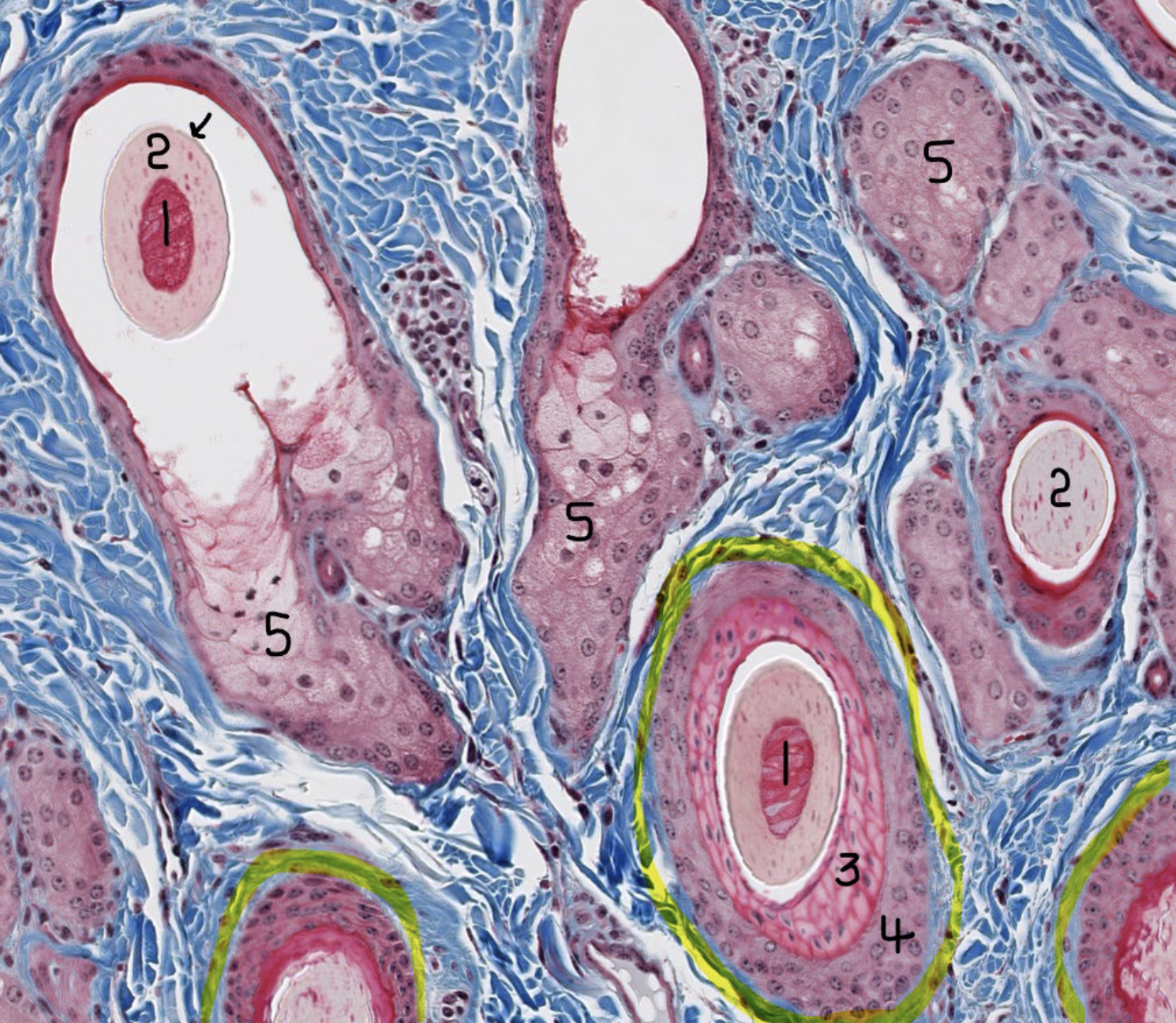
Image of haired skin, what is 2?
Hair cortex
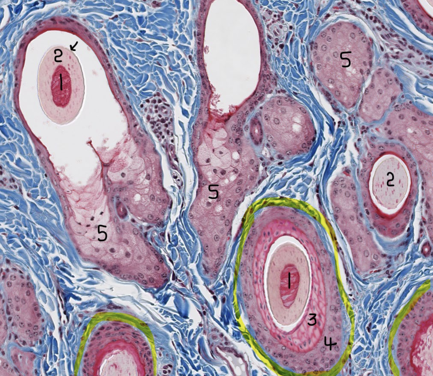
Image of haired skin, what is 3?
Inner/Internal root sheath
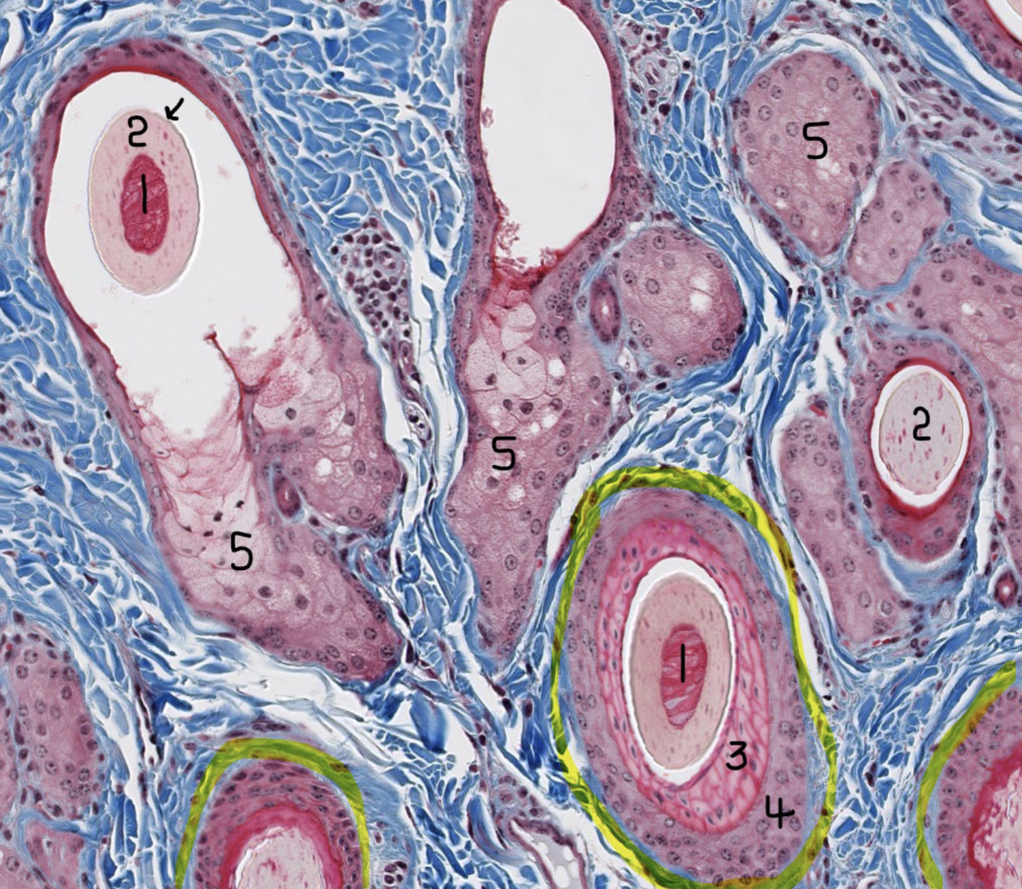
Image of haired skin, what is 4?
Outer/External root sheath
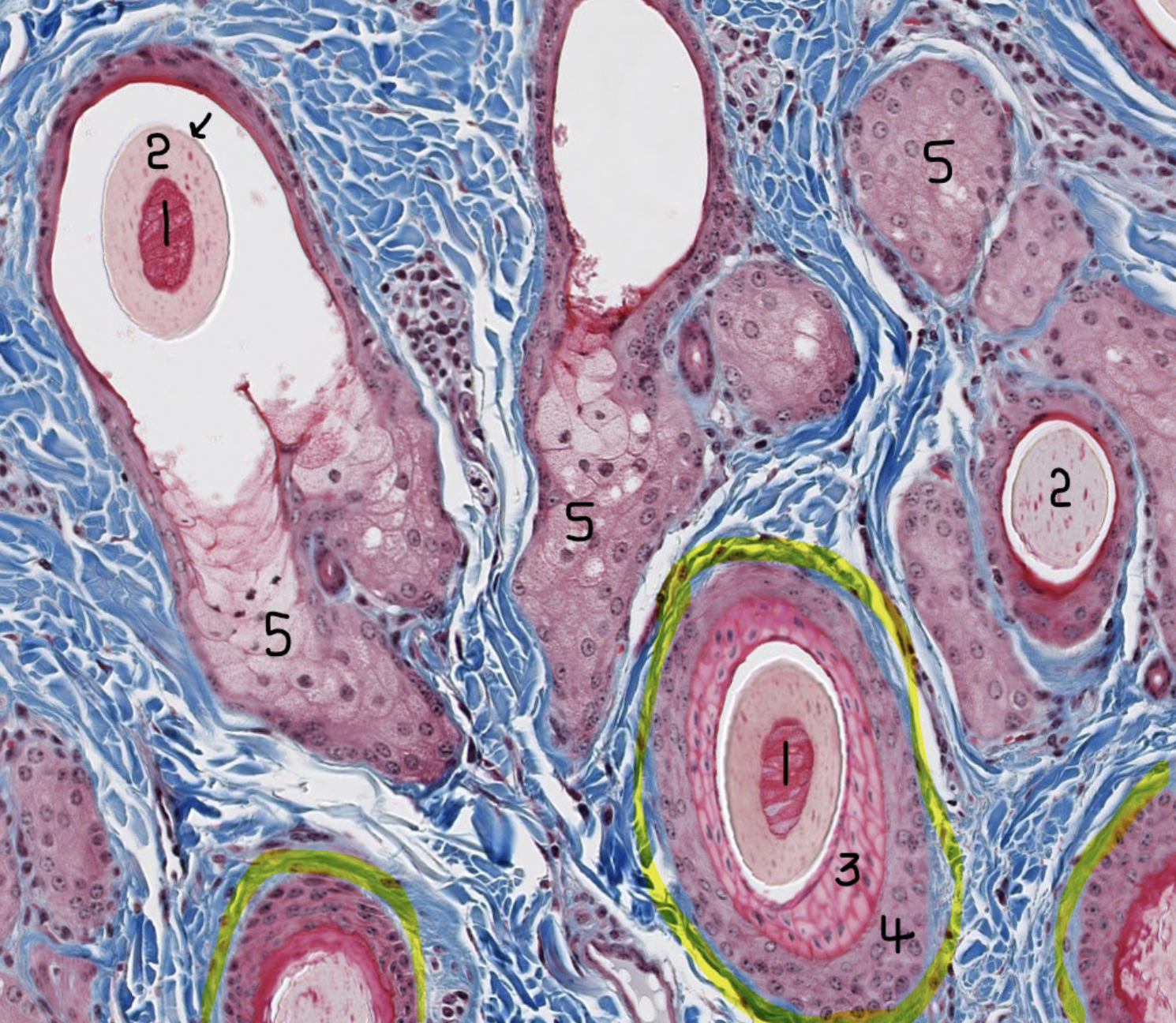
Image of haired skin, what is 5?
Sebaceous gland saccules
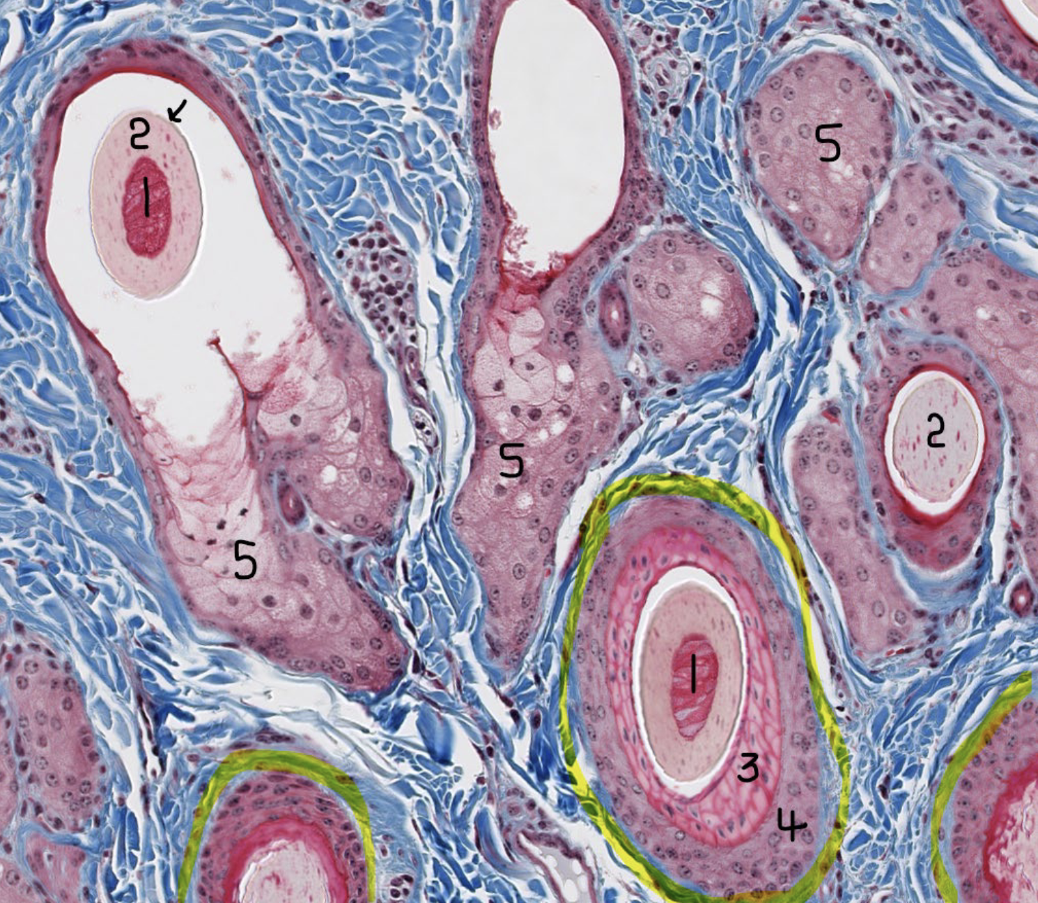
Image of haired skin, what is black arrow pointing at?
Hair cuticle
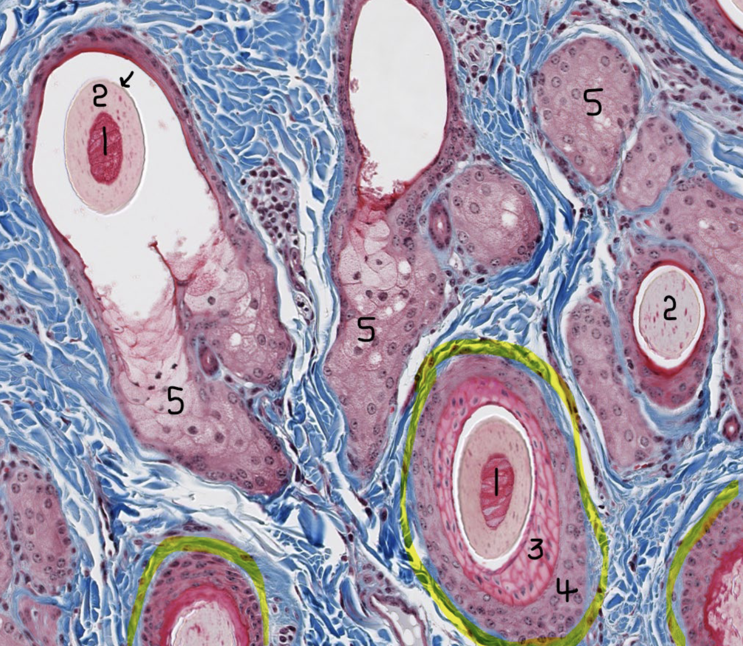
Image of haired skin, what is highlighted in yellow?
Dermal root sheath
What do sebaceous glands secrete?
Oily secretion called Sebum
What do sudoriferous glands secrete?
Sweat
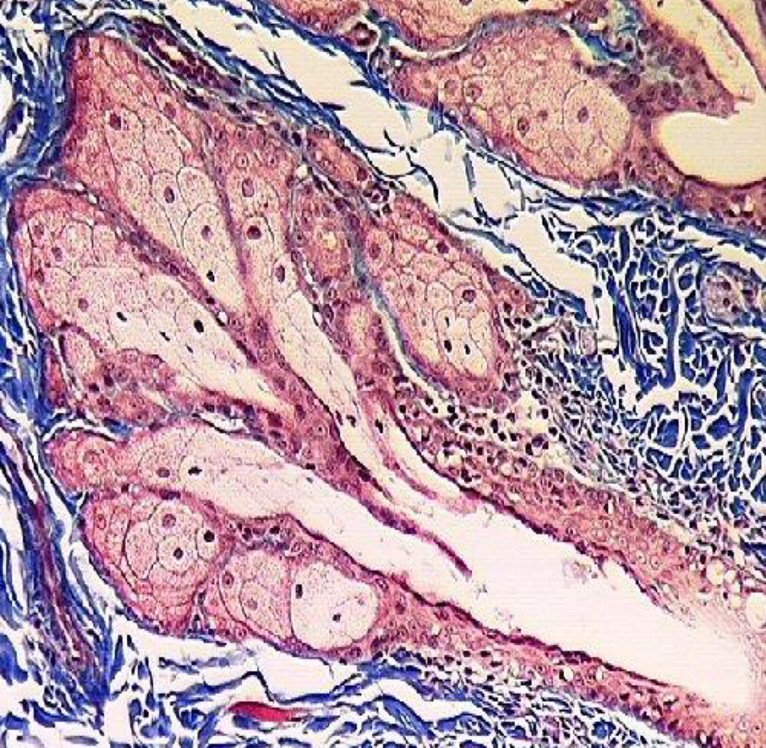
What type of gland?
Sebaceous gland
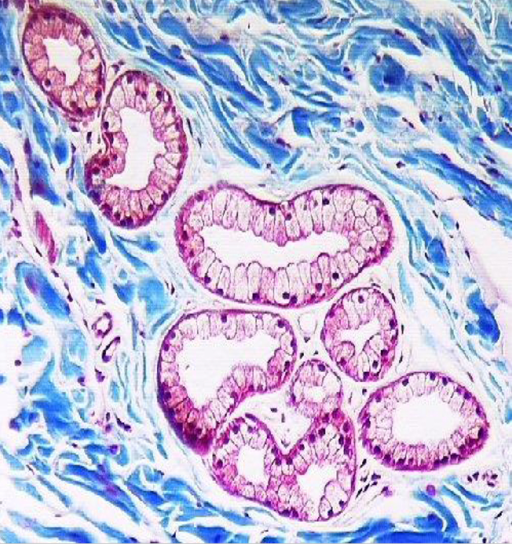
What type of gland?
Sudoriferous gland
Cerumen
(ear wax) is a combo of sebaceous and ceruminous gland secretions mixed with sloughed epithelium.
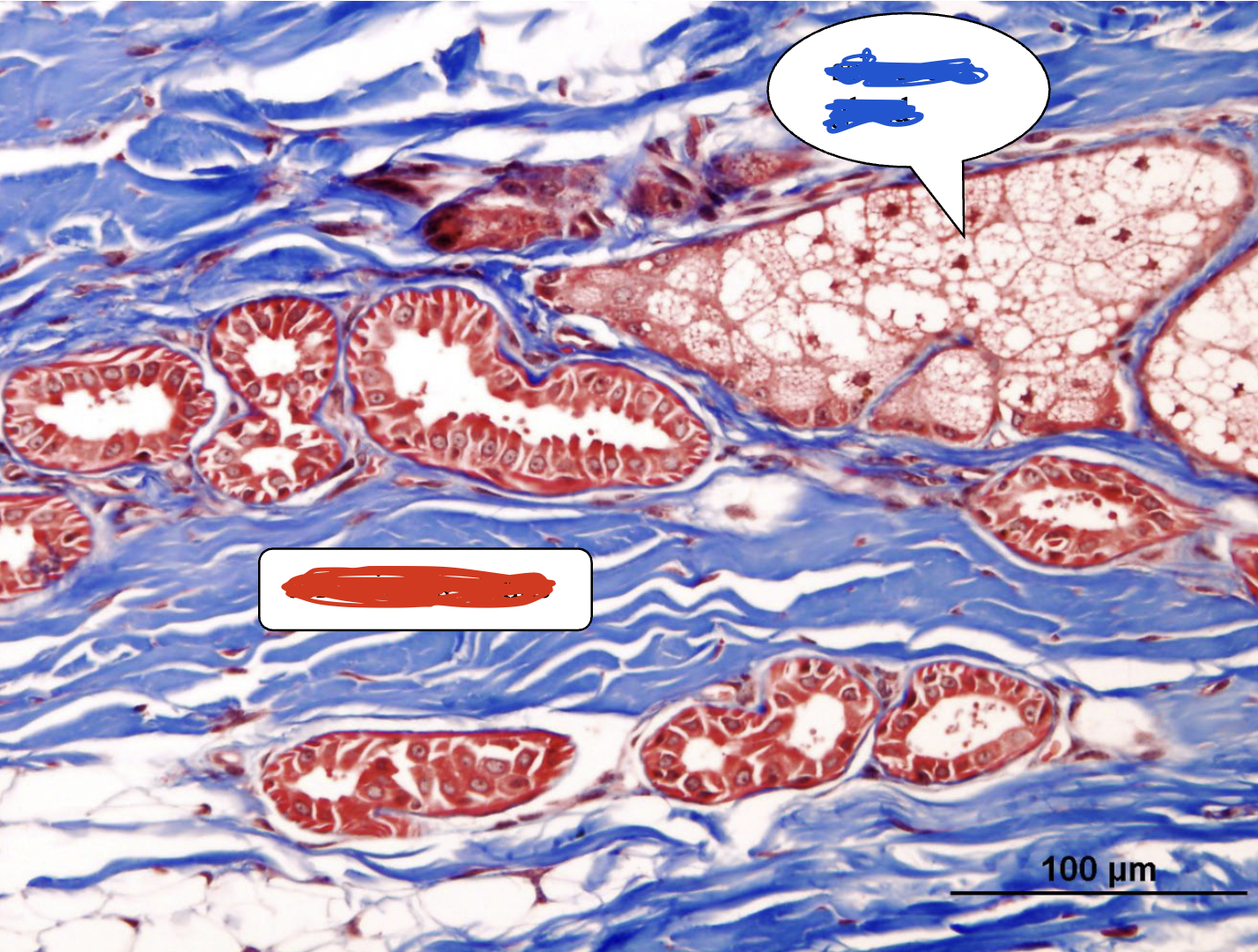
The following is a sample from the ear pinna. What structure is highlighted in blue and what structure is highlighted in red?
Sebaceous glands; Ceruminous glands
Meibomian glands
large, specialized sebaceous glands found in eyelids
Meibomian glands secrete what product?
An oily substance called meibum
What benefit does meibum possess?
Helps prevent evaporation of the tear film.
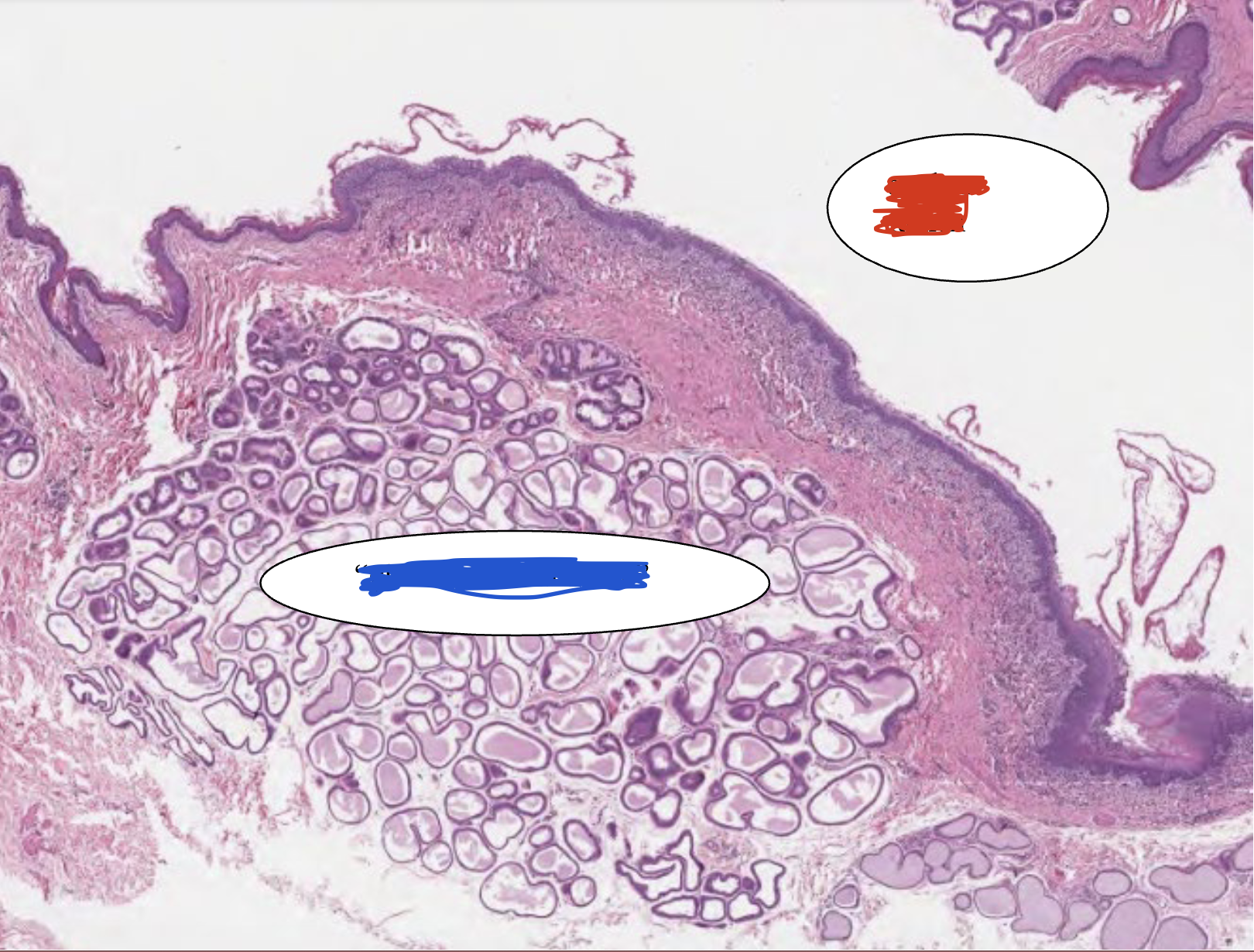
This is a portion of the anal sac. What is highlighted in blue and what is highlighted in red?
Glands of the anal sac; Anal sac lumen
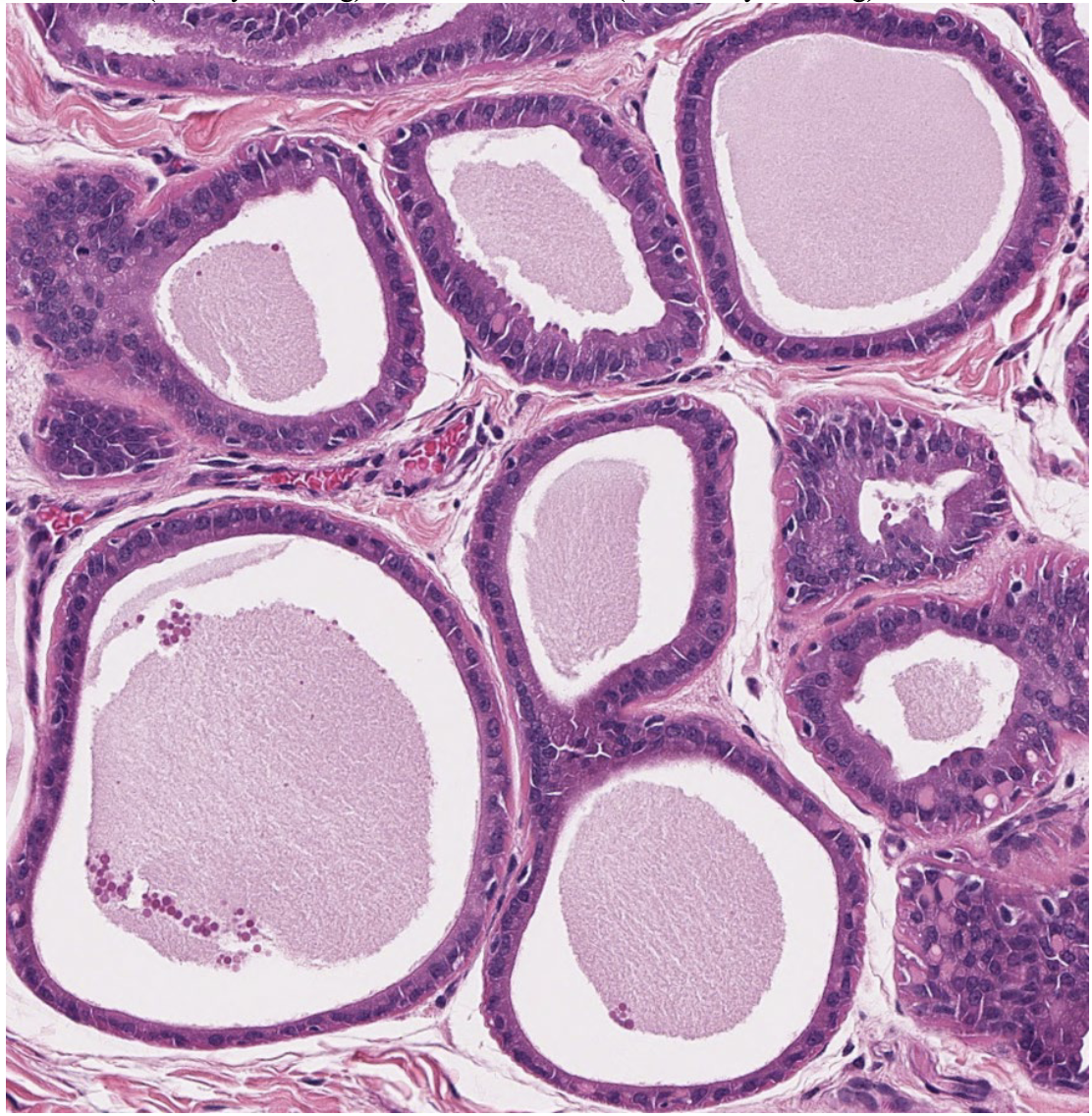
Pictured are the…
Glands of the anal sac
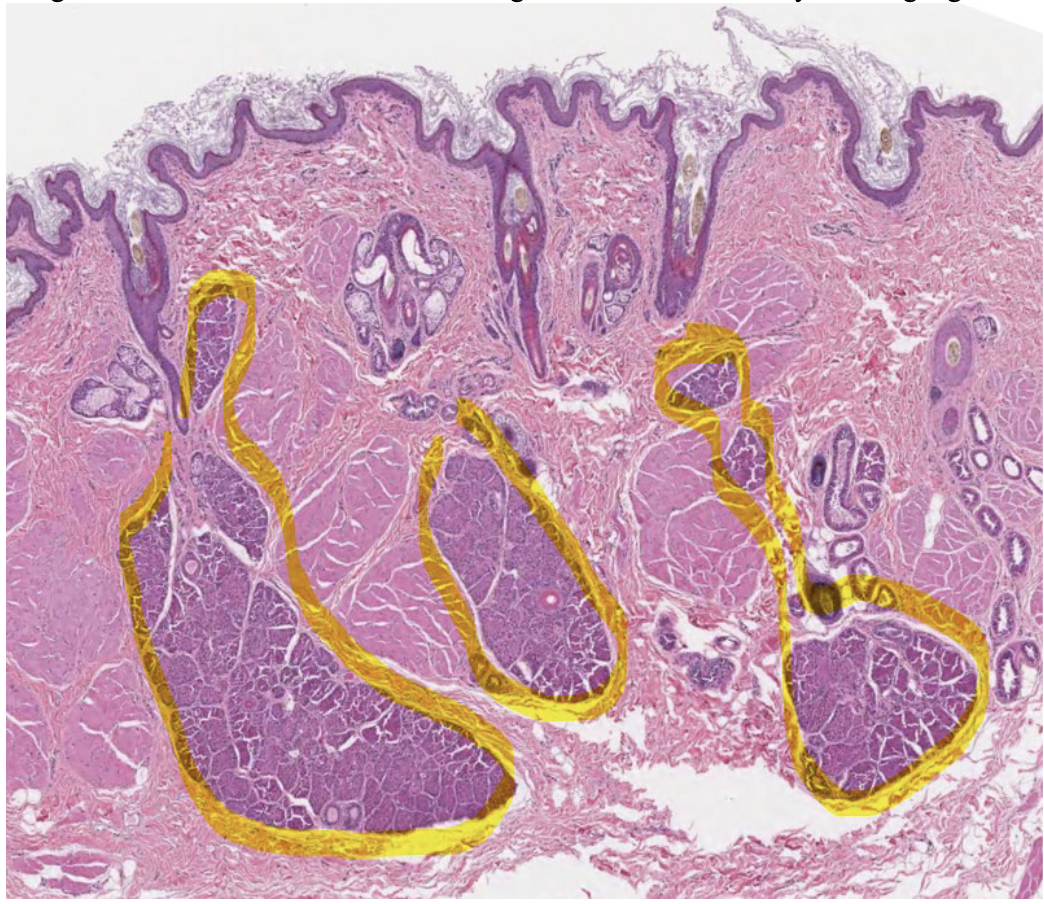
What is highlighted in yellow in this anal sac?
Circumanal glands
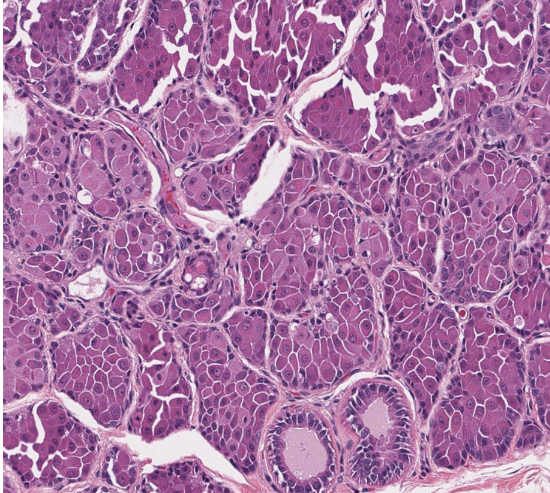
What gland is this?
Circumanal gland
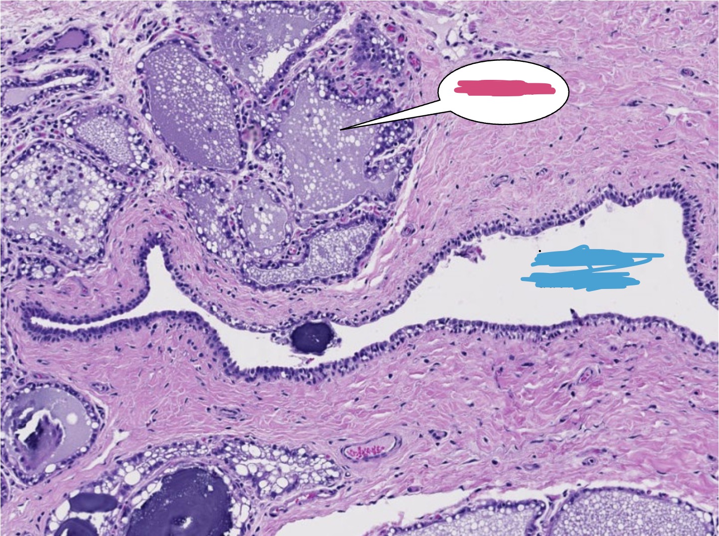
Pictured is a bovine mammary gland, what structure is highlighted in pink?
Alveolus
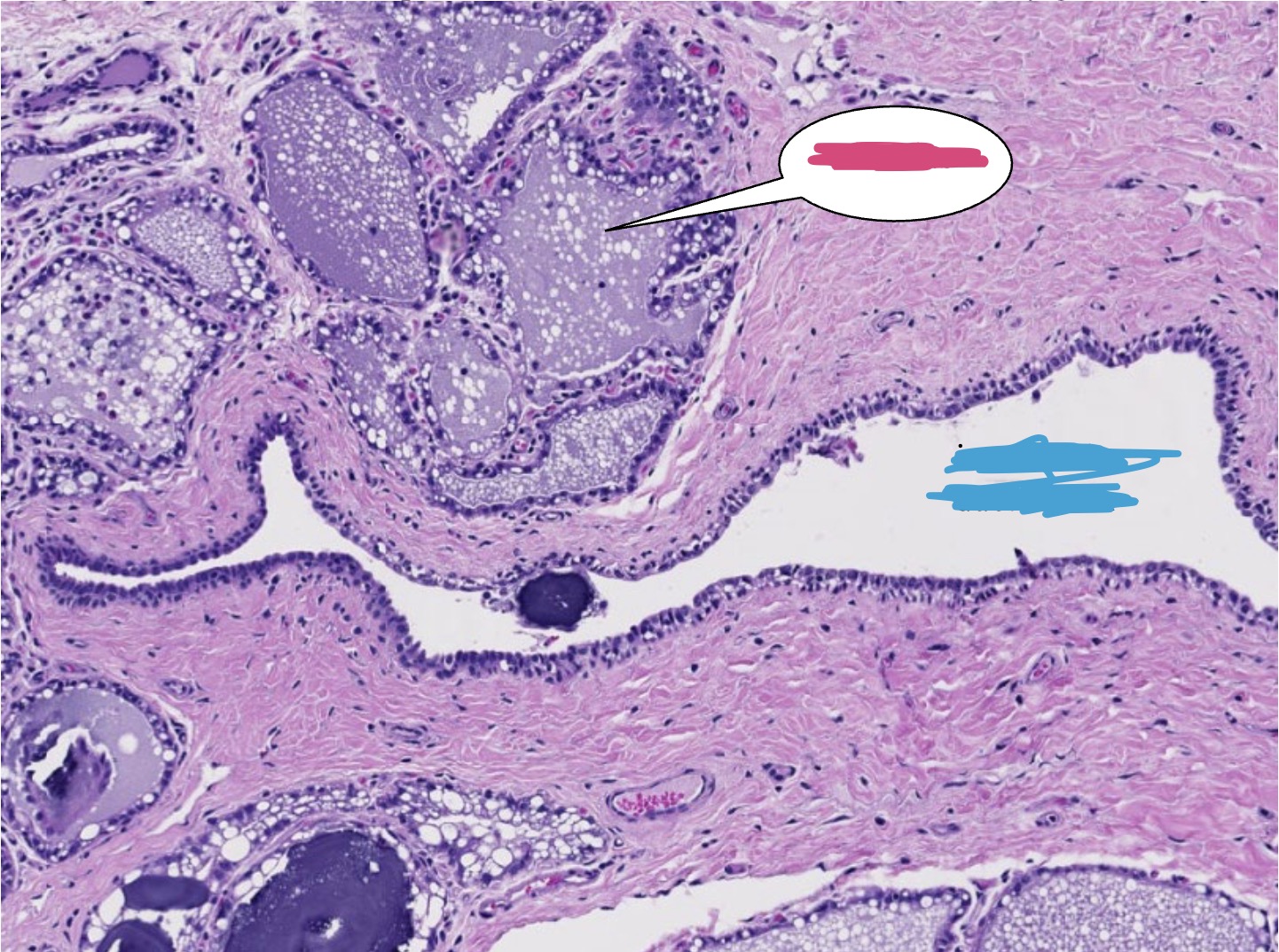
Pictured is a bovine mammary gland, what structure is highlighted in blue?
Interlobular duct lumen
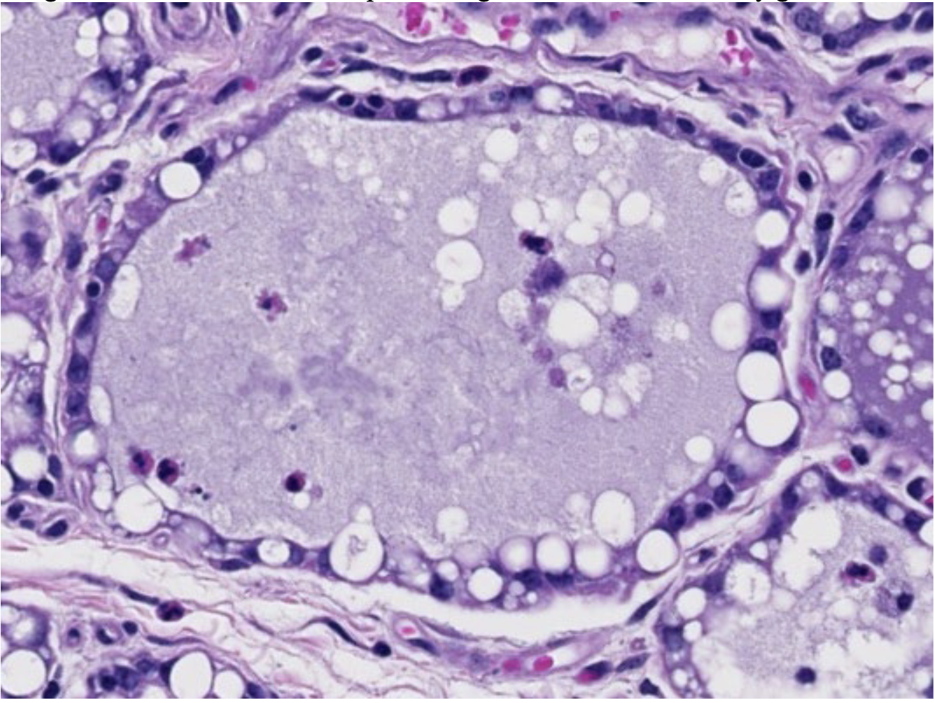
What is this?
Mammary gland alveolus
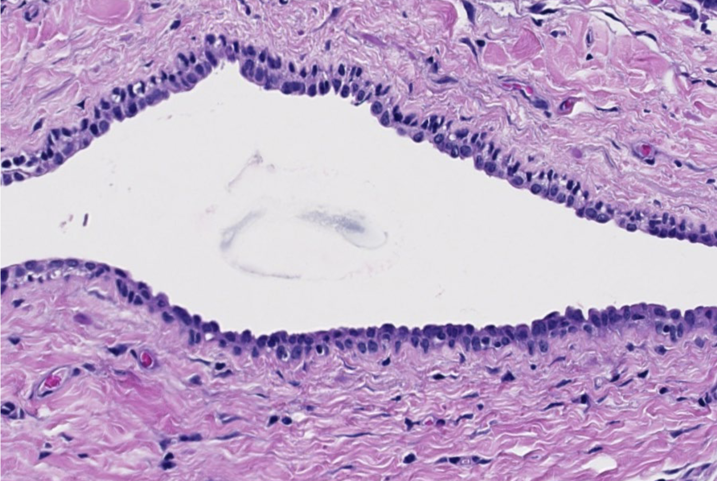
What is this?
Interlobular duct
Vacuoles
Large clear spaces within the cytoplasm
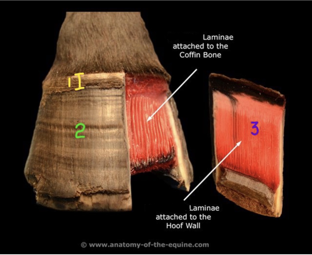
Which type of lamellae is attached to the coffin bone?
Sensitive
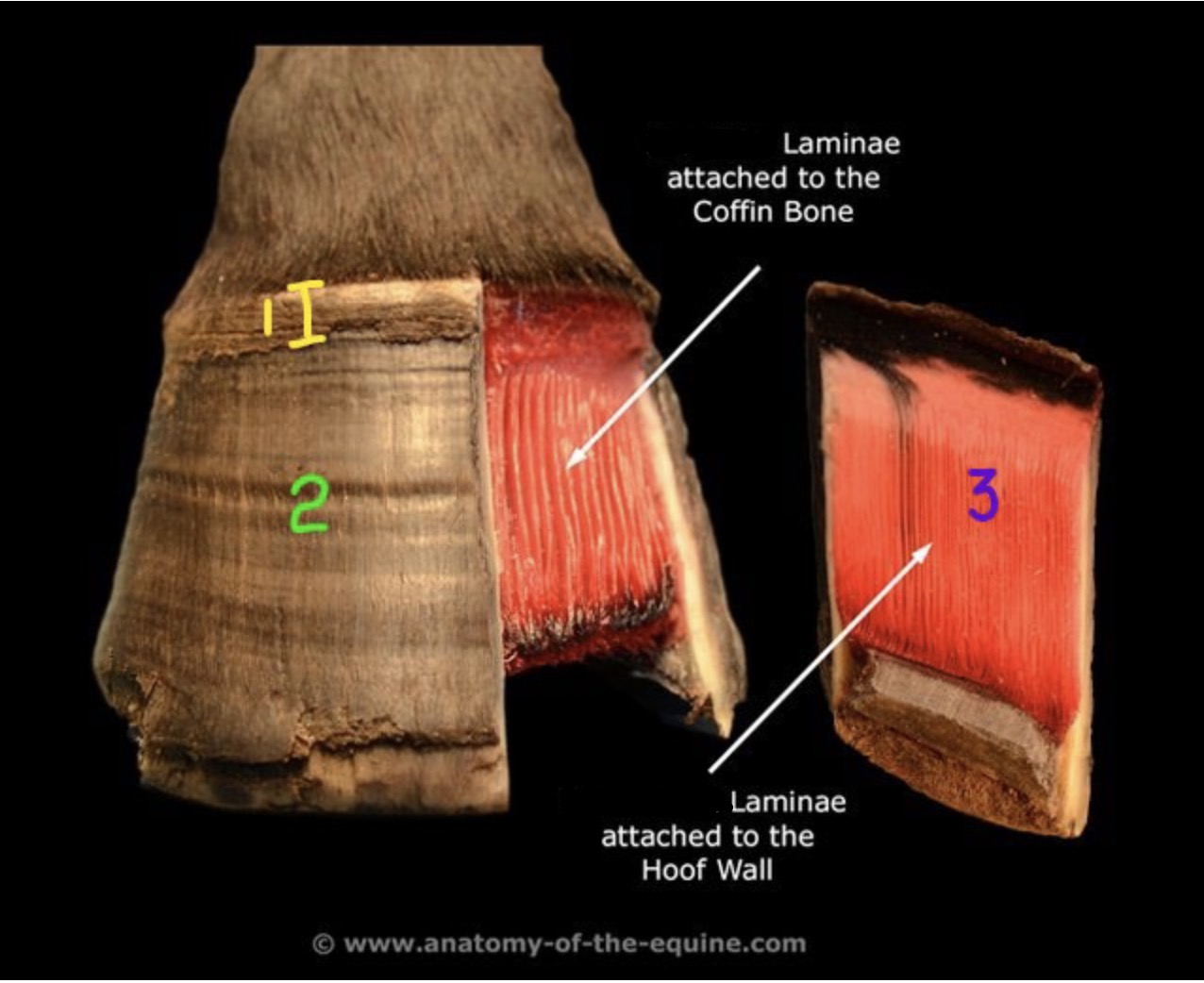
Which type of lamellae is attached to the hoof wall?
Insensitive
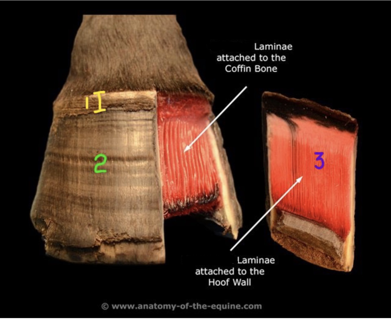
What is 1?
Stratum Externum
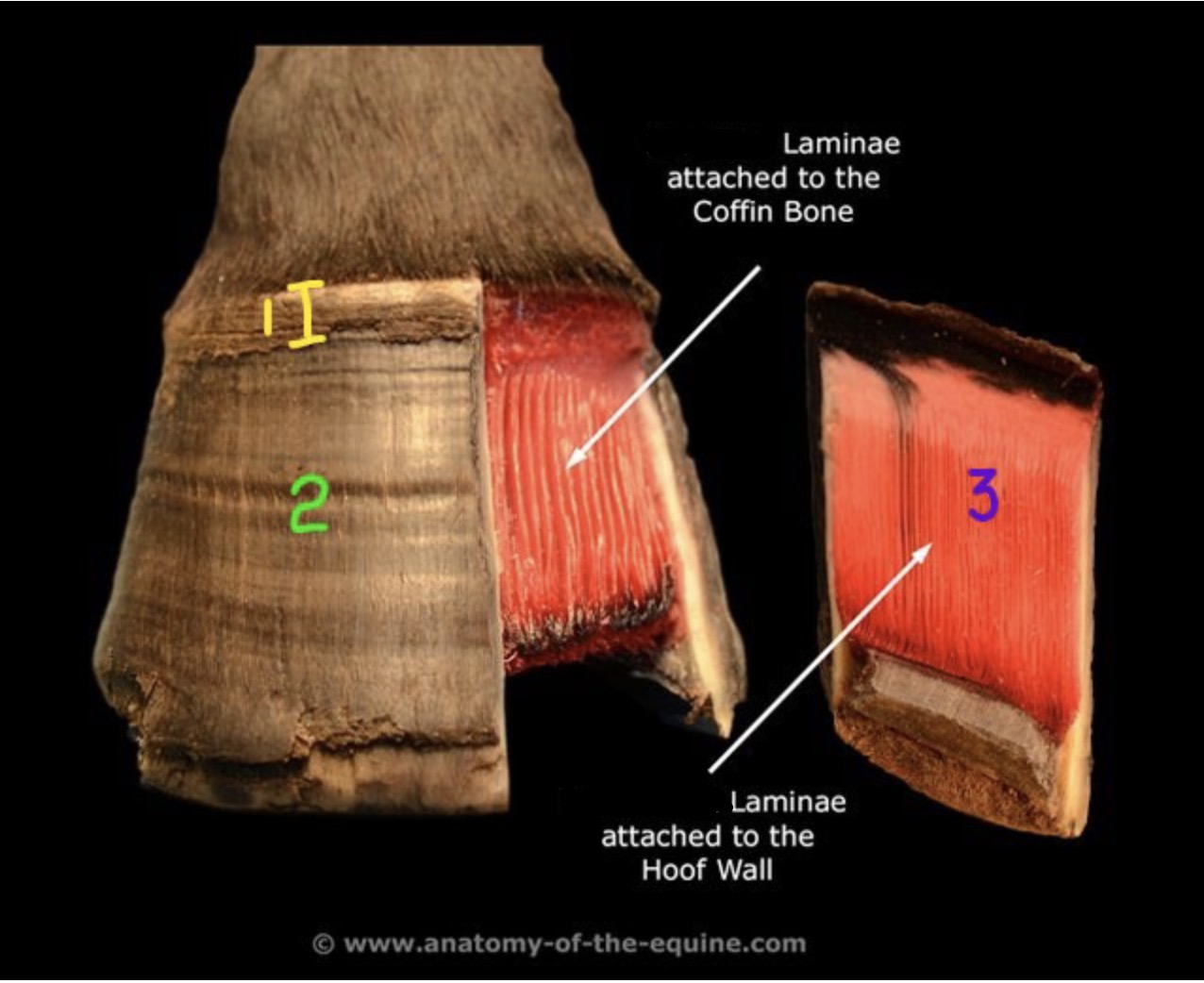
What is 2?
Stratum Medium
What is 3?
Stratum Internum
What are the sensitive layers of a hoof?
The corium (dermal structures), stratum basale & spinosm
What are the insensitive structures of the hoof?
The wall, the sole, and the frog
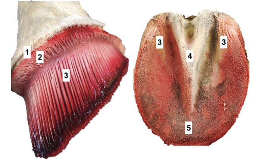
What is 1?
Perioplic groove
What is 2?
coronary groove
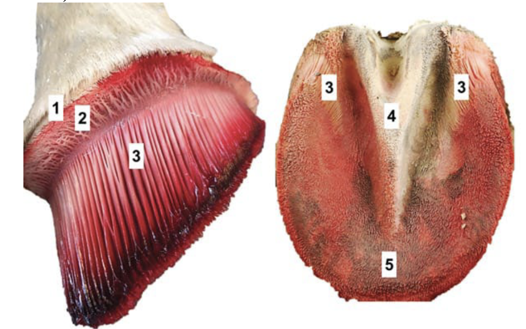
What is 3 (left image)?
stratum internum
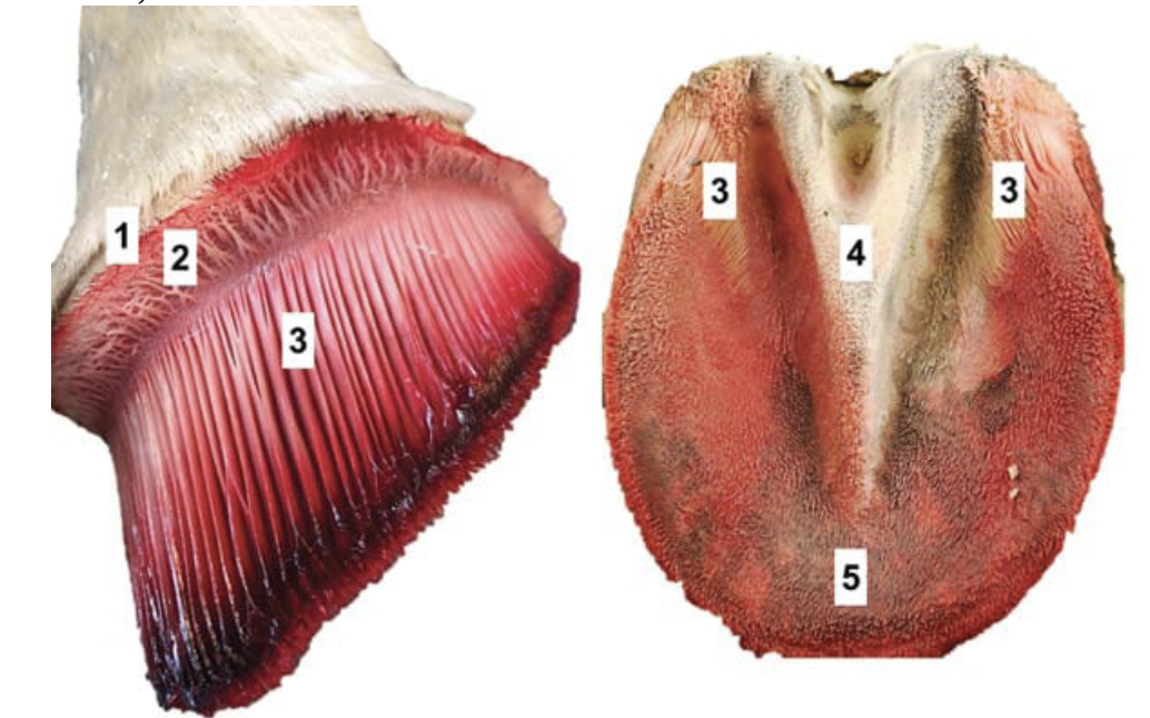
What is 3 (right image)?
laminar corium
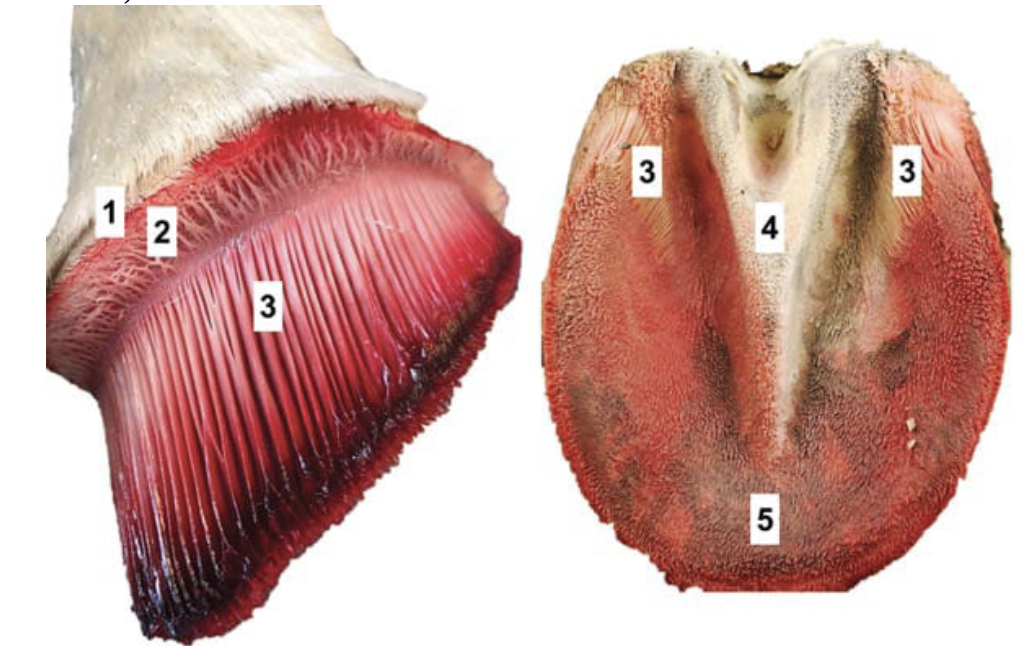
What is 4?
frog corium
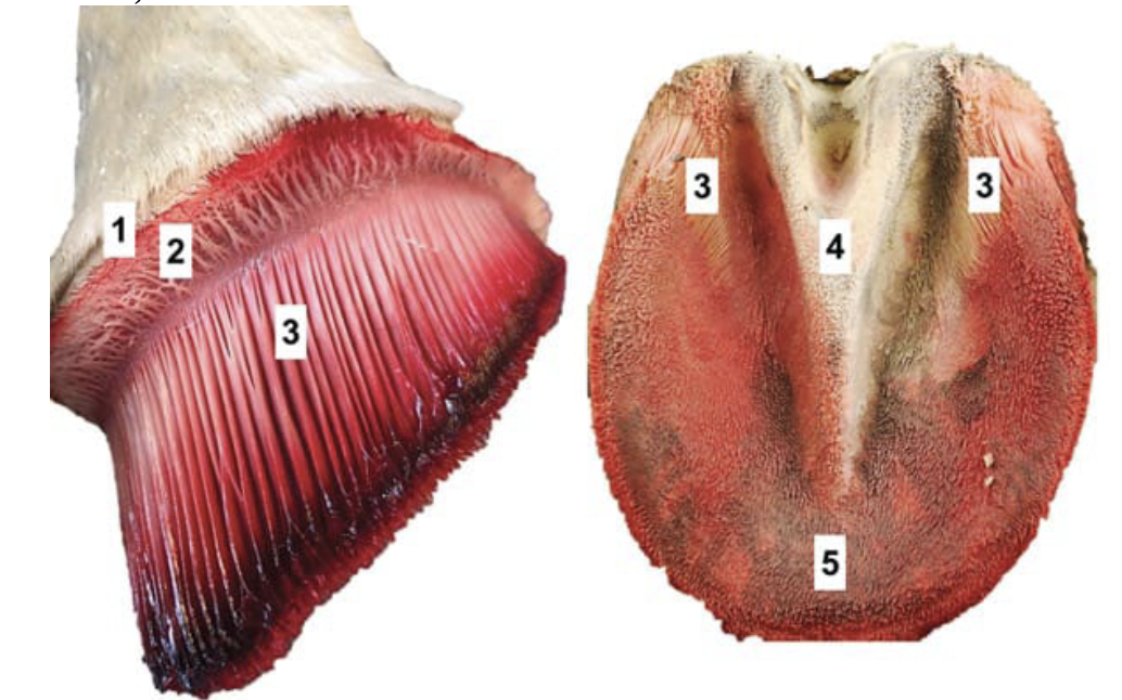
What is 5?
sole corium
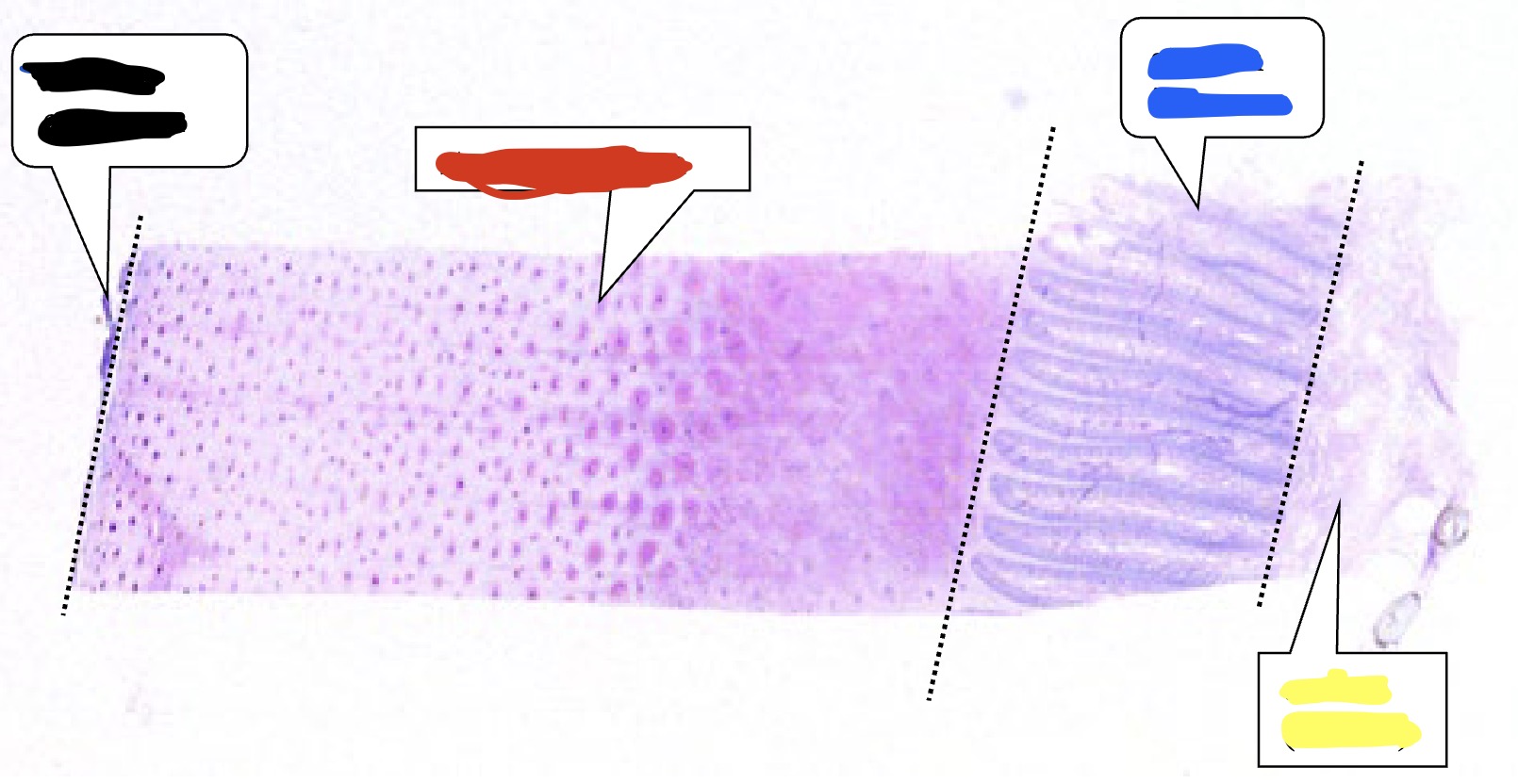
What is labeled in black?
Stratum externum
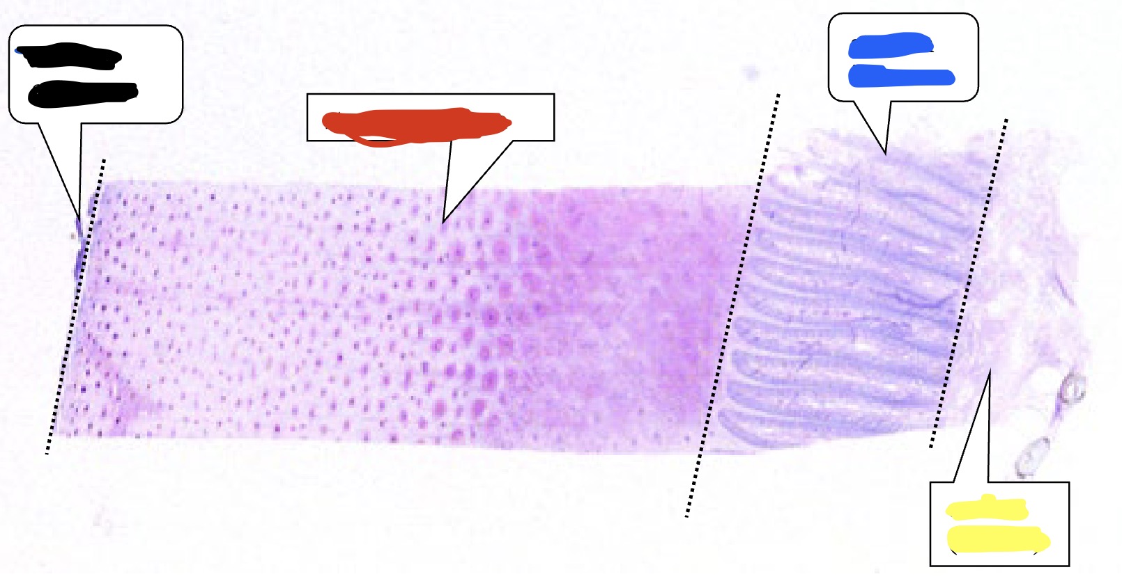
What is labeled in red?
Stratum medium
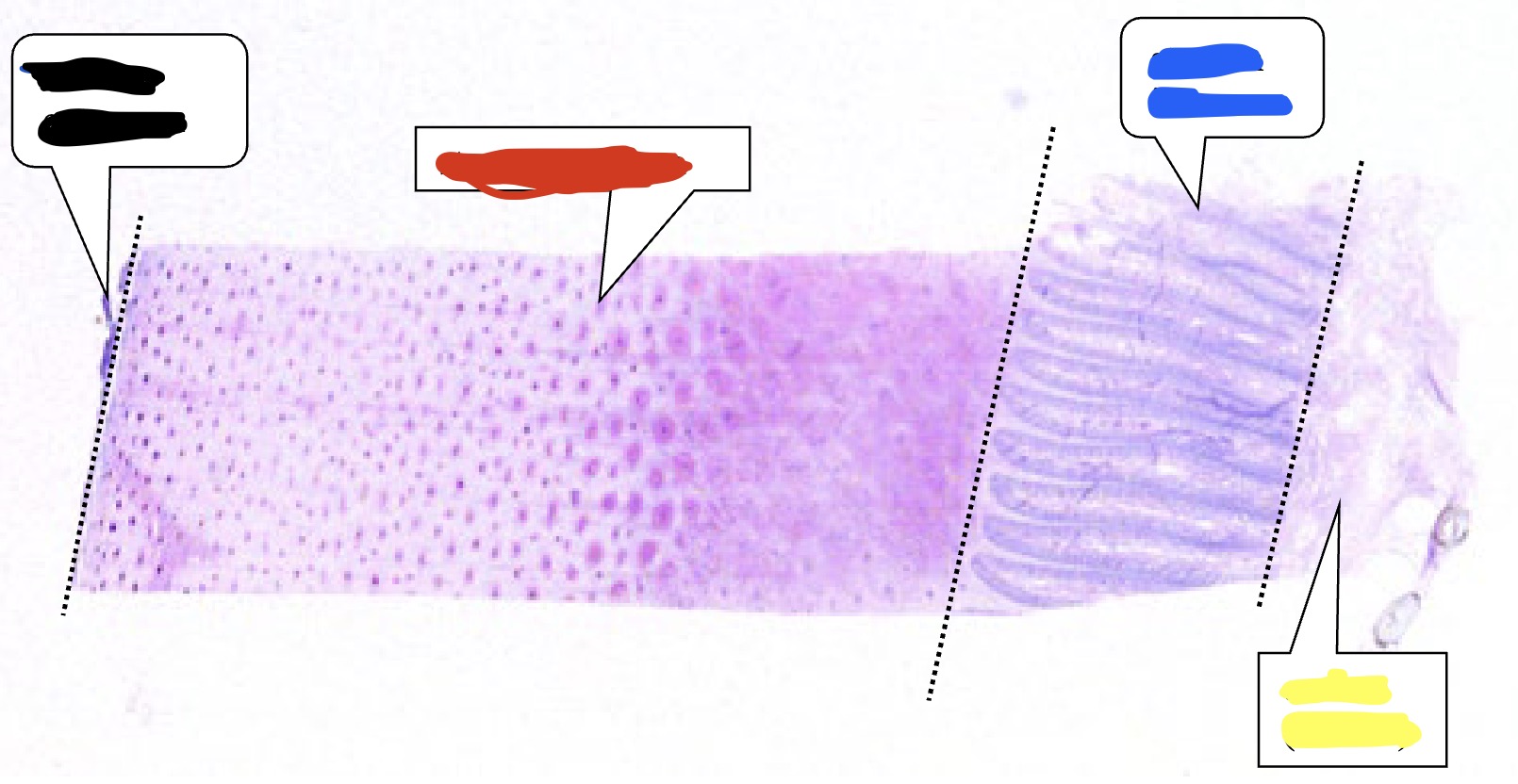
What is labelled in blue?
Stratum internum
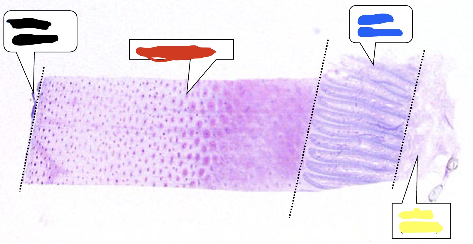
What is labelled in yellow?
Corium (dermis)
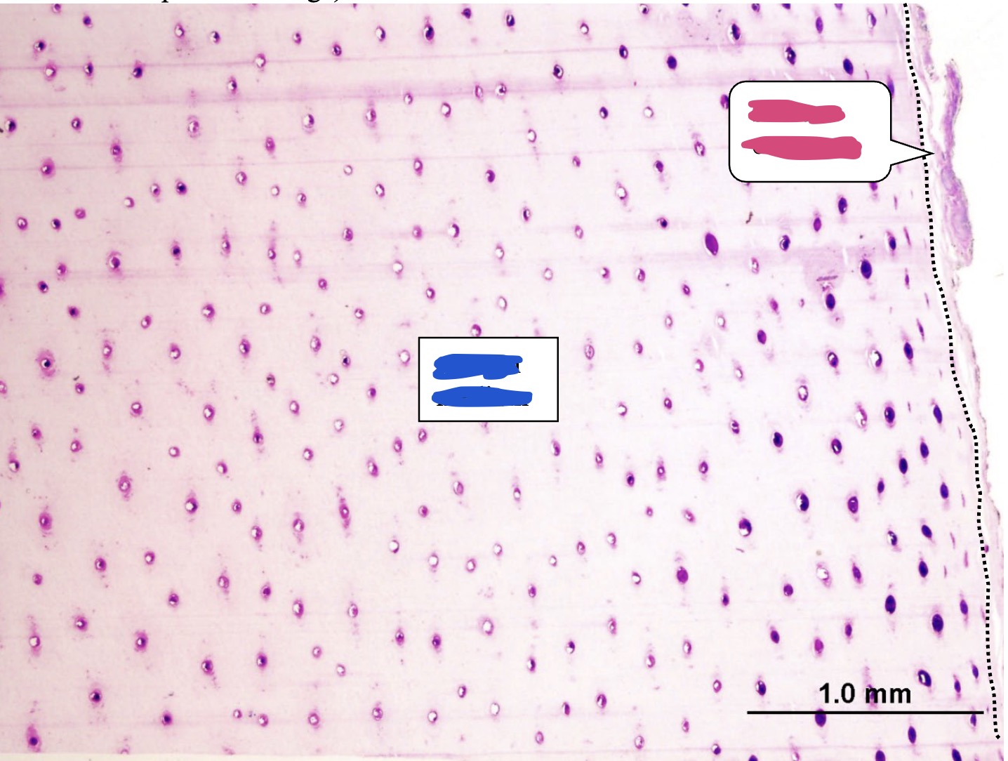
What is labelled in pink? (horse hoof)
Stratum externum
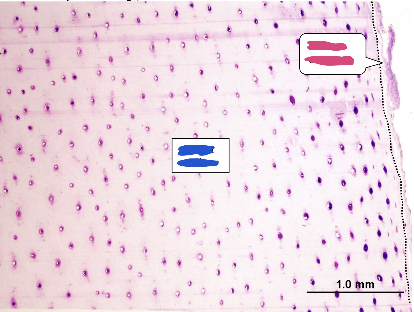
What is labelled in blue? (horse hoof)
Stratum medium
The stratum internum is composed of laminae what aspects of it are non-living?
Primary epidermal laminae (stratum corneum)