Anatomy and Embryology 1
 Call Kai
 Learn
Learn Practice Test
 Spaced Repetition
 Match
Match Flashcards
 Knowt Play
1/32

Earn XP
Description and Tags
Describe the development of different endocrine glands. Describe the gross structure, histological appearance and location of each gland with the following glands emphasized: pituitary gland; adrenal gland; thyroid gland; parathyroid gland; endocrine pancreas; pineal gland. Recognize the relationships between different endocrine glands. Describe the blood/nerve supply to each endocrine gland.
Name | Mastery | Learn | Test | Matching | Spaced | Call with Kai | Chat |
|---|
No analytics yet
Send a link to your students to track their progress
33 Terms
Pheromone
chemical which is released by an animal to be detected by another animal of the same species. Usually alters behaviours
Endocrine
chemical released by a tissue into the blood stream for action in another tissue.
Paracrine
§chemical released by a cell which targets nearby cells by diffusion.
Juxtacrine
§chemical produced (not released) by a cell which targets cells immediately in contact, usually forming ligands.
Autocrine vs Intracrine
Autocrine cell flags itself! chemical released by the cell and targets the same cell, where as intracrine works INSIDE the cell that releases it
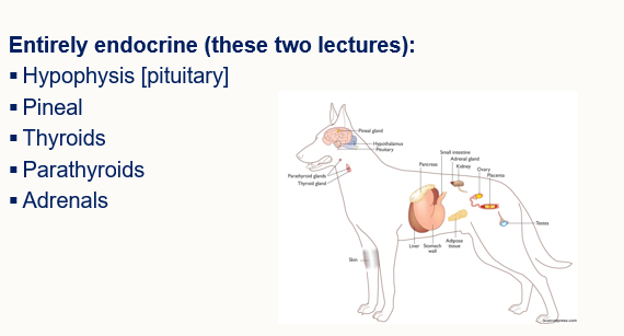
What are some examples of endocrine organs, i.e. organs which release a chemical into a tissue by the blood stream for action in another tissue…
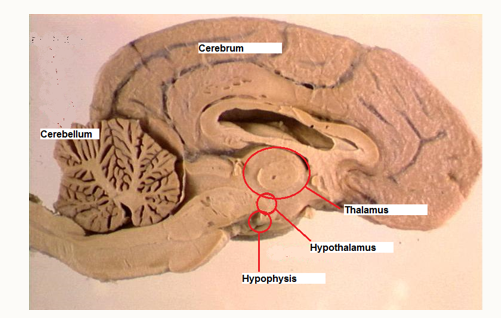
What are the components of the diencephalon
§Epithalamus (includes pineal gland).
§Thalamus.
§Hypothalamus.
§Hypophysis
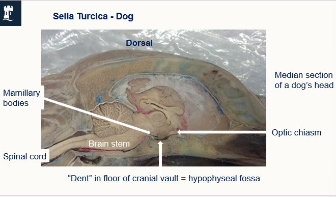
Where is the pituatory/ hypophysis located
Beneath the hypothalmas is a bony fossa
Where is the hypoohysis / pituarotye gland locataed -
hypophyseal fossa
Transverse section of a sheeps head

What is the function of the bbb
prevents large/ hydrophillic molcules in the plasma entering the extra cellular fluid. It is an extra layer of pericytes and astrocytes sounding blood capilaries in the cns.
What 2 regions of the CNS have no bbb and what do they have instead
Hypothatlamus + hypophysis (as sits just below the brain) dura mater here forms the diaphragma sellae. Instead this contrains fenestrated capillaries.
lack tight junctions, have fenerstrated capilaries so there can be free flow of hormones into circulation.
What are the anatomical parts of the pituatory gland / hypophysis
Neurohypophysis — posterior pituatory - 2 parts
Adenohypopysis - anterior pituatory - 3 parts

Adenohypohysis (low priority)
§Pars tuberalis – monitors melatonin levels. Forms a collar around the stalk.
§Pars inter
media – minor function in mammals. Surrounds neurohypophysis in equids and carnivores.
§Pars distalis – main secretory region. Collection of cells, each secrete a different hormone.

CLinical Significance of the pars intemedia in the pituatory gland..
Remenants of rathkes pouch
MSH produced in fetus - melanocyte stimulating hormone
seasonal coat colour changes
Has neuronal input from the hypothalamus + several neurotransmitters. Particularly dopamine. Adenomas of these cells can occur in aged horses.
What are the 2 parts of the neurohypophysis ( anterior pituatory)
§Infundibulum – the “stalk” attached to tuber cinereum of hypothalamus.
§Pars nervosa = neural lobe – contains projection axons from neurons in the hypothalamus.
Adenohypophysis embryology (anterius pituratoy)
Rathke’s pouch (from developing mouth)
grows upwards and cups around the neurohypophysis
(ectodermal origin means cells are epithelioid)
Neurohypophysis embryology
Down growth of ectoderm from forebrain, connects with the brain from the infundibulum.
Neurohypophysis - from brain
Adenohypophysis - from mouth

What happens when the 2 parts of the adeno + hypop squish together..
This forms the pituatory gland..
What is pituitary dwarfism
Where rathetkes pouch from the developingmouth does not differeniate fully into glandular tissue, therefore no anterior pituartory, so this causes the development of pituarory cysts. DOg remains a puppy and does not mature
How is the adenohypophysis/ pituartory gland linked to the hypothalamus
Linked hormonally via a capillary portal system. Releasing factors from the hypothalamu.
How is the neurohypopythis linked to the hypothalamus
neurologically, cell bodies + acons extend down the stalk
Pituatry blood supply
circle of willlis. portal set of blood vessels, 2x capillary systems
How does the anterior pituatory gland stimulate cell signalling
Hypothalamus produces releasing factors into the portal system. Specific target cells release stimulating hormones. I.e Releasing factor will bind to cells withthat specifc recepotror in the hypothalamus for it, which will then produce that specific hormone.
How does the neuro hypophysis stimulate cell signalling
like half a NMJ. released from supraoptic region of the hypothalamus. via infundibulum down axons directly to blood stream where it can diffuse in.
What are the only 2 hormones released by neurohypophysis
OXYTOCIN + ADH
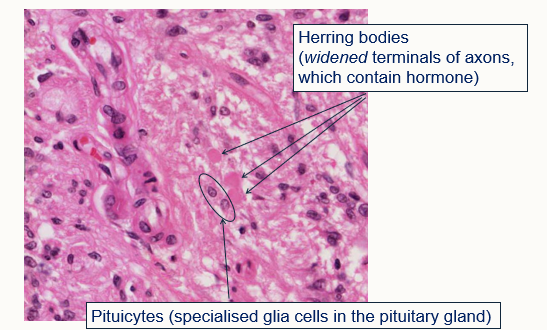
What can you see on histology of the neurohypophyseal histology 0 wide herring bodies which have hormones in the axons…
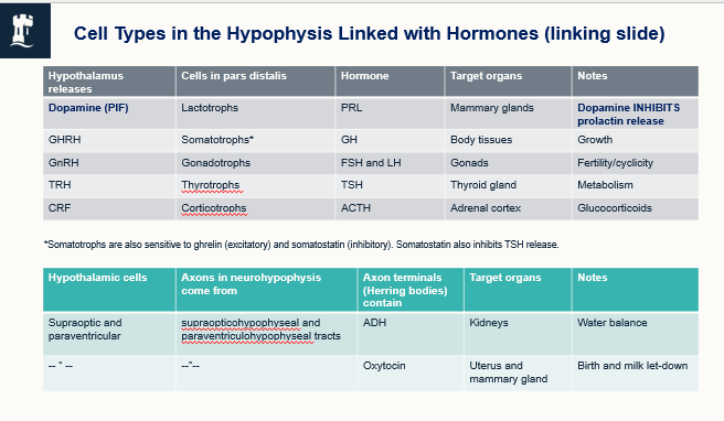
Cell types and hormone table (for referance)
OVerview and role of the pineal gland
Circaidian rythmns
+ breeding systems
functional eye in reptles and fish (not in crocadiles or armadillos)
Pineal gland location

Pineal gland how does it produce melatonin
secretes melatonin
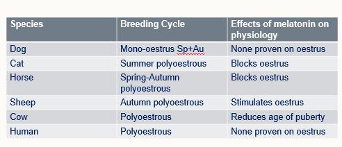
- different species responses
controls physiology and behaviour
can determine cycling depending on the day length
What is the difference between short and long day cyclers
Winter = less light = more melatonin.
Inhibits reproductive cycles in long-day breeders (horses).
Stimulates reproductive cycles in short-day breeders (sheep).
And vice versa.
Compartive melatonin cycles (for refereance)
dark = more melatonin - species specific affects