Control of eukaryotic gene expression
 Call Kai
 Learn
Learn Practice Test
 Spaced Repetition
 Match
Match Flashcards
 Knowt Play
1/29

There's no tags or description
Looks like no tags are added yet.
Name | Mastery | Learn | Test | Matching | Spaced | Call with Kai | Chat |
|---|
No analytics yet
Send a link to your students to track their progress
30 Terms
Control of eukaryotic gene expression
genes contained in nuclei of all somatic cells are exactly the same, but the cell types differ morphologically and functionally
differences between cell types are due to differential gene expression i.e. the expression of different sets of genes by cells with the same genome
regulation of gene expression gives a cell control over its structure and function —> allows cell differentiation to occur
only a fraction of genes in a eukaryotic cell are expressed at any one place (tissue specificity) and at any one time (temporal specificity)
gene expression, the generation of a protein or RNA product from a particular gene, is controlled/regulated by complex mechanisms in eukaryotes
Eukaryotes VS Prokaryotes
Eukaryotic DNA is organised into nucleosomes
genes must be in an active structure to be accessible to transcription factors and RNA polymerase —> during transcription
Eukaryotic genes are not organised into operons
genes encoding proteins that function together are usually located on different chromosomes
each gene needs its own regulatory sequence i.e. promoters, silencers and enhancers
The processes of transcription and translation in eukaryotes are separated by the nuclear envelope
eukaryotic pre-mRNA must be processed and translocated out of the nucleus before it is translated
Levels of control of eukaryotic gene expression
chromatin level: histone modification and DNA methylation
transcriptional level: initiation of transcription (control elements and proteins)
post-transcriptional level: 5’ capping, 3’ polyadenylation and splicing
translational level: half-life of RNA and initiation of translation
post-translational level: biochemical modification and protein degradation
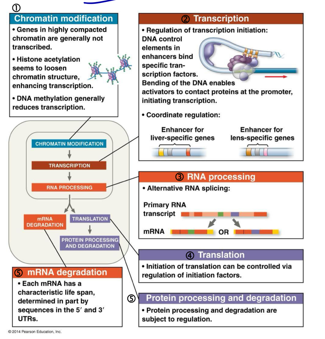
Control of eukaryotic gene expression: Chromatin level
a common and most important control point for gene expression is transcription
transcriptional control is often in response to signals coming from outside the cell e.g. hormones or other signalling molecules
includes a few regulatory mechanisms, namely regulation of gene accessibility (histone acetylation and DNA methylation) and initiation of transcription
Chromatin level: Regulation of gene accessibility
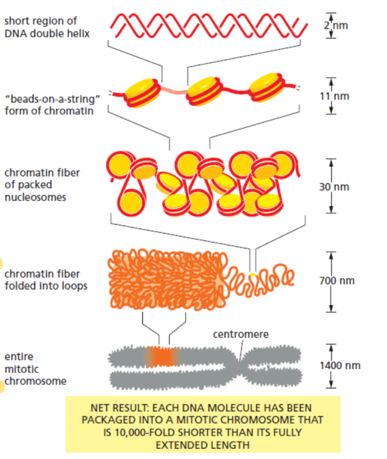
structural organisation of chromatin not only compacts DNA into a form that fits inside the nucleus but also important in control of gene expression
chromatin has the appearance of “beads on a string”
each bead is a nucleosome comprising DNA comped with a histone octamer
chromatin within a nucleus can be organised into
euchromatin, which is diffused
heterochromatin, which is highly condensed
genes found in euchromatin are available for transcription, whereas those in heterochromatin are transcription ally inactive
condensation prevents transcription factors and RNA polymerases from gaining access to the promoter of a specific gene, thus inactivating transcription of that gene
2 mechanisms (histone modification and DNA methylation) regulate a gene’s access to transcription factors and RNA polymerases
histone modification
DNA methylation
Chromatin level: Histone modification
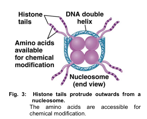
the N terminus of each histone in a nucleosome protrudes outwards from the nucleosome
for every nucleosome, 8 histone N-termini protrude outwards - protrusions known as histone tails
histone tails are rich in lysine residues, which are positively-charged
they interact strongly with the negatively-charged phosphate groups of the DNA backbone and increase the affinity of DNA for the nucleosome surface (bind more strongly)
these histone tails are accessible to various modifying enzymes, which catalyse the addition or removal of specific chemical groups
these chemical groups alter the tightness of DNA winding around the histone, thus altering the ease of transcription initiation
Chromatin level: Histone modification
the N terminus of each histone in a nucleosome protrudes outwards from the nucleosome
for every nucleosome, 8 histone N-termini protrude outwards - protrusions known as histone tails
histone tails are rich in lysine residues, which are positively-charged
they interact strongly with the negatively-charged phosphate groups of the DNA backbone and increase the affinity of DNA for the nucleosome surface (bind more strongly)
these histone tails are accessible to various modifying enzymes, which catalyse the addition or removal of specific chemical groups
these chemical groups alter the tightness of DNA winding around the histone, thus altering the ease of transcription initiation
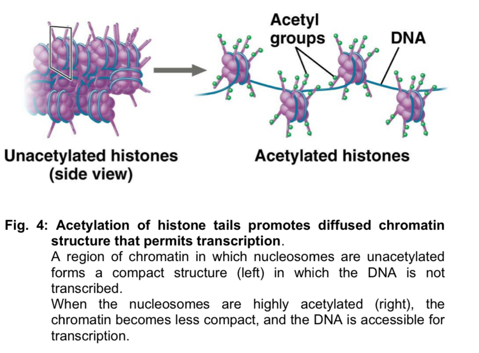
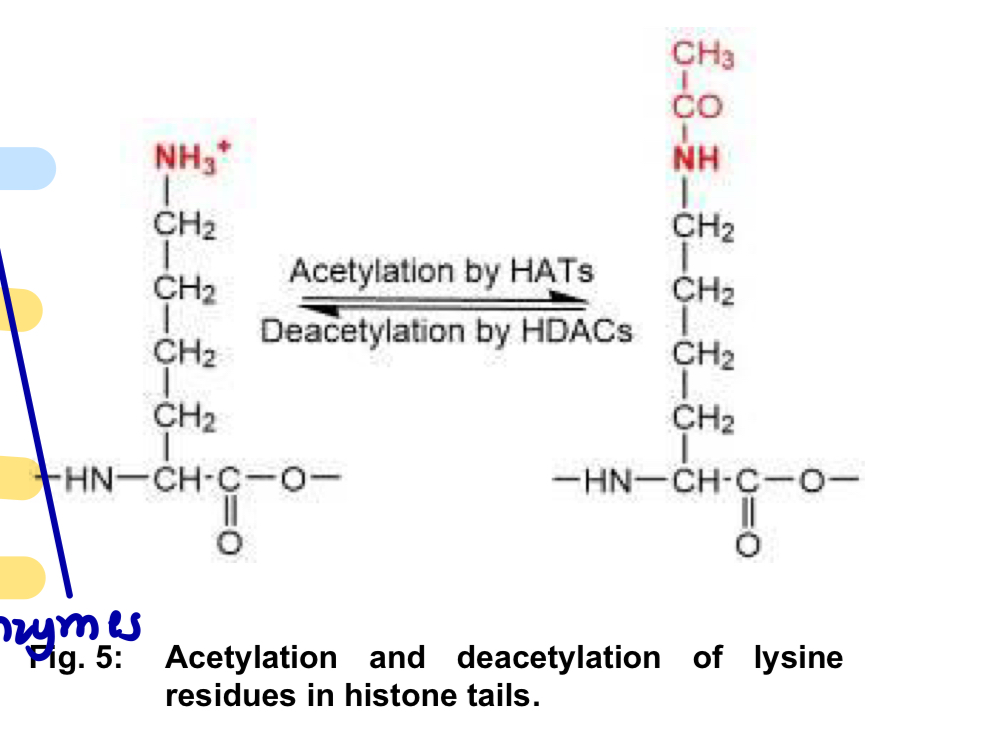
Histone modification - Deacetylation
Deacetylation
histone deacetylases (HDACs) catalyse the deacetylation of acetylated lysine residues in histone tails
lysine residues regain their positive charges, resulting in an increase in the affinity of the histone complex for DNA
consequently, the chromatin becomes more compact and prevents access of transcription factors and RNA polymerase to the control regions of genes
Regulation of gene accessibility - DNA Methylation
DNA can be covalently modified by the addition of methyl groups (CH3) to specific nucleotides after DNA replication
DNA methylation in vertebrate DNA is restricted to cytosine (C) nucleotides in the sequence 5’— CG — 3’ (known as CpG dinucleotides)
*CpG: cytosine linearly followed by guanine
this process is catalysed by DNA methyltransferases
*DNA methylation is only for cytosine nucleotides
2 mechanisms to explain how DNA methylation represses gene expression:
Methylation changes the 3D conformation of DNA and thus, prevents the binding of transcription factors to the promoter
transcription initiation is prevented
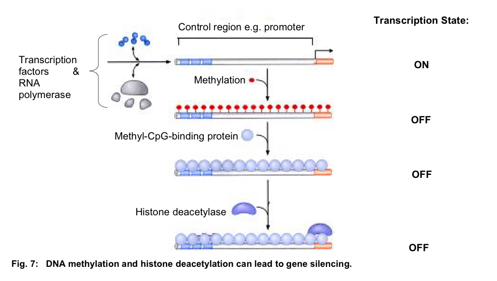
Methylated DNA serves as recognition signals for methyl-CpG-binding proteins (MeCPs) that, in turn, recruit other proteins such as histone deacetylases (HDACs)
HDACs modify chromatin in the region of the CpG island such that it becomes more condensed
this prevents binding of the transcription factors and RNA polymerase
transcription initiation is prevented
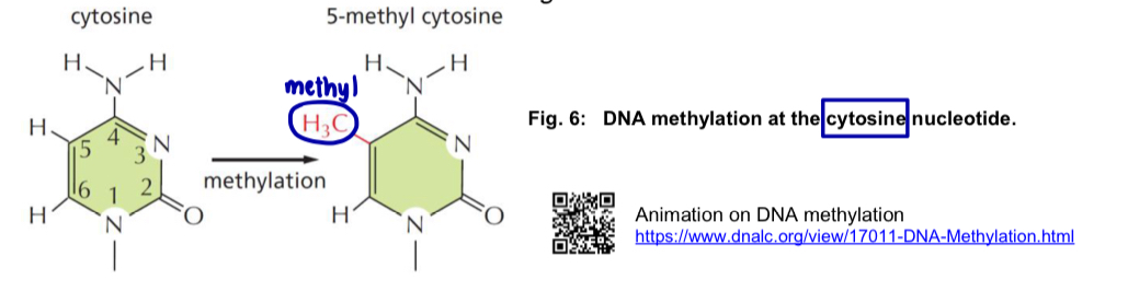
figure showing DNA methylation and histone deacetylation, can lead to gene slicing
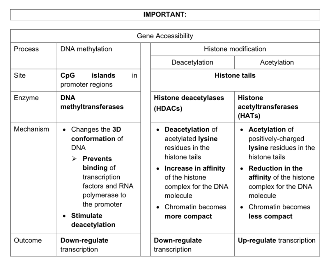
Summary table of DNA methylation and histone modification
Control of eukaryotic gene expression: Transcriptional level
control of the transcription initiation is the most importantly mechanism in determining
(i) whether or not genes are expressed
(ii) quantity of encoded mRNAs
(ii) quantity of proteins encoded (mRNA —> proteins)
for transcription of a gene to begin, general transcription factors and RNA polymerase must assemble at the promoter to form a transcription initiation complex (TIC)
general transcription factors are essential for the transcription of all genes
interaction of general transcription factors and RNA polymerase often initiate transcription at a “basal” rate i.e. only a very small number of RNA transcripts are produced
to achieve maximum transcription rate of a gene, the interaction of specific transcription factors and distal control elements e.g. enhancers or silencers with the GTFs and RNA polymerase to form a stable TIC is necessary
specific transcription factors control the transcription of specific genes in response to specific internal or external signals
Gene and intergenic DNA
Recall: a gene is a specific DNA sequence to direct the synthesis of protein or RNA
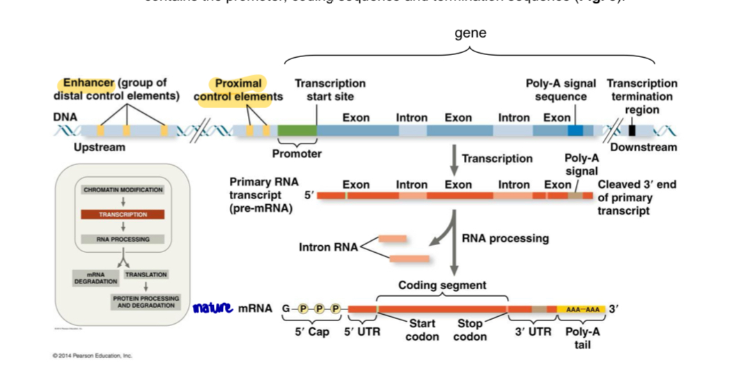
it contains the promoter, coding sequence and termination sequence
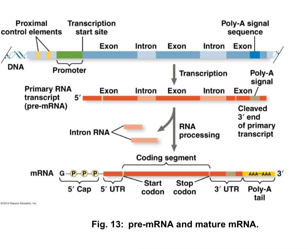
figure shows: A eukaryotic gene and its transcript. Each eukaryotic gene has a promoter, A DNA sequence where RNA polymerase binds and starts transcription, proceeding “downstream.” A number of control elements are involved in regulating the initiation of transcription; these are DNA sequences located near (proximal to) or far from *distal to) the promoter. Distal control elements can be grouped together as enhancers, one of which is shown for this gene. A polyadenylation (poly-A) signal sequence in the last exon of the gene is transcribed into an RNA sequence that signals where the transcript is cleaved and the poly-A tail added. Transcription may continue for hundreds of nucleotides beyond the poly-A signal before terminating. RNA processing of the primary transcript into a functional mRNA involves three steps: addition of the 5’cap, addition of the poly-A tail, and splicing. In the cell, the 5’ cap is added soon after transcription is initiated, and splicing occurs while transcription is still underway
Within a gene
there are coding sequences and non-coding sequences
coding sequences contain codons, which are translated to form polypeptides
non-coding sequences include regulatory sequences and introns
regulatory sequences are (i) the promoter, and (ii) the 5’ and 3’ untranslated regions (UTRs), which can affect the rate of gene expression
introns are non-coding sequences inserted between exons of eukaryotic genes
introns may contain regulatory sequence
Intergenic DNA
these are non-coding sequences that fall outside genes and are found in between genes
in eukaryotes, intergenic DNA constitutes a high proportion of the total genome sequence
important intergenic DNA sequences include origin of replication, centromeres and telomeres
Transcriptional level: Control elements
associated with most eukaryotic genes are control elements, which are non-coding DNA sequences that regulate transcription by binding transcription factors
control elements can be classified based on their locations
Promoter: this includes the TATA box that resides approximately 25-39 bp upstream of the transcription start site
in eukaryotic genes, the TATA box determines the start point of transcription
general transcription factors and RNA polymerase assemble at this region to form a transcription initiation complex
Proximal control elements: these are found between 100 to 200 bp upstream of the transcription start site
proximal control elements also serve as binding sites for general transcription factors
they are essential for efficient transcription
Distal control elements: these include enhancers and silencers that can exert their effects when located hundreds or even thousands of bp upstream or downstream of the transcription start site
they may also be located within an intron
enhancers and silencers bind specific transcription factors such as activators and repressors respectively
they can function in an orientation-independent fashion
enhancers greatly increase the transcription rate while silencers greatly decrease the transcription rate
*the control elements i.e. the promoter, proximal control elements and distal control elements, bind different transcription factors, which are proteins
*the particular combinations of control elements and transcription factors are specific to each gene and result in different transcription rates in different cell types i.e. spatial specificity or at different stages of development i.e. temporal specificity
Transcription factors
Transcription factor: regulatory protein that binds to DNA and affects transcription of genes
besides general transcription factors and RNA polymerase, specific transcription factors affect the expression of certain genes in response to specific signals
general properties of specific transcription factors include
they mediate response to a stimulus, which signals that one or more genes should be switched on or switched off
recognise and bind to enhancers or silencers
interact with components of the transcription machinery, either directly or indirectly
contain 2 binding domains in their structures
DNA binding domain - a part of the protein’s 3-dimensional structure that binds DNA and
one or more protein binding (aka activation) domains, which binds other regulatory proteins or components of the transcription machinery, facilitating the protein-protein interaction that result in gene transcription
specific transcription factors can be divided into 2 groups: activators and repressors
Activators (proteins) bind to enhancers (DNA sequence), triggering a series of interaction that results in an increased rate of transcription
Repressors (proteins) bind to silencers (DNA sequence), triggering a series of interaction that results in a decreased rate of transcription
Events leading to initiation of transcription
Activators bind their respective enhancers
General transcription factors bind to the promoter and mediate the binding of RNA polymerase, forming a transcription initiation complex
rate of transcription is basal
DNA-bending protein causes the looping of DNA
this allows activators bound to enhancers located far upstream or downstream to be brought close to the promoter
Activators interact with mediator proteins, which serve as adaptor molecules and facilitate the interaction of the activator with general transcription factors and RNA polymerase
this results in improved recruitment of general transcription factors and RNA polymerase to the promoter to form a stable TIC
activator also facilitates the proper positioning of the transcription initiation complex
activator also facilitates the proper positioning of the transcription initiation complex on the promoter to initiate transcription
rate of transcription is increased
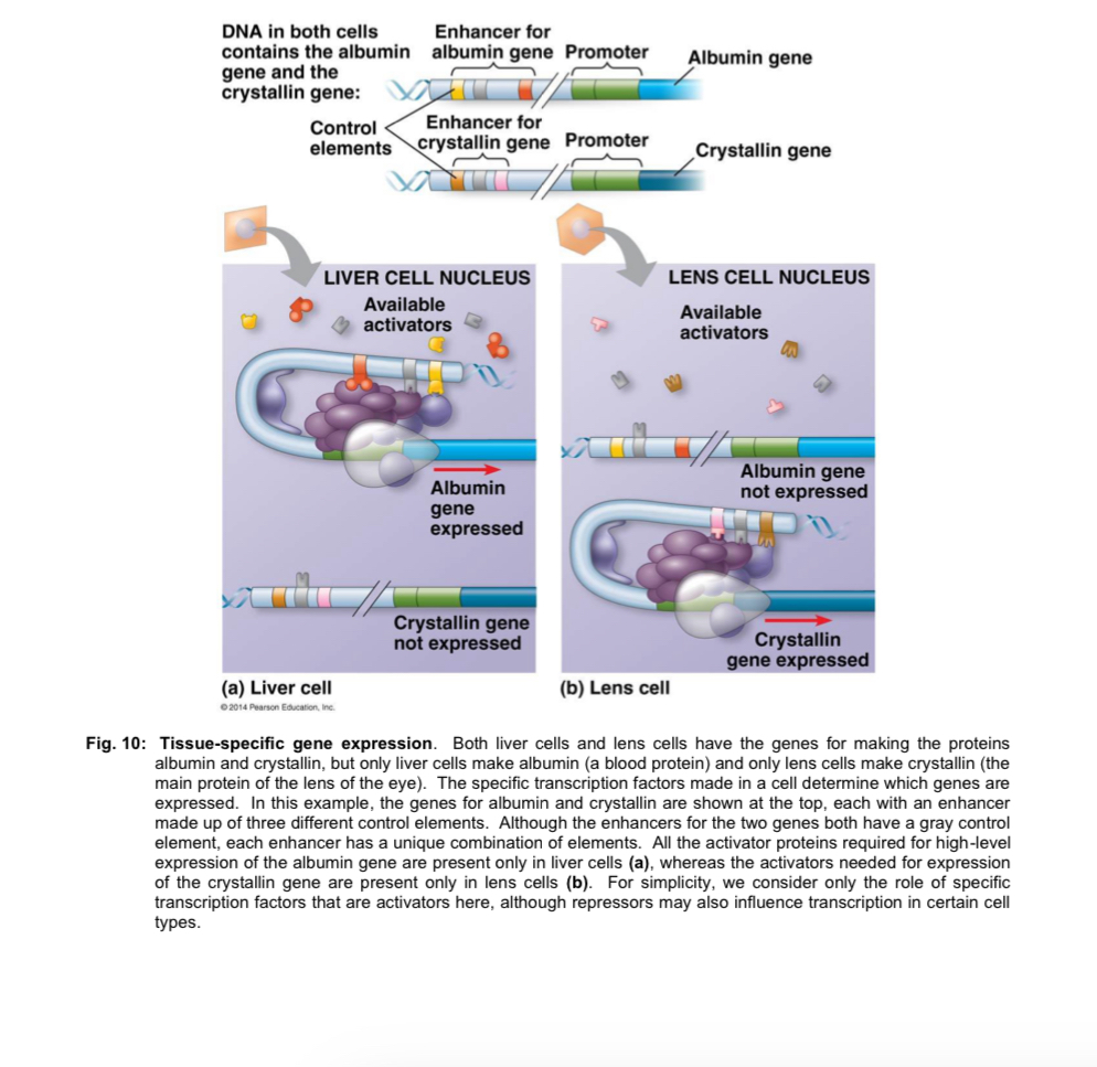
note: in eukaryotes, the precise temporal/spatial control of transcription depends largely on the binding of activators to their respective enhancers
the particular combination of enhancers associated with a a gene will be able to activate transcription only when the appropriate activators are present during precise timing of development or in a specific cell type like liver or lens cell
Eukaryotic repressor proteins
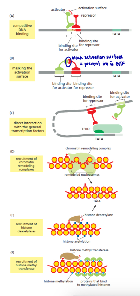
transcription repression can act through more than one mechanism at a given target gene, thereby ensuring efficient repression (shown in fig. 11)
gene repression is especially important to animals and plants whose growth depends on elaborate and complex developmental programmes
6 ways in which eukaryotic repressors proteins can operate to inhibit transcription:
Competitive DNA binding
Activator proteins and repressors proteins compete for binding to the same regulatory DNA sequence
Masking the activation surface
Both proteins bind DNA, but the repressors prevents the activator from interacting with the general transcription factors (GTFs)
The repressors blocks assembly of the GTF
The repressors recruits a chromatin remodeling complex
chromatin remodeling complex returns the nucleosome state of the promoter region to its pre-transcriptional form
The repressor attracts a histone deacetylase to the promoter
repressor reverses histone acetylation, thereby repressing transcription initiation
The repressor attracts a histone methyl transferase
histone methyltransferase modifies certain positions on histone by attaching methyl groups
the methylated histone, in turn, are bound by proteins that maintain the chromatin in a transcriptionally silent form
Control of eukaryotic gene expression: Post-transcriptional level
pre-mRNA is produced by transcription of a protein-coding gene
all pre-mRNA produced in the eukaryotic nucleus must undergo post-transcriptional modification to produce functional mature RNA molecules for export to the cytosol
Post-transcriptional modification involves
Modification of the 5’ end of the nascent RNA chain by capping with 7-methylguanosine triphosphate to form a 5’ cap
RNA splicing occurs after the release of pre-mRNA from RNA polymerase
defined as the removal of introns while the remaining exons are lighted together to form a mature mRNA
requires hydrolysis of ATP
splicing is carried out by spliceosome which is a large complex comprising of several subunits known as small nuclear ribonucleoproteins (snRNPs)
Modification of the 3’ end by polyadenylation
during this modification, a series of adenine (A) nucleotides are added to the 3’ end, by an enzyme called poly (A) polymerase
the resulting 3’ poly (A) tail is about 200 nucleotides long
Post-transcriptional level - mRNA splicing
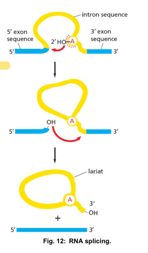
cleavage at the 5’ splice site and joining of the intron to a branch point within the intron
this reaction yields a lariat-like intermediate, in which the intron forms a loop
cleavage at the 3’ splice site and simultaneous ligation of the exons, resulting in an excision of the intron as a lariat-like structure
DNA sequences at the 5’ and 3’ ends of an intron serve as recognition sites for spliceosomes to bind
mature mRNA
consists of
5’ cap
5’ UTR (untranslated region)
additional sequence at the 5’ end preceding the start codon and does not code for protein
protein-coding region
consists of a series of codons representing the amino acid sequence of the protein, starting with the start codon (AUG) and ending with a stop codon (UAA, UAG, UGA)
3’ UTR: sequence following the stop codon and does not code for protein
3’ poly A tail
note: almost all pre-mRNA transcripts in eukaryotes undergo post-transcriptional modifications.
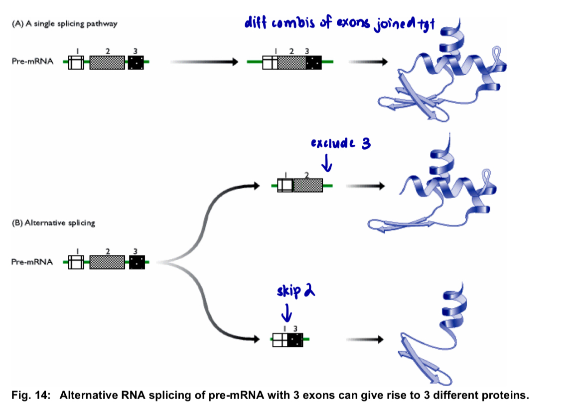
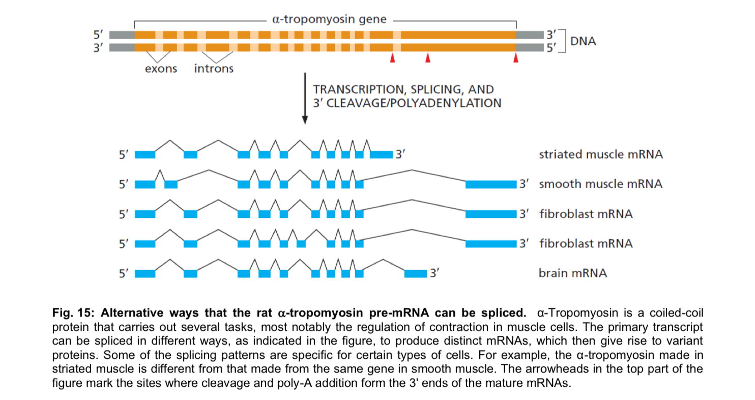
pre-mRNA transcribed from one gene can be processed in more than one way, leading to the formation of different mature mRNAs, each containing a different combination of exons = enables the synthesis of different polypeptides from a single gene
Alternative splicing
use of different splice sites i.e. a phenomenon known as alternative splicing, allows exons to be joined together in different combinations
this produces different mature mRNAs from the same pre-mRNA that in turn, generate different proteins
a substantial proportion of higher eukaryotic genes synthesises different proteins from one gene
Alternative ways that the rat alpha-tropomyosin pre-mRNA can be spliced
Control of eukaryotic gene expression: Translational level
once mRNA molecules have been exported from the nucleus to the cytoplasm, several control mechanisms are available to regulate the rate at which mRNA is translated not its polypeptide
Stability of mRNA
determines the duration for which translation can occur
less stable (i.e. the more rapidly an mRNA molecule is degraded) , the less time is available for it to be translated
mRNAs in eukaryotic cells are more stable than those present in prokaryotic cells, and thus have longer half-lives
half-life: time required for 50% of the initial amount of RNA to be degraded, varies among eukaryotic mRNAs
stability of mRNA is affected by:
length of poly(A) tail i.e. mRNAs with longer poly(A) tails tend to be more stable than mRNAs with shorter poly(A) tails
stabilising/destabilising sequences in the 3’ UTR i.e. these sequences contain binding sites for specific proteins that increase or decrease the rate of poly-A tail shortening
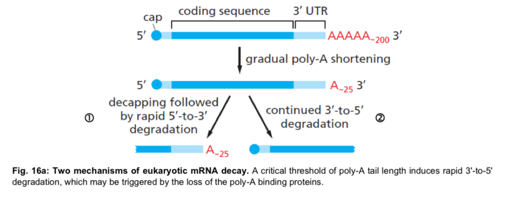
2 mechanisms exist for the eventual decay of eukaryotic mRNA
both begin with the gradual shortening of the poly-A tail by an exonuclease, a process that starts as soon as the mRNA reaches the cytosol
this poly-A shortening acts as a timer that counts down the lifetime of each mRNA
once the poly-A tail is reduced to a critical length (about 25 nucleotides in humans), the two pathways diverge
Pathway 1: the 5’ cap is removed (a process called decapping) and the “exposed” mRNA is rapidly degraded from its 5’ end
Pathway 2: the mRNA continues to be degraded from the 3’ end, through the poly-A tail, into the coding sequences
although 5’ to 3’ and 3’ to 5’ degradation are shown here on separate RNA molecules, these two processes can occur together on the same molecule
poly-A shortening controls the half-life of most eukaryotic mRNAs
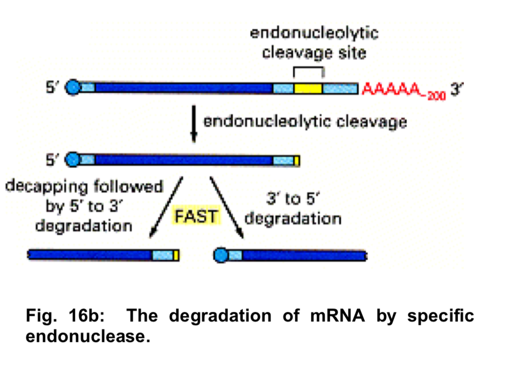
specialised mechanism to degrade mRNA
endonuclease (i.e. specific nucleases that cleave the mRNA internally) effectively decaps one end and removes the poly-A tail from the other so that both halves are rapidly degraded
the mRNAs that are destroyed in this way carry specific nucleotide sequences, often in the 3’ UTRs that serve as recognition sequences for these endonucleases
makes it especially simple to tightly regulate the stability of these mRNAs by blocking or exposing the endonuclease site in response to extracellular signals
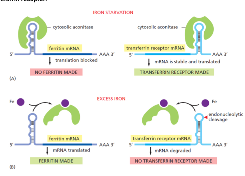
Case study: Uptake of iron by transferrin receptor
The addition of iron to cells decreases the stability of the mRNA that encodes the receptor protein that binds the iron-transporting protein transferrin, causing less of this receptor to be made. This effect is mediated by the iron-sensitive RNA-binding protein aconitase. Aconitase can bind to the 3ʹ UTR of the transferrin receptor mRNA and increase receptor production by blocking endonucleolytic cleavage of the mRNA. On the addition of iron, aconitase is released from the mRNA, exposing the cleavage site and thereby decreasing the stability of the mRNA
Fig. 17: Uptake of iron by transferrin receptor.
(A) During iron starvation: The binding of aconitase to the 5ʹ UTR of the ferritin mRNA blocks translation initiation; its binding to the 3ʹ UTR of the transferrin receptor mRNA blocks an endonuclease cleavage site and thereby stabilizes the mRNA.
(B) Excess iron: In response to an increase in iron concentration in the cytosol, a cell increases its synthesis of ferritin in order to bind the extra iron and decreases its synthesis of transferrin receptors in order to import less iron across the plasma membrane.
Both responses are mediated by the same iron-responsive regulatory protein, aconitase, which recognizes common features in a stem-loop structure in the mRNAs encoding ferritin and the transferrin receptor.
Aconitase dissociates from the mRNA when it binds iron. But because the transferrin receptor and ferritin are regulated by different types of mechanisms, their levels respond oppositely to iron concentrations even though they are regulated by the same iron-responsive regulatory protein.
Cytoplasmic poly(A) tail addition
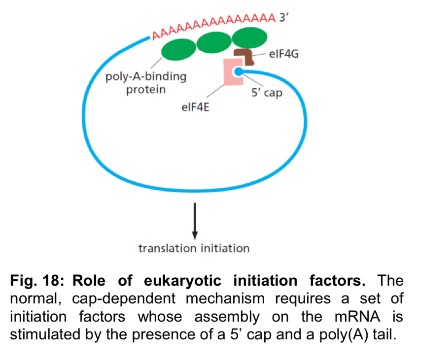
modifications can be made to specific mRNAs present in the cytoplasm to promote translation
this is achieved by lengthening their poly(A) tails
for example, in oocytes (unfertilised eggs), mRNAs have shortened poly (A) tails, and they are not translated
these mRNAs are stored in the cytoplasm until at specific times during oocyte maturation, when proteins encoded by these mRNAs are required, the poly(A) tails become extended by cytoplasmic poly(A) polymerases
known as cytoplasmic poly(A) tail addition
Initiation of translation - eukaryotic initiation factors
Eukaryotic initiation factors
initiation of translation in both eukaryotes and prokaryotes is dependent on translation initiation factors in addition to the small and large subunits of the ribosome
in eukaryotes, these factors are termed eukaryotic initiation factors (eIFs)
eIFs are involved in scanning the mRNA for the start codon AUG, locating the binding site of initiator tRNA to the AUG codon and forming the translation initiation complex at the 5’ mRNA region
by varying the abundance and activity of these factors, it is possible to affect the rate of translational initiation
however, this has a global effect on overall translational activity, rather than on the rate of translation of specific mRNAs
Initiation of translation - Translational repressors
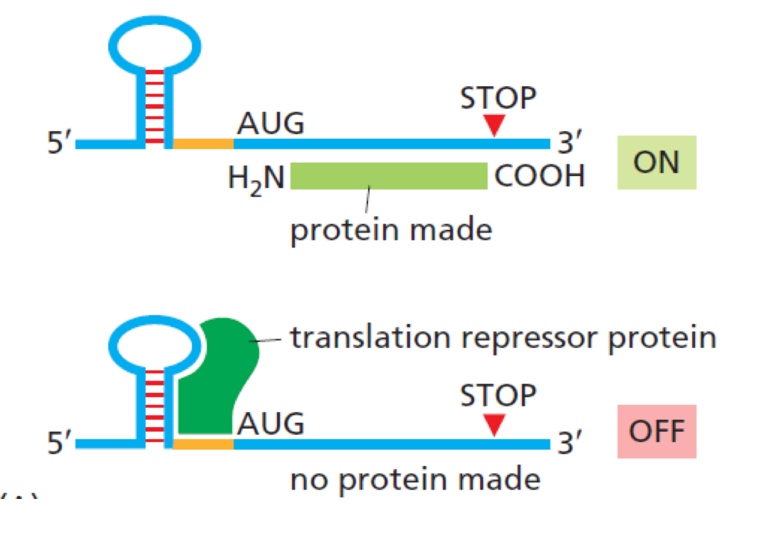
translational repressors can decrease the rate of translational initiation
bind to various regions of the mRNA molecule, usually the 5’ or 3’ UTRs, and interfere with the initiation of translation by blocking the attachment of ribosomes or other translation initiation factors
Alternative translation initiation sites
may be initiated at sites other than the first AUG codon in the sequence of an mRNA molecule, including
use of second or subsequent AUG for translation initiation
eukaryotic translation usually begins at the first AUG start codon as defined by Kozak rules (note: Kozak rules are not in syllabus) downstream of the 5’ UTR
however, the small ribosomal subunit could skip the first AUG codon and use the second or subsequent AUG to initiate translation
“leaky scanning” and results in proteins that vary in their N-terminal sequence
initiation of translation in the middle of mRNA
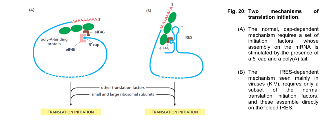
eukaryotic translation usually begins at the 5’ end of the mRNA molecule, since 5’ cap recognition is required for the assembly of teh initiation complex
internal ribosome entry site (IRES) is a specialised nucleotide sequence that allows for translation inhibition in the middle of an mRNA sequence in a cap-independent manner, since the need for a 5’ cap structure is bypassed
a protein with a different primary structure is produced with this mechanism
RNA interference
RNA molecules perform many critical tasks in the cell besides serving as intermediate carriers of genetic information
A group of short non-coding RNAs carry out RNA interference
this group can be classified into 3 types of RNA, one of which is microRNAs (miRNAs)
all 3 types of short RNAs locate their targets through RNA-RNA base pairing and they generally cause reductions in gene expression
Action of miRNA
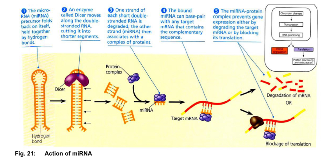
RNA transcripts (synthesised by transcribing miRNA-coding genes) fold back on themselves, forming a hairpin structure held together by hydrogen bonds
They are processed by an enzyme called Dicer, which cuts the double-stranded RNA into smaller fragments.
3. One strand of the double-stranded RNA fragment is degraded by a protein complex known as RNA-
inducing silencing complex (RISC). The remaining strand binds to RISC to form miRNA-protein complex.
4. This miRNA strand then binds to mRNA molecules that have the complementary sequence.
5. The miRNA-protein complex then inhibits translation by blocking formation of the translation
initiation complex or degradation of the mRNA
Post-translational modification
*Read with eukaryotic gene expression pg 39
Protein activation
After translation is complete, some polypeptides require alterations before they becomefunctional.
These alternations are known as post-translational modifications (Refer to: Section 7 ofEukaryotic Gene Expression Notes).
By controlling the timing of such protein modifications, the rate of functional (active) protein formation can be controlled.
Protein Breakdown
Many proteins have limited lifespan. Some of the proteins that trigger metabolic changes in cells are broken down within a few minutes or hours. The proteasomes break down proteins. This regulation allows a cell to (i) adjust the kinds and amounts of its proteins in response to
changes in its environment; and (ii) to maintain its proteins in working order.