Adv EP Lesson 3 - Tachycardia Mechanisms
 Call Kai
 Learn
Learn Practice Test
 Spaced Repetition
 Match
Match Flashcards
 Knowt Play
1/28

There's no tags or description
Looks like no tags are added yet.
Name | Mastery | Learn | Test | Matching | Spaced | Call with Kai | Chat |
|---|
No analytics yet
Send a link to your students to track their progress
29 Terms
cardiac muscle cell fast action potential
membrane potential reaches a threshold
sodium and calcium channels open and enter cell
potassium channel open and they shift outward causing repolarization
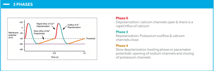
why are pacemaker cells depolarized slower
presence of calcium channels and absence of sodium
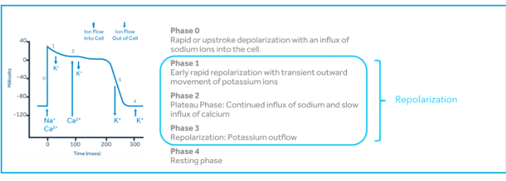
cardiac cell action potential
phase 4 resting phase - potassium channels open > spontaneous depolarization
phase 0 depolarization - influx of sodium and calcium ions
phase 1 to 3 repolarization - 1: sodium channels close and potassium open causing outward flow of K+
2: plateau; inward of Ca++ and calcium induced Ca++ release occurs from the sarcoplasmis reticulum causing cellular contraction
3: potassium channels open and repolarization is triggered > closes sodium and calcium channels
pacemaker cell action potential
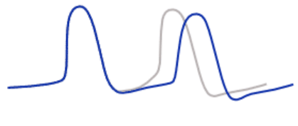
ERP
occurs in phases 0-3
depolarization cannot be initiated no matter how strong the impulse, because it follows the initial depolarization too closely
RRP
following the ERP; cardiac cell can be stimulated to depolarize, but only by a stimulus or impulse that is stronger than what is normally required for depolarization
late phase 3
mechanisms of tachycardia
abnormal automaticity
triggered activity
reentry
focal tachycardias
abnormal automaticity or triggered activity
reentrant tachycardias
reentry
abnormal automaticity
development of spontaneous phase 4 depolarization in cardiac muscle cells from accelerated phase 4 sodium leakage into cardiac cells
reentry
trigger activity
A second action potential arising during repolarization is called an early after-depolarization (EAD), while one arising after repolarization is called delayed after-depolarization (DAD)
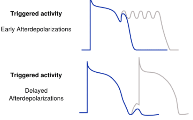
triggered activity cause
electrical instability in the myocardial cell membrane during bradycardia, hypokalemia, hypoxia, or drug effects
EADs are integral to initiation of
torsades de pointes and atrial fibrillation and long QT syndrome
reasons for abnormal automaticity
ischemia stretch, electrolyte imbalance, high sympathetic tone
EADs can initiate and maintain atrial fibrillation T/F
F because a reentrant circuit must be present to maintain the tachycardia
range of resting potentials for spontaneous activity
-70to -30 mV
DAD cause and example
digoxin toxicity
RVOT ventricular tachycardia
examples of arrhythmias caused by abnormal automaticity
inappropriate atrial tachy cardia, multifocal atrial tachycardia, premature atrial contractions
torsades de pointes
prolonged QT interval
fast heart rhythm starting in the heart's lower chambers that can lead to sudden cardiac arrest
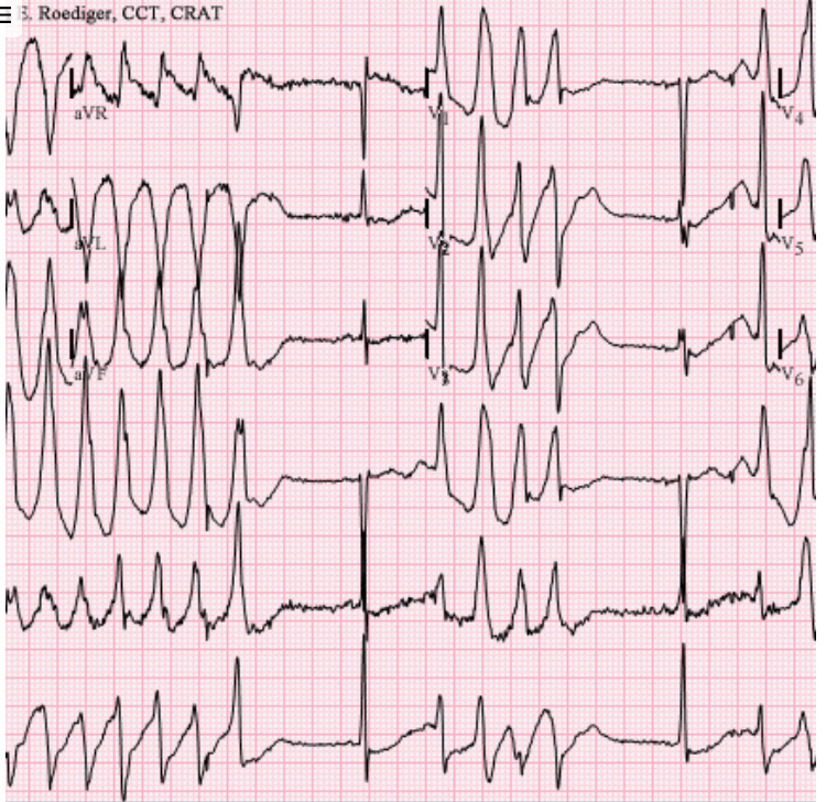
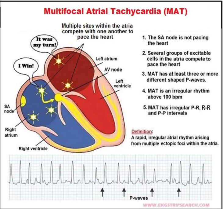
multifocal atrial tachycardia
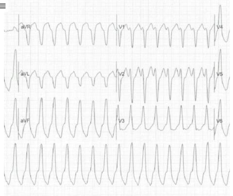
RVOT Ventricular tachycardia
where fast electrical signals begin in the RVOT near the pulmonary valve.
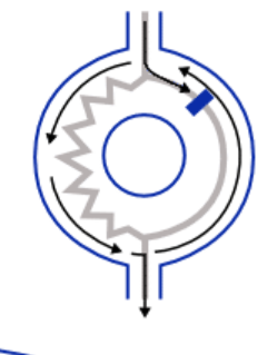
reentry
continuous propagation of a wavefront that perpetually re-excites the tissue.
requirements for reentry
two pathways that form a circuit (fast and slow)
two pathways with differing refractory pathways (slow and fast)
unidirectional block in one of the pathways because of a premature beat often initiates reentry
examples of reentrant atrial tachycardia
atrioventricular reentrant tachycardia (AVNRT), atrioventricular reciprocating tachycardia (AVRT), atrial flutter,atrial tachycardia
AVNRT
pwave hidden in QRS in V1
AH jump >50ms which indicates conduction changes from fast to slow
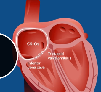
Atrial flutter
reg conduction of the fast path and the isthmus between tricuspid valve and inferior vena cava called the CTI cavo tricuspid isthmus
travels CC around tricuspid annulus
sawtooth pattern 2:1, 3:1, or 4:1
atypical flutter: conducts Clockwise or in the LA
AVRT
accessory pathway present on the left around mitral valve or on the right side around the tricuspid valve
accessory pathway conducts antegrade > delta wave > WPW (antidromic/overt)
accessory pathway conducts retrograde > RP interval is shorter than the PR interval > P wave hidden in T or within ST >