Lab 1: Anatomical Terms, Histology, and the Integumentary System
 Call Kai
 Learn
Learn Practice Test
 Spaced Repetition
 Match
Match Flashcards
 Knowt Play
1/130

Earn XP
Description and Tags
Biology 223
Name | Mastery | Learn | Test | Matching | Spaced | Call with Kai | Chat |
|---|
No analytics yet
Send a link to your students to track their progress
131 Terms
Anatomical Position
Body viewed from anterior surface with palms facing forward
Cephalic
Head Region
Cervical
Neck
Axillary
Armpit
Brachial
arm
Thoracic
chest
Abdominal
stomach
Dorsal
Upper back
Lumbar
lower back
Pelvic
hips/pelvis
Transverse/Cross Sectional Plane
Horizontal cut of body
Frontal/Coronal Cross Sectional Plane
Vertical horizontal length cut of body (back side/ front)
Sagittal Cross Sectional Plane
vertical cut between eyes all the way down the body (symmetric)
Anterior/Ventral
front
Superior
top/going up
Cranial/Cephalic
towards head
Medial
towards midline/belly button
Proximal
closer to ____
Superficial
towards surface
Posterior/Dorsal
back (hint:dorsal fin)
Inferior
bottom/going down
Caudal
towards tail
Lateral
away from midline/belly button
Distal
further from ____
Deep
further in
superficial
Your epidermis is ______ to your dermis
distal/inferior
your feet are ______ to your knee
Apical
exposed/top surface/not attached
Basal
attached/not exposed
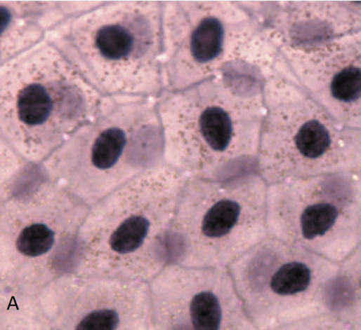
pink image with black dots
Simple Squamous Epithelium

purple dots in purple
Stratified Squamous Epithelium
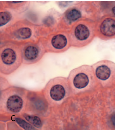
red cubes with black dots
Simple Cuboidal Epithelium

big pink circles with purple dots
Stratified Cuboidal Epithelium
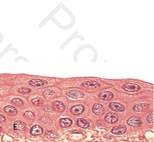
squished pink with white above
Transitional Epithelium - Stretched State
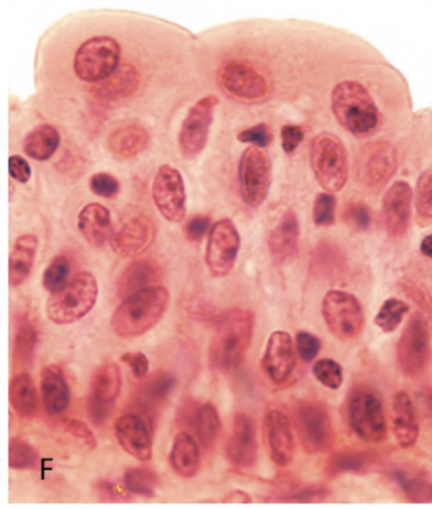
peach and red dots
Transitional Epithelium - Relaxed State
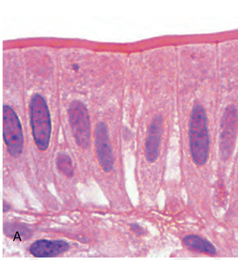
purple and pink with hot pink line on top
Simple Columnar Epithelium

dark purple and pink with wavy on top
Psuedostratified Ciliated Columnar Epithelium
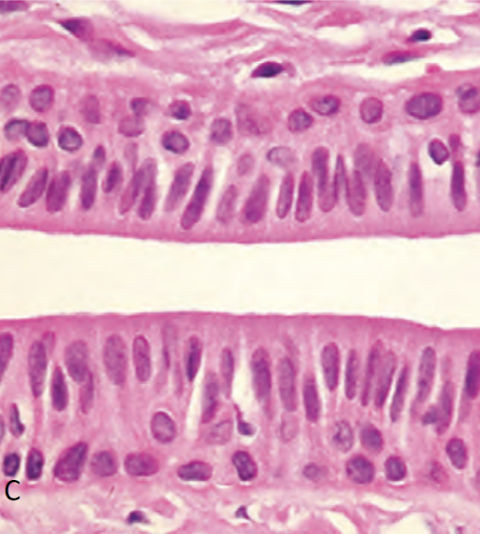
white line in the center
Stratified Columnar Epithelium
Villi
pink line on top of Simple Columnar Epithelium (structure)
Cilia
wavy pink line on top of Pseudostratified Ciliated Columnar Epithelium (structure)

Glandular Epithelium
tissue made of cells that produce and release chemicals

Endocrine Glands
release products into interstitial fluid/blood stream (pituitary/hormones)
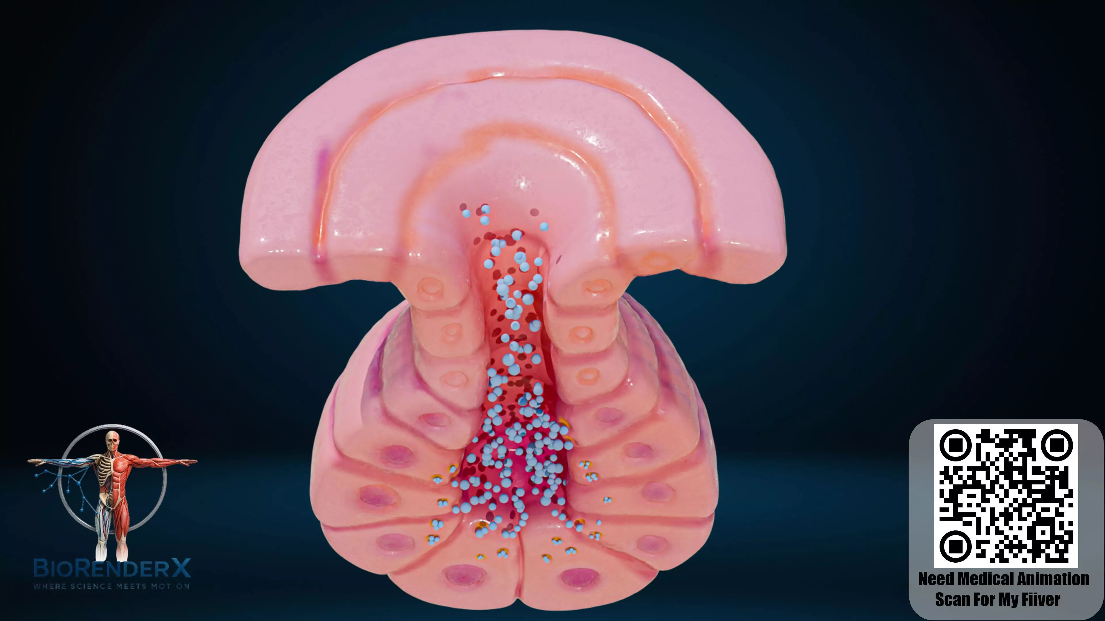
Exocrine Glands
release products through ducts on apical surfaces (sweat glands)

pink spider webs
Areolar Loose Connective Tissue
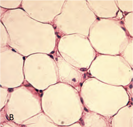
yellow fatty spheres
Adipose Loose Connective Tissue

green and yellow mush
Reticular Loose Connective Tissue

hortizontal hot pink
Dense Regular Connective Tissue
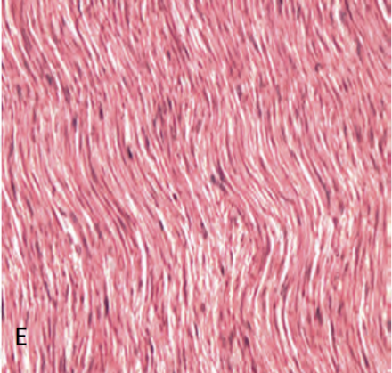
pink and White Sea of waves
Elastic Connective Tissue

gray and purple swirls
Dense Irregular Connective Tissue
Tendon
muscle to bone elastic connective tissue
Liagment
bone to bone elastic connective tissue
aponeurosis
muscle to muscle elastic connective tissue
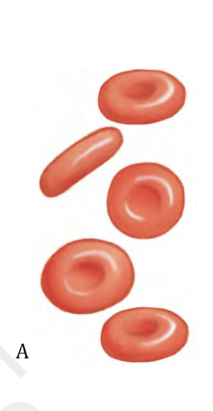
Erythrocytes
Formed Element of Blood (RBC)
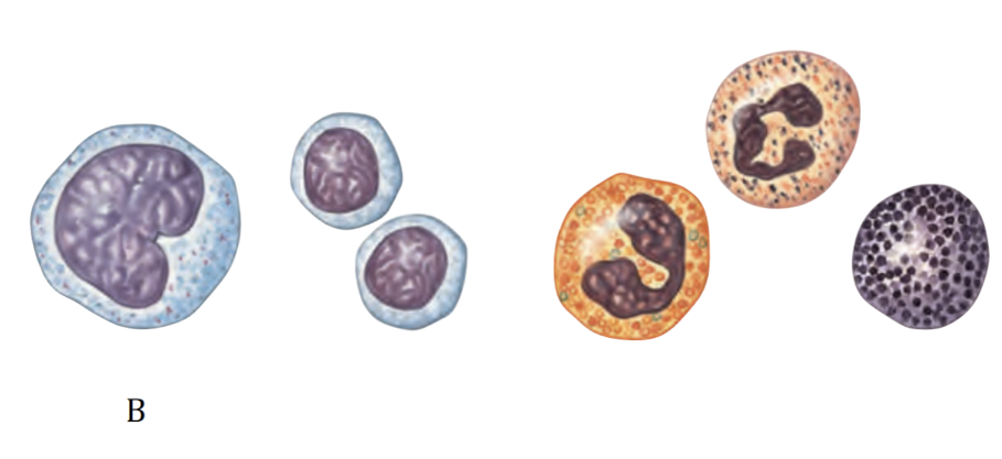
Leukocytes
Formed Element of Blood (WBC)

Platelets
Formed Element of Blood (cell fragments)
Plasma
everything in the blood besides the 3 formed elements

pink ombre with purple/white dots
Hyaline Cartilage Supporting Connective Tissue

pink and purple dots
Elastic Cartilage Supporting Connective Tissue

light pink with hot pink swirls
Fibrocartilage Supporting Connective Tissue

brown with dots and hole
Bone/Osseous Supportive Tissue

Osteocytes
Red dots on superficial surface of bone that help rebuild
Osteoblasts
produce fibers and matrix of bone
Osteoclasts
dissolves fibers and matrix of bone
Canaliculi
red lines around osteocytes that are passage ways
Periosteum
dense fibrous membrane that covers outer surface of bone
Compact Bone
hard bone on outside
Spongy Bone
inside of hard bone
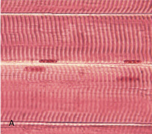
pink vertical lines with 4 dots
Skeletal Muscle Tissue - Voluntary
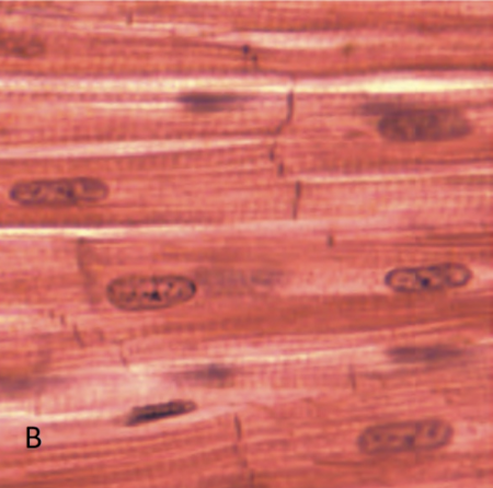
vertical red and white
Cardiac Muscle Tissue - Involuntary

basically all red with black dots
Smooth Muscle Tissue - Involuntary
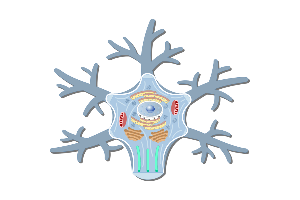
bulk of neuron
Cell Body (structure)

nucleus
Dot in Cell Body (structure)
Perikaryon
fluid inside the cell body
Dendrite
hard branches outside cell body
Axon
tail at the end of cell body (structure)
Myelin Sheath
insulation surrounding axon (layer)
Internode
everything that’s not a node on the axon (structure)
Nodes of Ranvier
squish between axons (structure)
Axon Hillock
side of cell body before axon (structure)
Telodendria/Terminal Branches
pink squish by dendrites
Synaptic Terminals/Knobs
thick bottom of telodendria
Synaptic Cleft
little gap between the cell body and Synaptic Terminal
Postsynaptic Cell
cell that receives information from pre-synaptic cell
Pre-Synaptic cell
sending end (dendrite end)
Neuroglia/Glial Cells
helper cells
Oligodendrocyte
glial cells within CNS
Schwann Cell
glial cells within parasympathetic NS
Central NS
Oligodendrocytes
Parasympathetic NS
Schwann Cells
C.O.P.S
Stratum Corneum
first two layers of epidermis (most superficial)
Stratum Lucidum
3rd layer of epidermis
Stratum Granulosum
4th layer of epidermis
Stratum Spinosum
5th layer of epidermis
Stratum Germinativum/Basale
6th layer of epidermis S
Basal Lamina
7th layer of epidermis
Dermal Papillae
8th layer of epidermis (most deep)
C.L.G.S.G.B.D
Cut
Lab
Get
Some
German
Beer
Dude
Artery
red lines (accessory structure)
Vein
blue lines (accessory structure)
Papillary Plexus
a mixed bundles of vein and arteries in one area (accessory structure)
Nerve
yellow lines (accessory structure)