Anatomy Lab Exam 1
 Call Kai
 Learn
Learn Practice Test
 Spaced Repetition
 Match
Match Flashcards
 Knowt Play
1/144

There's no tags or description
Looks like no tags are added yet.
Name | Mastery | Learn | Test | Matching | Spaced | Call with Kai | Chat |
|---|
No analytics yet
Send a link to your students to track their progress
145 Terms
Head of femur
Round, ball and socket joint, medial on femur
Fovea Capitis
Hole on head of femur
Neck of femur
Attached to head of femur, posterior and anterior
Greater trochanter of femur
Superior protrusion on top of femur
Lesser trochanter of Femur
Inferior, protrusion on top of bone
Intertrochanteric crest of femur
Seam between the two trochanters on posterior side
Intertrochanteric line of femur
Line between two trochanters on anterior side
Trochanteric fossa of femur
indention below greater trochanter of femur
Spiral line of femur
Medial line that is a spiral on the posterior side
Gluteal tuberosity of femur
Lateral location where gluteal muscles attach
Linea aspera of femur
Where spiral line and gluteal tuberosity come together on posterior side
Lateral lip of linea aspera of femur
Lateral side of linea aspera on posterior side
Medial lip of linea aspera of femur
Medial side of linea aspera on posterior side
Medial Condyle on femur
Condyle onsame side as head
Lateral Condyle of femur
Condyle on opposite side of head
Medial epicondyle of femur
Outer edge of medial condyle
Lateral epicondyle of femur
Outer edge of lateral epicondyle
Medial supracondylar line on femur
Medial split of linea aspera above medial Condyle on posterior
Lateral supracondylar line on femur
Lateral split of linea aspera above lateral Condyle on posterior
Adductor tubercle of femur
Notch above medial condyle on posterior of femur
Adductor tubercle of femur
indention between condlyes on posterior side of femur
Popliteal Surface of Femur
in between condycular lines on posterior of femur
Patellar Surface of Femur
Where the patella sits on the anterior side of the femur
Medial Articular Surface for Tibia
Smooth surface where lateral side of tibia sits on the bottom of the femur
Lateral Articular Surface for Tibia
smooth surface where the lateral side of tibia on the bottom of the femur
Base of Patella
top of patella
Apex of Patella
bottom, point of patella
Medial Facet of patella
medial side, bigger surface of patella
Lateral facet of Femur
lateral side, smaller surface of patella
Medial Condyle of Tibia
medical circular end of tibia
Lateral Condyle of Tibia
lateral circular end of Tibia
Medial Superior Articulate Surface on Tibia
surface of medial condyle of tibia
Lateral Superior Articulate Surface of Tibia
surface of lateral condyle on tibia
Medial intercondylar eminence of tibia
medial bump in between condyles on tibia
Lateral Intercondylar eminence of tibia
lateral bump in between condyles on tibia
Anterior intercondylar fossa of tibia
anterior surface between condyles on tibia
Posterior Intercondylar eminence of tibia
posterior surface between condyles
Tibial Tuberosity
bump on anterior of tibia
Articular surface for head of fibula on tibia
flat spot on posterior of tibia on proximal side
Soleal Line of Tibia
curved line on posterior side of tibia
Tibial Crest
line down from tibial crest on anterior side
Interosseous crest of tibia
line on lateral side where interosseous membrane is
Medial Malleolus of tibia
structure on bottom of bone that sticks outward of tibia
Fibular Notch of tibia
where fibula sits on dorsal sideAr
Articular surface for talus on tibia
where talus meets tibia
Obturator Foramen
hole on the bottom of the bone
Iliac Crest of ilium
top curve
Iliac Fossa of ilium
on medial side, indented surface, more anterior on ilium
Auricular surface of ilium
smaller face on medial side of iliuma
anterior superior iliac spine of ilium
on anterior side, jut out near iliac crest
anterior superior iliac spine of ilium
on anterior side, jut out below iliac crest
anterior inferior iliac spine of ilium
on posterior side, jut out near auricular surface on ilium
posterior inferior iliac spine of ilium
on posterior side, jut out near auricular surface
Greater Sciatic Notch of ilium
large indention on ilium
Anterior Gluteal Line of ilium
line from ASIS, line across
Posterior Gluteal Line of ilium
lines up with both posterior spines
Inferior Gluteal Line of ilium
above circular joint
Ischial tuberosity
rounded bone sticking downward from ischium
Ischial Spine
sticking out up by greater sciatic notchL
Lesser Sciatic Notch
notch below ischial spine
Ischial spine
bridge from ischium to pubis
body of pubis
chuck of bone sticking out medially on pubis
surface for pubic symphysis
flat surface where two pubic bones connect
superior ramus of pubis
on top of pubic bodyin
interior ramus of pubis
bellow pubic body
Lunate Articular surface
half moon strucutre
Acetabular notch
notch between sides of lunate surface
Acetabular Fossa
right inside of joint
Head of fibula
circle at top of fibula
articular facet for tibia of fibula
flat area on head of fibula
neck of fibula
area below head of fibula
interosseous crest of fibula
opposite the lateral malleolus facing tibia where membrane forms
Lateral Malleolus of fibula
smooth surface on lateral side of fibula that sicks out distally
Articular Facet for talus of fibula
flat surface on anterior side
Malleolar Surface of fibula
large indention on posterior side of fibula
Talus
where tibia and fibula join onto the foot, large bone of foot
Trochlea of Talus
top portion, very smooth of talus
Head of Talus
where the talus meets the navicular
Navicular of foot
rectangular shaped bone near talus, medial
Calcaneus of foot
heel bone
Calcaneal Tuberosity of Calcaneus on foot
bump of the bottom of the calcaneus
Sustentaculum tali of Calcaneus on foot
outward projection on lateral side of foot, below the talus
Cuboid of foot
just next to the navicular, looks like a large cube, on lateral side
Medial (#1) cuneiform of foot
farthest cuneiform from cuboid
Intermediate (#2) cuneiform of foot
in between other cuneiforms
Lateral (#3) Cuneiform of foot
closest Cuneiform to cuboid
Order of metatarsals on foot (#1-#5)
medial (big toe) to lateral (pinky toe)
Base of metatarsals of foot
first bone past cuneiforms on foot, most proximal part of bone
shaft of metatarsals of foot
long portion of metatarsals
head of metatarsal of foot
first bone past cuneiforms on foot, most distal part of bone
How many phalanges are on the big toe
2; proximal and distal
How many phalanges are on 4 other toes that aren’t the big toe
3; proximal, middle, distal
Base of phalanges of foot
bone after the metatarsals, most proximal portion
Shaft of phalanges of foot
bone after the metatarsals, longest protion
head of phalanges
bone after the metatarsals, most distal portion of bone
Vastus lateralis

Vastus medialis
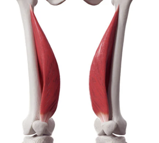
Vastus intermedius

Rectis femoris
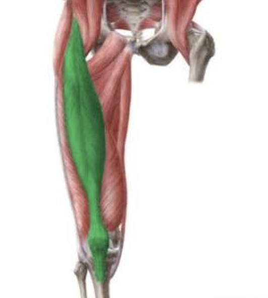
Sartorius
